Vol. 135 (2019) ACTA PHYSICA POLONICA A No. 5
Special Issue of the 8th International Advances in Applied Physics and Materials Science Congress (APMAS 2018)
Bioactivity of Hydroxyapatite Produced
from Sea Snail
Turritella Terebra
Z. Orman
a,∗, S. Yucel
a, Y.M. Sahin
b,c, O. Gunduz
d,eand F.N. Oktar
e,faYildiz Technical University, Department of Bioengineering, 34349, Istanbul, Turkey bIstanbul Arel University, Department of Biomedical Engineering 34349, Istanbul, Turkey
cArelPOTKAM (Polymer Technologies and Composite Application and Research Center), 34349, Istanbul, Turkey dMarmara University, Department of Metallurgical and Material 34349, Istanbul, Turkey
eMarmara University, Nanotechnology and Research and Implementation Centre, 34349, Istanbul, Turkey fMarmara University, Department of Bioengineering, 34349, Istanbul, Turkey
In this study, hydroxyapatite was produced via mechanochemical method using the sea snail Turritella terebra as a calcium source at 1200◦C followed by sintering. FT-IR, SEM/EDX, BET, XRD, ICP-OES analyses were ap-plied for complete characterization. Biodegradability test in tris-buffer solution and bioactivity tests were carried out. In vitro bioactivity tests showed hydroxyapatite formation when sample was immersed in simulated body fluid (SBF) and high rate of cell viability determined based on MTT assay after 24 hours and 7 days incubation. Degradation of the samples was evaluated via pH changes for 7 days. Results exhibited that produced hydroxyap-atite has ideal pore size and properties supporting bone tissue growth and cell proliferation. Therefore, it can be a good candidate for clinical applications owing to low production cost and natural–biological origin.
DOI:10.12693/APhysPolA.135.1089
PACS/topics: bioactivity, bioceramic, bone regeneration, hydroxyapatite, sea shell
1. Introduction
Hydroxyapatite [HA, Ca10(PO4)6(OH)2] is a
natu-ral component of bones and teeth; thus hydroxyapatite is employed in bone defects and voids as coatings on implant materials. Furthermore their applications in biomarkers, scaffold and controlled drug delivery are spe-cial interest due to its biocompability, bioactivity and osteoconductivity and high loading capacity. Therefore hydroxyapatite is crucial in fields of implantology, stom-atology, and regenerative medicine Last decades, biologi-cal resources that deposit biologi-calcium such as eggshells, clam shells, sea shells (such as european sea bass [1], tiger cowrie [2]), bovine bone, corals etc. have been utilized to produce calcium phosphate apatites in use of bone graft with the addition of phosphorus source. Natural biocom-posites composed of 95–96 wt% mineral mostly as arag-onite, 4 wt% organic matter and less than 1% other ox-ides [3]. Furthermore studies reported their biocompat-ibility, in vitro and in vivo osteoinductive features. Wu et al. [4] produced HA powders with small amounts of DCPA (dicalcium phosphate dihydrate) through plane-tary ball milling after heat treatment derived from oyster shell as a calcium source. Turritella terebra is a species of sea snail that can be found in Indo-West Pacific: from East Africa, including Red Sea, to Melanesia; north to Taiwan Province of China and south to central Queens-land [5] In this study Turritella terebra was utilized as
∗corresponding author; e-mail: [email protected]
Ca source to produce hydroxyapatite at 1200◦C sinter-ing temperature via high reproducibility and low cost mechanochemical method.
2. Materials and equipment
Empty local Turritella terebra sea snail shells were bought in Istanbul. They were crushed in an agate mor-tar by hand grinding and then processed in a high energy ball mill for an hour to obtain the size of a powder fol-lowing sieving with a sieve to reduce the size of less than 100 µm. According to Ca content in the raw shell, P con-centration was calculated to evaluate the exact H3PO4.
Stoichiometry ratio was set to Ca:P ratio of hydroxya-patite which is 1.67. Mixture of powder sea shell was prepared in a distilled water. The temperature was set to 80◦C for 15 min. Equivalent amount of H3PO4 was
added drop by drop to the mixture and the reaction kept to evolve for 2h and a half under continuous stirring. Chemical Eq. (1) of the reaction was shown below:
10CaCO3+ 6H3PO4→
Ca10(PO4)6(OH)2+ 8H2O + 10CO2. (1)
After completed drying, powders were heat treated us-ing an oven with a 5◦C of increase per minute and sus-pended at the sintering temperature 1200◦C for 4 hours. Sample was obtained by this procedure was coded as TT1200. Phase was identified by comparing the experi-mental XRD patterns to standards complied by the Joint Committee on Powder Diffraction Standards (JCPDS) using the card 09–0432 for hydroxyapatite. The sample were degassed at 100 ◦C for 2 h under nitrogen purg-ing prior to BET measurements. In vitro bioactivity of
1090 Z. Orman et al. the sample was tested via SBF. Then sample immersed
in falcon tubes at 37◦C containing SBF and was stored in an oven at temperature of 37◦C. SBF was renewed on 7th, 14th days and samples were stored. Inductively coupled plasma/optical emission spectrometer ICPE9000 was used to obtain the level of calcium and phosphorus ion releases during certain periods in SBF solution. Sam-ples were immersed in tris-buffer solution at pH 8 and at 37◦C to analyze the effects of sintering temperature in dissolution. pH were measured at exact time per day for 7 days and data registered. Cell viability study was conducted according to the MTT assay.
3. Results and discussion
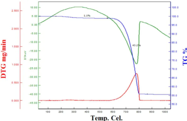
3.1. TG/DTA analysis and X-ray diffraction analysis CaCO3 amount of samples was determined by
TG/DGA indicate mass% loss of CaO corresponding to some heat induced processes data (Fig. 1). At 800◦C ac-companied by a 56.8% mass loss denoted the decomposi-tion of CaCO3and CO removal. Concentration of H3PO4
solution was calculated in the ratio of Ca:P = 1.67. Sharp peaks of the diffractogram and the straight baseline can be attributed to well-crystalized structure (Fig. 2). Peaks of HA was found at 25.8◦, 28.8◦,
31.7◦, 32.8◦, 34◦ for sample TT1200 [6].
Fig. 1. DTA analysis graphic of Turritella terebra.
Fig. 2. XRD patterns of TT1200.
3.2. BET analysis
Table I demonstrates specific surface area, pore volume and average pore and particle diameter of samples.
TABLE I Specific surface area, pore volume and average pore di-ameter of Turritella terebra samples sintered at 1200◦C.
Specific surface area [m2/g] Pore volume [cm3/g] Average pore diameter [nm] (4 V/A by BET) Average particle diameter [mm] 0.4189 0.000057 0.54 0.045
3.3. Fourier transform infrared spectroscopy (FT-IR) analysis
FT-IR spectrum of before and after heat treatment of the Turritella terebra shell powder at 1200◦C was shown in Fig. 3a. In Fig. 3b for TT1200 CO3 bands
at 1465.9, 858.32, 711.73 cm−1 disappear, PO4 bands
at 1085.92, 1016.49, 962.48 cm−1 appear and hydroxyl band at 3568.31 cm−1are present. TT1200 does not dis-play characteristic bands of the functional groups of shell powder.
Fig. 3. (a) FT-IR spectra of before and after heat treatment of the Turritella terebra shell powder (b) TT1200: before and after soaking in SBF.
Bioactivity of Hydroxyapatite Produced from Sea Snail Turritella Terebra 1091 Figure 3b displays FT-IR spectra of samples before and
after SBF. After a week of immersion in SBF, TT1200 showed CO3bands present at around 1500 cm−1. HPO2−4
peak was shown at 671.23 and 873.75. Superficially de-posited layer of Ca-deficient HA appears when the fol-lowing reaction occurs;
(H++ PO3−4 )solution→ (HPO2−4 )solid. (2)
OH band was revealed at 3641.6 cm−1 and broad hy-droxyl peak at around 3400 cm−1 that is a characteristic peak of HCA These peaks confirm that HCA (hydroxyl carbonate apatite layer) formed on the bioceramic which leads to bone-like apatite formation. Also, for TT1200 after 14 days of immersion in SBF peak intensities are relatively less comparing to 7 days of immersion in SBF. HCA layer formation needs enough saturation of Ca and P ion concentration in the solution. Ca+2 and
HPO2−4 concentrations in SBF accelerate apatite forma-tion on the sample. The ions accumulate on the surface and initiate the process of spontaneous growth of crystals followed by heterogeneous nucleation.
3.4. ICP-OES analysis of SBF after sample soaking and SEM analysis
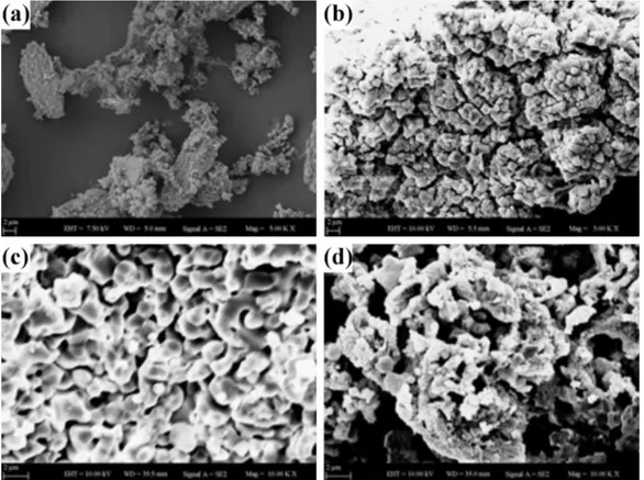
Ca level of TT1200 increased from 100.03 to 290.02 ppm and afterwards decreased to 213.62 ppm in 15 days (Fig. 4a). Phosphorus level decreased from 28.8 to 7.59 ppm (Fig. 4b). Decrease of Ca and P level in the solution can be attributed to aggregation of the ions on the sample surface. SBF results and SEM analysis
re-vealed aggregation of HA after immersing of TT1200 in SBF. After 1 and 2 weeks, surface of TT1200 was covered with HA. Figure 5a is SEM image of sea shell Turritella terebra, Fig. 5b is SEM image of sea shell after heat treat-ment. Figure 5c shows sample after being immersed in SBF for a week and Fig. 5d shows immersion in SBF for two weeks.
Fig. 4. (a) Ca ion content of TT1200 in SBF (b) P ion content of samples in SBF.
Fig. 5. SEM images of TT1200 produced from sea shell Turritella terebra before and after soaking in SBF (a) sea shell (b) after sintering at 1200◦C (c) after 1st week in SBF (d) after 2nd week in SBF (5000×).
1092 Z. Orman et al. 3.5. pH changes in tris-buffer solution
pH of the tris-buffer solution changes due to dissolution reactions and H+ and OH ions release in the tris-buffer
solution. pH stabilized in 7 days for TT1200 which can indicate that hydroxyapatite is not soluble in tris-buffer solution. Figure 6 indicates pH changes of TT1200 in tris-buffer solution.
Fig. 6. pH changes of TT1200 in a tris buffer solution in 7 days.
3.6. Cell viability test
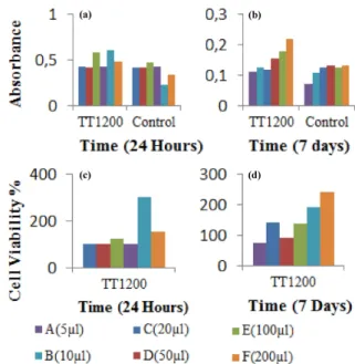
Samples were seeded to SAOS-2 osteoblast like cells at different concentrations. Figure 7 indicates cell viabil-ity of TT1200 samples. ISO10993-5 standard represents that if the viability of treated human cell line culture is more than 70% samples do not have toxic effect on the cells. Samples have the ability to cell adhesion, spread-ing, increased viability over time. Highest cell viability was obtained in the concentration of 100mm in 24 hours. Figure 7a shows absorbance levels of samples in 24 hours, Fig. 7b shows absorbance levels of samples in 7 days. Figure 7c shows cell viability of samples in 24 hours and Fig. 7d shows cell viability of samples in 7 days.
4. Conclusion
In this study sea snail Turritella terebra was used as a source of calcium to produce calcium phosphate ceramics via mechanochemical method. Bioactivity and biodegra-dation tests are promising for further investigations and clinical applications. SEM images and SBF studies con-firmed that samples were covered with HCA after soaking in SBF. Cell viability tests revealed that samples do not have toxic effect on SAOS-2 osteoblast cells.
Fig. 7. Absorbance levels of the samples in (a) 24 hours, (b) 7 days; cell viability (c) 24 hours and (d) 7 days.
Acknowledgments
This study was supported by the Scientific Research Project of Yildiz Technical University (Project No. 3119).
References
[1] Y.M. Sahin, O. Gunduz, A. Ficai, N. Ekren, A. Tuna, A.T. Inan, F.N. Oktar, Key Eng. Mater. 696, 60 (2016).
[2] Y.M. Şahin, O. Gündüz, B. Bulut, L. Özyeğin, H. Gökçe, D. Ağaoğulları, J. Chou, E. Kayalı, B. Ben-Nissan, F.N. Oktar, Acta Phys. Pol. A 127, 1055 (2015).
[3] A.F. Lemos, J.H.G. Lemos, S.S.F. Quaresma, S. Kan-nan, F.N. Oktar, S. Agathopoulos, J.M.F. Ferreira, J. Eur. Ceram. Soc. 26, 3639 (2006).
[4] S.C. Wu, H.C. Hsu, S.K. Hsu, C.P. Tseng, W.F. Ho, Adv. Powder Technol. 28, 1154 (2017).
[5] Turritella terebra (Linnaeus, 1758) screw turret, Sea-lifebase.
[6] B.L. Jolliff, J.M. Hughes, J.J. Freeman, R.A. Zeigler, Am. Mineral. 91, 1583 (2006).