RNASEL G1385A VARIANT AND
BREAST CANCER SUSCEPTIBILITY
A THESIS SUBMITTED TO
THE DEPARTMENT OF MOLECULAR BIOLOGY AND GENETICS AND
THE INSTITUTE OF ENGINEERING AND SCIENCE OF BILKENT UNIVERSITY
IN PARTIAL FULFILLMENT OF THE REQUIREMENTS FOR THE DEGREE OF MASTER OF SCIENCE
BY AKIN SEVİNÇ AUGUST, 2003
I certify that I have read the thesis, and that in my opinion it is fully adequate, in scope and in quality, as a thesis for the Master of Science.
____________________________________ Assoc. Prof. Dr. Uğur ÖZBEK
I certify that I have read the thesis, and that in my opinion it is fully adequate, in scope and in quality, as a thesis for the Master of Science.
____________________________________ Asst. Prof. Dr. Işık G. YULUĞ
I certify that I have read the thesis, and that in my opinion it is fully adequate, in scope and in quality, as a thesis for the Master of Science.
____________________________________ Assoc. Prof. Dr. Tayfun ÖZÇELİK
Approved for the Institute of Engineering and Science
____________________________________ Prof. Dr. Mehmet BARAY
ABSTRACT
RNASEL G1385A VARIANT AND
BREAST CANCER SUSCEPTIBILITY Akın SEVİNÇ
M.Sc. in Molecular Biology and Genetics Supervisor: Assoc. Prof. Dr. Tayfun ÖZÇELİK
August 2003, 128 pages
RNASEL (MIM# 180435) encodes for the ubiquitously expressed ribonuclease L
(RNase L), which mediates the antiviral and pro-apoptotic activities of the 2-5A system. Recently, RNASEL Arg462Gln (G1385A) variant is shown to be implicated in up to 13% of prostate cancer cases. Furthermore, RNASEL mutations segregating with disease within hereditary prostate cancer (HPC) families, and loss of heterozygosity (LOH) in tumor tissues have been reported. RNase L has been proposed to suppress the development of cancer through its ability to degrade RNA and initiate a cellular stress that leads to apoptosis. Analysis for allelic losses at the long arm of the chromosome 1 suggested that inactivation of a gene(s) on 1q23-32, which encompasses the RNASEL locus, might contribute to the genesis of breast cancer. Based on chromosomal location and function of RNASEL, and pleitropic effects of cancer associated mutations, we south to investigate the hypothesis that Arg462Gln variant of RNASEL is associated with breast cancer risk. The homozygote mutant (odds ratio (OR) = 0.75, 95% CI= 0.49-1.14), heterozygote (OR=1.02, 95% CI= 0.76-1.37), or the genotype having at least one mutant allele (OR= 0.94, 95% CI=0.72-1.24) was found to be not associated with the breast cancer risk. The adjustment of the data with age, menopausal, smoking status, body-mass-index, age at menarche, age of 1st pregnancy, number of children, and family history of breast cancer did not change the results (homozygote mutant (OR= 0.72, 95% CI= 0.46-1.12), heterozygote (OR= 0.95, 95% CI= 0.70-1.29), or genotype having at least one mutant allele (OR= 0.89, 95% CI= 0.66-1.18)). In conclusion, our study
ÖZET
RNASEL G1385A MUTASYONUNUN
MEME KANSERİ İLE İLİŞKİSİ Akın SEVİNÇ
Moleküler Biyoloji ve Genetik Yüksek Lisansı Tez Yöneticisi: Doç.Dr. Tayfun ÖZÇELİK
Ağustos 2003, 128 sayfa
RNASEL (MIM# 180435), tüm dokularımizda sentezlenen ve 2-5A sisteminin
antivirütik ve pro-apoptotik aktivitelerini gerçekleştiren, “ribonükleaz L” (RNase L) enzimini kodlar. RNASEL Arg462Gln (G1385A) varyantının, prostat kanserli hastaların %13’ ünde etkili olduğu saptanmıştır. Ayrıca, RNASEL mutasyonlarının prostat kanserli ailelerde hastalık ile kalıtıldığı ve tümör dokularında heterozigotluğun kaybı (Loss of heterozygosity, LOH) rapor edilmiştir. RNase L’in, hücresel stress yaratarak programlanmış hücre ölümünü başlattığı, ve kanser oluşumunu engellediği önerilmiştir. 1. kromozomun kısa kolundaki allel kayıplarının incelenmesi ile, RNASEL lokusunu da içeren 1q23-32 bölgesindeki gen veya genlerin işlevlerini yitirebilecekleri ve meme kanserinin oluşumunda etkili olabilecekleri gösterilmiştir. RNASEL’in kromozomal lokalizasyonu ve fonksiyonu, ve kanserle ilişkili genlerin “pleiotropik” etkilerini göz önüne alarak, RNASEL Arg462Gln varyantının meme kanseri ile ilişkisinin olduğu hipotezini kurduk. Çalışmamız, homozigot mütant (odds ratio (OR) = 0.75, 95% CI= 0.49-1.14), heterozigot (OR=1.02, 95% CI= 0.76-1.37), veya en az bir mutant allele sahip genotiplerin (OR= 0.94, 95% CI=0.72-1.24) meme kanseri oluşumuna bir etkisinin olmadığını göstermiştir. Verilerin yaş, menaposal, sigara içme durumları, vücud-kütle endeksleri, menarche ve ilk hamilelik yaşları, çocuk sayıları, ve ailelerindeki kanser hikayelerine göre ayarlanmaları da sonucu değiştirmedi (homozigot mütantlarda (OR= 0.72, 95% CI= 0.46-1.12), heterozigotlarda (OR= 0.95, 95% CI= 0.70-1.29), veya en az bir mütant allele sahip genotiplerde (OR= 0.89, 95% CI= 0.66-1.18)). Sonuç olarak,
ACKNOWLEDGEMENT
I gratefully acknowledge my advisor, Assoc.Prof.Dr. Tayfun ÖZÇELİK, for teaching me how to be a scientist.
I also would like to thank Assist.Prof.Dr. Işık G. YULUĞ, Prof.Dr. Mehmet ÖZTÜRK and all researchers of our department for providing us a scientific environment to work.
I also gratefully acknowledge my undergraduate advisor, Prof.Dr. Feride SEVERCAN for her invaluable and unconditioned support and help. She guided me throughout during my undergraduate education and on the following years, as well. I would also like to thank to Ayşegül GÖZEN for her help.
I extend my thanks to our collaborators (Betül BOZKURT and Ömer CENGİZ from Ankara Numune Research and Teaching Hospital; Ali Esat KARAKAYA, Semra SARDAŞ and Neslihan Aygün KOCABAŞ from Gazi University, Faculty of Pharmacy; Güven LÜLECİ, Taner ÇOLAK and Esra MANGUOĞLU from Akdeniz University, Faculty of Medicine; Drakoulis YANNOUKAKOS and Irene KONSTANTOPOULOU from National Center for Scientific Research Demokritos Moelcular Diagnostic Laboratory; Voutsinas GERASSIMOS and George NASIOULAS from National Center for Scientific Research Demokritos Institute of Biology; Eirene PAPADOPOULOU from Molecular Biology Research Center “HYGEIA”, Molecular Biology and Cytogenetics Center; Lina FLORENTIN from Alfalab; Elena KONTOGIANNI from IVF&GENETICS) for kindly providing us the samples used in our study, and Atilla Halil Elhan for his help in statistical analysis.
I would also like to thank all Bilkent-MBG family for their help and friendship, including our group members Cemaliye AKYERLİ, Elif UZ, and Gülsün Sevgi BAĞIŞLAR for their help, and Banu SÜRÜCÜ, Suha KOÇOĞLU for their friendship.
I would like to also thank my close friends Ziya Haktan KARADENİZ, Burcu DÜZEN, Hande ERSOY, Sevgi ADAK, Bengü SEVİL, Ayşe AYYILDIZ, İlter SEVER and Gülsen ÇOLAKOĞLU for always being with me for all the time I know them.
Lastly, but most importantly, I extend my most sincere gratitude to my family for their unconditional support.
TABLE OF CONTENTS
SIGNATURE PAGE ………...…....i
ABSTRACT ……….………..….ii
ÖZET ...………..………..…iii
ACKNOWLEDGEMENT………...…....iv
TABLE OF CONTENTS ………....vi
LIST OF TABLES ………....………..…ix
LIST OF FIGURES ……….………...…...xi
ABBREVATIONS ………..……....xiii 1. Introduction………...………..……1 1.1. Introduction to cancer………...…...………..……1 1.1.1. History of cancer………...………..…..1 1.1.2. Epidemiology of cancer……….……...………...……….…2 1.1.3. Conceptualizing cancer………...………..4
1.1.4. Cancer and related genes………...…………...………6
1.1.4.1. Tumor suppressor genes…….………….……….……….8
1.1.4.2. Oncogenes……….…………..……….………..9
1.1.4.3. Genomic variations at a glance……….…….………...10
1.1.5. Molecular profiling of cancer……….…….…………12
1.1.6. Inherited predisposition……….….………….13
1.1.6.1. Strong predisposition………..….………….15
1.1.6.2. Weak predisposition……..……….………..18
1.2. Breast Cancer………..……….…….…20
1.2.1. Setting the stage………..……….………20
1.2.2. Genetics of breast cancer…………..……….……..26
1.3. RNASEL………...………32
1.3.1. Prostate cancer……….32
1.3.2. RNase L………..……….35
1.3.3. RNASEL………..……….40
1.4. Our Aim………..………..43
2. Materials and Methods………..………44
2.1. Materials………..……….44
2.1.1. DNA samples and collaborators…………..………44
2.1.2. Study population………..45
2.1.2.1. Patients……….………46
2.1.2.2. Control group………...……….46
2.1.3. Primers………..………...50
2.1.4. Chemicals and reagents………..…….51
2.1.5. PCR materials………..………51
2.1.6. Standard solutions and buffers…………..………..52
2.2. Methods………..………..54
2.2.1. Amplification Refractory Mutation System (ARMS)…..………...55
2.2.1.1. Polymerase Chain Reaction (PCR) for ARMS…………..………..56
2.2.2. Agarose gel electrophoresis………..……….…….56
2.2.3. Genotyping of individuals………..……….57
2.2.4. Statistical analysis………..………59
2.2.4.1. Chi-square (χ2) test………...……….…...59
2.2.4.2. P-value calculation………..60
2.2.4.3. Odds ratio calculation………..………...61
2.2.4.4. Mutlivariate adjusted odds ratio calculation…….………...63
3. Results………...………65
3.1. Cohort information………...………65
3.2. Genotyping of RNASEL and genotype distributions…………...……….66
3.3. Statistical analysis………75
3.3.1. P-value calculation………..75
3.3.2. Results of odds-ratio calculation (Crude)……….………...76
3.3.3. Results of odds-ratio calculation (Adjusted)………….………...77
3.3.4. Results of power calculation for our study………..78
3.3.5. Further possible stratification of the Turkish group data………78
3.3.5.1. Stratification according to body-mass-index……….………...78
4. Discussion……….………80
5. Conclusion and Future Perspectives……….85
6. References……….………87
6.1. Articles……….87
LIST OF TABLES
Table 1.1.
Inherited predisposition to cancer……….14 Table 1.2.
List of familial cancer genes and syndromes……….17 Table 1.3.
TNM staging………...25 Table 1.4.
Hereditary cancer syndromes that feature breast cancer………28 Table 1.5.
Summary of RNASEL sequence variants implicated in patients with HPC.…..42 Table 2.1.
Selected characteristics of our study population………48 Table 2.2
List of primers for the amplification reactions………..50 Table 2.3.
Table 2.4.
Sample 2x2 Table for Odds Ratio Analysis………..63 Table 3.1.
Characteristics of participants in our study………67 Table 3.2.
Distribution of RNASEL G1385A genotypes and
breast cancer risk in the age matched controls and breast cancer patients……70 Table 3.3.
The allele frequencies and
sample odds ratios in subgroups according to menopausal status………..71 Table 3.4.
P-values………...75
Table 3.5.
LIST OF FIGURES
Figure 1.1.
Hallmarks of cancer………5 Figure 1.2.
Genetic alterations in progression of cancer………...7 Figure 1.3.
Caretakers and gatekeepers……….9 Figure 1.4.
DNA damage, repair and consequences……….11 Figure 1.5.
Breast cancer susceptibility genes………..19 Figure 1.6.
Mammary gland and its development………21 Figure 1.7.
Main anatomic structures of breast………22 Figure 1.8.
Summary of factors influencing breast carcinogenesis………..23 Figure1.9.
A hypothetical multi stage model of breast carcinogenesis………..24 Figure 1.10.
Possible role of RNase L in prostate carcinogenesis……….34 Figure 1.11.
Figure 1.12.
RNase L protein structure……….36 Figure 1.13.
Functional model for the activation of RNase L by 2-5A………37 Figure 1.14.
The pro-apoptotic role of RNase L………...39 Figure 1.15.
RNASEL………40
Figure 2.1.
Cohort facts………...45 Figure 2.2.
“Hasta Anket Formu”………...49 Figure 2.3.
pUC Mix Marker, 8………...52 Figure 2.4.
Mass Ruler………53 Figure 2.5.
Schematic representation of RNASEL genotyping………58 Figure 3.1.
ABBREVATIONS 2-5A 2’,5’-linked oligoadenylates
A Adenine nucleotide
APC Adenopolyposis Coli
Arg Arginine
ARMS Amplification Refractory Mutation System ASPCR Allele Specific Polymerase Chain Reaction
AT Ataxia telangiectasia
ATM Ataxia telangiectasia mutated
BMI Body Mass Index
Bp Base pairs
BRCA1 Breast Cancer Susceptibility Gene 1 BRCA2 Breast Cancer Susceptibility Gene 2 Cdk Cyclin dependent kinase
CHEK2 Cell cycle checkpoint kinase 2
CI Confidence interval
DNA Deoxyribonucleic Acid
dNTP Deoxynucleotide triphosphate E Expected (in statistical calculations) EDTA Ethylene diamine tetra acetic acid
F Forward primer
G Guanine nucleotide
G6PD Glucose-6-phosphate dehydrogenase
Gln Glutamine
GTP Guanosine Triphosphate
HPC Hereditary Prostate Cancer IBC Inflammatory Breast Cancer
IFN Interferons
LOH Loss of Heterozygosity M Molar mg Milligram min Minutes ml Milliliter MLH1 Mut H Homolog 1 MSH2 Mut S Homolog 2 mM Millimolar µl Microliter µg Microgram
O Observed (in statistical calculations)
OAS 2-5A synthetases
OR Odds Ratio
PASA Polymerase chain reaction amplification of specific alleles
PC Prostate Cancer
PCR Polymerase Chain Reaction PIN Prostate intraepithelial neoplasia
pmol Picomol
R Reverse primer
RB Retinoblastoma
RNA Ribonucleic Acids
rpm Revolutions per minute
s second(s) T Tymine
TBE Tris Borate EDTA
TP53 Tumor Protein p53
UV Ultraviolet light
w/v Weight per volume
WHO World Health Organization
1. Introduction
1.1. Introduction to cancer
“Cancer” is accepted as a group of diseases characterized by uncontrolled cellular growth and the spread of abnormal cells, which is believed to be dictated by a series of genetic alterations.
It is now well recognized that, cancer is one of the most common and severe problems of human population. According to World Health Organization (WHO), more than 1.2 million people worldwide are diagnosed with breast cancer annually. An estimated 211,300 new cases of invasive breast cancer are expected to occur among women in the United States during 2003, being the most commonly diagnosed non-skin cancer in women. In addition, 1,300 cases of male breast cancer are also predicted. An estimated 40,200 deaths (39,800 women and 400 men) are anticipated from breast cancer this year in U.S. only (American Cancer Society, “http://www.cancer.org”). In order to completely understand the concept of cancer, we must also know the history of today’s problem.
1.1.1. History of cancer
Although the ancient origin of the word “cancer” is uncertain, it is believed to be derived from Latin for “crab”, presumably because the cancer “adheres to any part that it seizes upon in an obstinate manner like the crab”.
Incidents of breast cancer have been documented back to the early Egyptians, when the popular treatment was “cautery” of the diseased tissue. Surgery was practiced, but it was an extremely radical treatment since anesthesia or antisepsis was not available. The reason for the disease was suggested to be melancholia (by the Greek physician Caudius Galen, 130-200 AD), where the suggested treatments were special
Vesalius, a Flemish anatomist of Renaissance). Due to lack of detailed records, the level of success associated with these archaic treatments is not known.
After the mid 1800’s, surgeons first began to keep detailed records, which provides us the information that the patients treated with mastectomy had a high rate of recurrence within eight years – especially when the glands or lymph nodes were affected. Nevertheless, the common therapy was removal of the breast and the surrounding glands in an effort to stave off any further tumor development, which shows us the belief, that breast cancer is a systemic disease and could spread and affect other parts of the body. The cure was based only on a “three-year survival rate”. Although it is hardly for today, such a survival rate was acceptable at those times.
The treatment improvements were noticeable between the 1930’s and 1950’s, because of a better classification of the stage and progression of the tumors. Therefore, the survival rates increased dramatically during the 1900’s (ten year survival rate, 10% in the 1920’s to roughly 50% in the 1950’s).
It was not before 1975, that the role of the accumulation of the genetic variations was shown in the development of cancer. Following that discovery, scientists identified approximately 70 genes that can spur cancerous growths and at least a dozen genes that should deter such growth but do not (Breast Cancer Society of Canada, “http://www.bcsc.ca”).
1.1.2. Epidemiology of cancer
Knudson’s “two-hit” hypothesis and its molecular confirmation in retinoblastoma focused attention in certain rare cancers (Knudson, 1971), and the contribution of “genetic susceptibility” (Macleod, 2000).
Before 1980s, the origins of common cancers were dominantly viewed as “environmental”. This was because of the studies performed in 1960s and 1970s. The varying frequencies of cancer types observed in different populations and the
“environmental view” (Peto, 2001). The transition of the cancer pattern of an immigrant population from their original to the pattern of their new country was another supporting evidence (Balmain et al., 2003). The transition of cancer pattern was also verified among the Turks residing in Germany (Zeeb et al., 2002). The results of these studies led scientists to conclude that most cancers are in principle preventable and many could be avoided by a suitable choice of life style and environment.
By the early 1980s, many important clues about the causes of cancer were identified and this increased the emphasis on the role of genetic predisposition in the common cancers (Peto, 2001, and Balmain et al., 2003).
After a quarter century of rapid advances, cancer research has generated a rich and complex body of knowledge, underlining the involvement of dynamic changes in the genome (Hanahan et al., 2000). Besides the genetic susceptibility, many other factors have been identified. The most important ones being;
1. Oncogenic viruses: Identification of the carcinogenic effects of infectious pathogens was one of the most important discoveries of the past two decades (Peto, 2001).
2. Smoking: The identification of the effect of tobacco in cancer development was one of the most important hallmarks in history of cancer epidemiology (Peto, 2001). Now it is well understood that incidences of many cancer types are increased by tobacco use, i.e. lung cancer, esophageal cancer, stomach cancer, liver cancer. Tobacco use cause 13% (and will probably cause 33%) of deaths in men (Liu et al., 1998).
3. Reproductive and hormonal factors: The impact of reproductive and hormonal factors was first verified on breast and ovarian cancer (Peto, 2001, and Baselga et al., 2003).
4. Obesity: Up to a third of cancers of breast, colon, kidney, and digestive tract were shown to be due to obesity (Josefson et al., 2001). Although the impact of obesity is subject to change among populations, it is clearly stated for the post-menopausal breast cancer and cancer of the endometrium, gall-bladder
1.1.3. Conceptualizing cancer
The word “cancer” does not refer to a single disease. Actually it is used to name a great variety of diseases characterized by masses of growth in an uncontrolled manner. The growth of the mass of the cells is autonomous, uncontrollable, increasingly malignant, and if untreated, invariably fatal. A tumor is formed by a parenchyma of proliferating cells, with a stroma of connective tissue and blood vessels (Thompson, 1991, p365). There are three main forms of tumors,
1. Sarcomas, in which the tumor has arisen in mesenchymal tissue, 2. Carcinomas, which originate in epithelial tissue,
3. Hematopoetic and lymphoid malignancies, such as leukemias and lymphomas. Within the major groups, tumors are classified by site, tissue type and degree of malignancy (Thompson, 1991, p365).
The presence of “uncontrolled growth” is gained through the accumulated variations in the genetic materials of the cells. It was suggested that, the vast catalog of cancer cell genotypes is a manifestation of six essential alterations in cell physiology that collectively dictate malignant growth: self-sufficiency in growth signals, insensitivity to growth-inhibitory (antigrowth) signals, evasion of programmed cell death (apoptosis), limitless replicative potential, sustained angiogenesis, and tissue invasion and metastasis (Hanahan et al., 2000). These alterations are summarized in Figure 1.1, where the crab is the cancer, and the six legs are the acquired capabilities of cancer.
Figure 1.1. Hallmarks of cancer.
Genomic integrity may be disrupted in many ways. It may be sporadic, because of environmental factors (i.e. ionizing radiation), lifestyle (i.e. smoking, diet), or hereditary (i.e. germ line tumor-suppressor gene mutations).
Up to now, the only environmental exposure proven to induce breast cancer is ionizing radiation (Grover et al., 2002). The reactive oxygen species (ROS) produced upon radiation exposure causes the genomic damage.
Documentation of family history in different types of cancers has shown that some individuals are more susceptible to cancer because of their genomic heritage. More than a century ago, Paul Broca described four generations of breast cancer in his wife’s family which underlined, probably for the first time, contribution of the hereditary factors in tumorigenesis (Lynch et al., 1994).
Population-based epidemiological studies showed the familial pattern of some cancers. This supported the implementation of genetic models rather than the environmental ones. Furthermore, it was also shown that, genetic alterations might account for a substantial fraction of cancer incidence without necessarily causing evident familial clustering. The demonstration of genetic linkage in breast cancer (Hall et al., 1990) by the use of DNA sequence polymorphisms dispelled the contribution of genetic susceptibility (Balmain et al., 2003).
1.1.4. Cancer and related genes
All cancers are found to be the result of abnormalities in DNA sequence. During its life, genetic material is subject to changes, and these changes are repaired by the sophisticated genome maintenance mechanisms. If these changes can not be repaired, they may result in the stable alteration of a critical gene, possibly providing a growth advantage to the cell in which it has occurred and result in the emergence of an expanded clone, derived from this cell (Figure 1.2) (Futreal et al., 2001).
With few exceptions, cancers are derived from single somatic cells and their progeny (Ponder, 2001). This clonal nature of cancer is supported by many evidences. The original evidence came from the study of tumors in women heterozygous for the linked enzyme glucose-6-phophate dehydrogenase (G6PD). Due to the process of X-inactivation, only one pair of a pair of X-linked allele in a female heterozygote is expressed in a somatic cell. Cell lines derived from tumors in these women expressed one or the other G6PD allele, but not both, indicating that each tumor had grown from a single cell. Some other chromosomal deformations also occur in the same way. All of the evidences indicate that these malignancies are of single-cell origin (Thompson, 1991, p366).
Genetic instability has long been hypothesized to be a cardinal feature of cancer. A huge body of evidence also strengthened the proposal that mutational alterations conferring instability occur early during tumor formation. The ensuing genetic instability drives tumor progression by generating mutations in oncogenes and tumor-suppressor genes. These mutant genes provide the cancer cells the selective advantages (Cahill et al., 1999).
Figure 1.2. Genetic alterations in progression of cancer.
Studies of inherited and sporadic colorectal cancer have demonstrated that in the overwhelming number of cases the primary mutation targets a single signal transduction pathway (Bienz et al., 2000).
After the initial promoting mutation in the primary cell of the tumor clone, additional mutations in the relevant target genes, and consequent waves of clonal expansion, produce cells that invade surrounding tissues and metastasize (Futreal et al., 2001). It is obvious that, any alterations in any gene will show its effect through the protein product of this gene. Mostly, this altered protein product is found to be involved in important cellular processes. Some critical ones are,
- Transcription factors in breast cancer development (reviewed in, Benz ,1998), - Telomerase in breast cancer (reviewed in, Herbert et al., 2001),
- Centrosome abnormalities in carcinogenic progression (reviewed in, Duensing et al., 2001),
While considering the genes in the progression of cancer, we may classify them into two broad groups, tumor-suppressor genes and oncogenes. The two classes have opposite effects on tumor development in their activated forms. Tumor-suppressor genes block tumor development, and oncogenes facilitate malignant transformation. So, cell proliferation and cell death are essential yet opposing cellular processes. Crosstalk between these processes promotes a balance between proliferation and death, and it limits the growth and survival of cells with oncogenic mutations (Guo et al., 1999).
1.1.4.1. Tumor suppressor genes
Tumor suppressor genes encode for the proteins that block the abnormal growth and malignant transformation. These proteins are generally involved in the growth regulatory or differentiation pathways. They generally contribute to malignancy when both alleles are lost. So, the mutations in these genes are told to be “recessive” at the cellular level.
The identification of cancer-susceptibility genes has revolutionized our understanding of cancer. Most of these genes were originally thought to control cellular proliferation directly, acting as “gatekeepers”. But afterwards it became clear that genes that maintain the integrity of the genome (“caretakers”) may be even more frequent causes of inherited predisposition to cancer (Kinzler et al., 1997). So the tumor suppressor genes are divided into two categories: gatekeepers and caretakers. By definition, the genes whose mutation or altered expression disrupts the cell-cycle control and cell division, death or life-span, promoting the outgrowth of cancer cells are termed “gatekeepers” (e.g. Rb). And those, which cause genomic instability, increase the frequency of alteration in gatekeeper genes are defined as “caretakers” (i.e. MLH1,
Figure 1.3. Caretakers and gatekeepers (Adapted from Kinzler et al., 1997).
1.1.4.2. Oncogenes
Oncogenes encode for the proteins that dictate cell growth and development. “Proto-oncogene” is the name used for the unaltered form of these genes. The protein products of these genes are generally involved in the regulation of cell cycle, cell division, and differentiation. If a proto-oncogene is altered or over expressed (that is, become an oncogene), the cell undergoes uncontrolled growth, and eventually become malignant.
Oncogenes exhibit a “dominant” phenotype at the cellular level, and activation of one copy of oncogenes is enough to result in gain-of-function. The activation is gained through several different ways; a point mutation due to a small change, partial deletions and chromosomal translocations as large scale changes. These changes may occur in the exons of the gene (protein coding sequences) or in the sequences controlling the expression levels of the gene. Another way to achieve high expression levels may be the presence of extra copies of the gene, due to gene amplification events. Oncogenes may be transmitted from generation to generation when the mutation is present in the germ-line.
1.1.4.3. Genomic variations at a glance
It is widely accepted that cancer results from the accumulation of mutations in the genes that directly control cell birth or cell death. But the way a cell acquires these changes is a subject of continuing debate. It is suggested that an underlying “mutator phenotype” is required to create the rest of the mutations (Lengauger et al., 1998). However, the opposite argument claims that normal rates of mutation along with the clonal nature of the cancer are enough to dictate malignant transformation (Heoijmakers, 2001).
Cells must guard the integrity of their genome to avoid both the inheritance of deleterious mutations and the accumulation of mutations in genes that control cell proliferation. Although cells employ many safeguards to protect their genomic integrity, cellular DNA is constantly bombarded by mutagens from endogenous and exogenous sources. DNA repair and cell-cycle checkpoints must all interlink to promote cell survival following DNA damage and preserve the integrity of chromosomes (Levitt et
al., 2002).
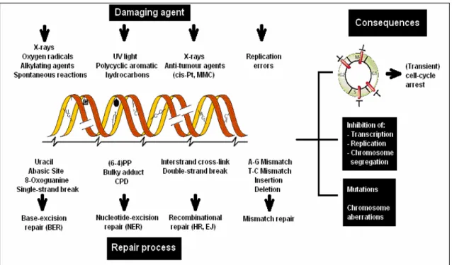
There are three main types of causes leading to the formation of DNA lesions that may lead to mutations if they are left unrepaired (Figure 1.4).
First type is the environmental agents such as ultraviolet (U.V) component of the sunlight, ionizing radiation, and numerous genotoxic chemicals.
Second type is the (by) products of normal cellular metabolism. These include the reactive oxygen species (superoxide anions, hydroxyl radicals and hydrogen peroxide) derived from oxidative respiration and products of lipid peroxidation.
Figure 1.4. DNA damage, repair and consequences (Adapted from Heoijemakers, 2001).
Finally, some chemical bonds in DNA tend to spontaneously disintegrate under physiological conditions. For example, hydrolysis of nucleotide residues leaves non-instructive abasic sites. Spontaneous or induced deamination of cytosine, adenine, guanine or 5-methylcytosine converts these bases to the miscoding uracil, hypohxantine, xanthine and thymine, respectively. Figure 1.4 summarizes some of the most common types of DNA damage and their sources (Heoijmakers, 2001).
1.1.5. Molecular profiling of cancer
Categorization of the tumors has been performed on the basis of histology. But, it is now clearly known that the staining patterns of cells viewed under the microscope is not sufficient to reflect the underlying molecular events that drive the neoplastic process. But, using today’s technology, reading the molecular signature of an individual’s tumor by surveying thousands of genes at once –using DNA arrays– is possible (Liotta et al., 2000). So the variations in the gene expression profiles will be beneficial to fully understand different cancers. It is generally accepted that four to seven rate limiting genetic events are required for the development of the common epithelial cancers (Rennan et al., 1993). It is noteworthy that the patterns of genetic alterations differ between cancers of different types and even of the same type. But fortunately, the patterns are not random (Liotta et al., 2000 and Suzuki et al., 2000).
The main aim of the recent use of DNA arrays (also protein arrays) is to be able to understand the sophisticated disease mechanisms and treatment targets (Liotta et al., 2000). So, the identification of the molecular signatures of the tumors in genomic alterations or expression profiles will enable us to understand the possible mechanisms involved in tumor development, which may also enable us to obtain valuable information about clinics (Suzuki et al., 2000).
1.1.6. Inherited predisposition
Family based studies led scientists to recognize the inherited predisposition to cancer. Since, cancer is a common disease; some families may contain several cases only due to chance. But there is a spectrum of probability that a given family history reflects inherited predisposition from near-certainty of strong predisposition in the rare inherited cancer syndromes, to the possibility of weak effects in familial clusters (Table 1.1) (Ponder, 2001).
Contribution of genetic factors to the development of cancer phenotype can be in varying degrees. Some genes may confer a high cancer risk to the individual but some not.
So, the concept of “inherited predisposition” must be investigated under two sections of “strong predisposition” and “weak predisposition”. For example, germ-line mutations in BRCA1 and BRCA2 genes confer a high risk of breast cancer (Bertwistle et
al., 1998; Ozdag et al., 2000, and Manguoğlu et al., 2003), whereas mutations in other
cancers such as GSTM1 do not confer a high breast cancer risk.
Ironically, the frequencies of these two types of mutations are inversely related to their penetrances. The mutations, conferring a high risk are generally rare in the populations, whereas the mutations conferring a low risk are generally more frequent.
Table 1.1. Inherited predisposition to cancer.
Contribution to overall cancer
incidence Clinical feature Frequency of predisposing alleles Effect on individual risk Inherited cancer syndromes
1-2% at most Rare/unusual cancers or combinations of cancers. Sometimes with
associated developmental defects or non-neoplastic phenotype. Mendelian dominant inheritance.
Rare (nearly
1:1,000 or less) Strong: lifetime risks of cancer up to 50-80% Familial
cancers
Up to 10% depending on definition Families with several cases of common cancers that fall into a recognized pattern of cancer types. Spectrum from families with multiple cases at young age to two or three cases at older ages: many of the latter will be due to chance or to
combinations of weaker genes. Generally show pattern consistent with dominant inheritance.
Uncommon to common Moderate to weak Predisposition without evident familial clustering
No precise figure possible.
Distribution of risk within population may result in substantial fraction of cancer incidence within predisposed minority.
Single cases of cancer at any site, some with one or two affected relatives. The distribution of these cases in the population is probably determined by the combined effects of multiple genetic and non-genetic risk factors.
Multiple common
1.1.6.1. Strong predisposition
A number of relatively rare, high-risk genes have been identified which predispose to common cancers such as breast, colon, and melanoma (Goldgar, 2002). The human inherited cancer syndromes and their transgenic mouse counterparts have been extensively studied. List of familial cancers and related genes are summarized in Table 1.2. As a result of these studies it was clearly seen that the strong predisposition to cancer results either through inheritance of one of the events on the cancer “pathway”, or through effects on DNA repair of genome stability (Ponder, 2001).
The tissue specificity and variability of expression are two important features of strong predisposition. All inherited predisposition to cancer seems to show a considerable degree of tissue specificity, even in the case of defective DNA repair. The mechanism governing tissue specificity is still unknown. There may also be considerable variation in the age at onset of cancer and in the specific types of cancer that predominate not only within a given syndrome, but also within a single family. Some of this variation is due to different germ-line alleles of the main predisposing gene, and some is environmental or chance. But much of the within-family variation is probably attributable to the effects of genetic modifiers (Ponder, 2001).
Some other characteristics of strong predisposition are the vertical and not sex-specific transmission of the cancer predisposition, sex-specific clinical characteristics (early age of diagnosis, presence of two or more primary cancers) (Ponder, 2001).
The first predisposing genes were identified as rare, mutated alleles. These mutated genes result in multiple cases of the disease in families. They were identified using genetic linkage and positional cloning. The prototypic gene associated with familial cancer syndromes is the retinoblastoma gene (RB1), which has turned out to be one of the most important hubs of cellular signaling. Other key signaling molecules such as p53 (encoded by TP53) were initially identified as important targets of viruses or somatic mutations in tumors and were subsequently found to function as germline-inherited tumor predisposition genes (Balmain et al., 2003).
High penetrance alleles have provided many fundamental and unexpected insight into various aspects of cancer biology, including identification of the adenomatosis polyposis coli (APC), β-catenin and Tcf-4 pathway, and the phosphatase PTEN, which is implicated in Cowden syndrome and in the development of a variety of tumor types (Balmain et al., 2003).
It is important to consider that, most of the genes whose altered forms are found to be involved in “strong predisposition”, encode for the proteins of DNA damage repair or related pathways (i.e. BRCA1 and BRCA2) (Heojimakers et al., 2002). This is obviously due to the high number of studies investigating the impact of DNA damage or related pathway genes. But, considering the variety of the pathways in the cellular metabolism, other pathways and genes must also be studied.
The explanation provided by the investigations on the high penetrance genes for how cancers develop is very incomplete. For example, we still have no mechanisms for the tissue specificity of many of the inherited cancer syndromes (Balmain et al., 2003).
Table 1.2. List of familial cancer genes and syndromes (Adapted from National Cancer Institute web site; “http://www.cancer.gov”).
Gene Cancer syndrome Location Discovery
APC Familial polyposis of colon 5q21 1991
BRCA1 Hereditary Breast/Ovarian cancer 17q21 1994
BRCA2 Hereditary Breast/Ovarian cancer 13q12.3 1995
CDH1 Familial gastric sarcoma 16q22.1 1998
CDK4 Hereditary Melanoma 2 11q14 1996
CDKN2A Cutaneous malignant melanoma 9p21 1994
CDKN1C Beckwith-Weideman syndrome 11p15.5 1995
CYLD Familial cylindramotosis 16q12-q13 2000
EXT1 Multiple exostoses type 1 8q24.1 1995
EXT2 Multiple exostoses type 2 11p12 1996
MADH4 Juvenile polyposis 18q21.1 1996
MEN1 Multiple endocrine neoplasia type I 11q13 1997
MET Hereditary Papillary Renal Carcinoma 7q31 1997
MLH1 Hereditary non-polyposis colon cancer 3p21.3 1994
MSH2 Hereditary non-polyposis colon cancer 2p21 1993
NF1 Neurofibromatosis type 1 17q11.2 1990
NF2 Neurofibromatosis type 2 22q12.2 1993
PMS1 Hereditary Non-polyposis Colon Cancer3 2q32 1994
PMS2 Hereditary Non-polyposis Colon Cancer4 7p22 1994
PRKAR1A Cancer complex 17q23-q24 1996
PTCH Nevoid basal cell carcinoma 9q22.3 1996
PTEN Cowden’s syndrome 10q23.3 1997
RB1 Familial retinoblastoma 13q14.1 1986
RET Multiple endocrine neoplasia MEN2A, MEN2B and
medullary thyroid carcinoma 10p11.2 1993
SDHD Familial paraganlioma 11q23 2000
SMARCB1 Rhabdoid predisposition syndrome 22q11 1996
TP53 Li-Fraumeni syndrome 17p13.1 1990
TSC1 Tuberous sclerosis 1 9q34 1996
TSC2 Tuberous sclerosis 1 16p13.3 1993
STK11 Peutz-Jegers syndrome 19p13.3 1997
VHL Von Hipple-Lindau syndrome 3p25 1993
1.1.6.2. Weak predispositon
Weak predisposition to cancer may in principle result from weak alleles of the pathway or caretaker genes. The study of weak predisposition is of interest both for its possible public-health implications and because just as the study of inherited cancer syndromes identified “pathway” genes, so weak predisposition may point to a wider range of processes that are relevant to cancer development, and to interactions between them.
The risk attributable to the effect of pathway genes and the low penetrance genes is more than the risk due to subtle sequence variants or polymorphisms (which can be associated with small to moderately risk for cancer). Low penetrance gene candidates are found in many pathways such as environmental carcinogen detoxification, steroid hormone metabolism and DNA damage repair. There is no doubt about the future of genetic medicine will allow us to identify more genes in several other pathways (Chakravarti, 2001).
Such factors modifying the probability are of extreme importance in sporadic cancers. They may be strongly associated either with disease susceptibility or with some other aspects of the disease phenotype, such as the treatment response of survival (Cardon et al., 2001).
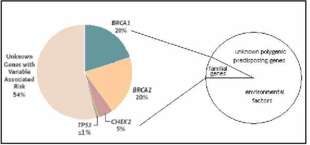
Predisposition by combinations of weak genetic variants may be of much greater significance to public health than the marked individual risks seen in inherited cancer syndromes (Pharoah et al., 2002). Population based epidemiological studies have shown that only 15-20% of the observed familial clustering of breast cancer occurs in families that carry a strongly predisposing BRCA1 or BRCA2 mutation (Figure 1.5) (Balmain et
al., 2003). In principle, the remaining 80-85% of familial risk might have a genetic or
environmental origin, but evidence from studies of breast cancer in twins (Peto, 2001) and the pattern of inheritance in families suggests that genetic factors predominate (Balmain et al., 2003).
1.2. Breast cancer
Breast cancer is the most commonly diagnosed cancer among women, after nonmelanoma skin cancer. It is the second leading cause of cancer deaths after lung cancer.
An estimated 211,300 new cases are expected to occur among women in United States during 2003. About 1,300 new male breast cancer cases are also expected. This year, 40,200 deaths from breast cancer will occur in United States only. It becomes the first leading cause of death among women with age between 15-54 (Atlanta, 2002).
In recent years, improved diagnostic tools have made it possible to detect breast cancers at early, even pre-invasive stages leading to a significant decrease in breast cancer mortality rates over the past decades. Despite all these improvements in the diagnostic tools of breast cancer, approximately a quarter of breast cancer patients die of their disease (Polyak, 2001).
1.2.1. Setting the stage
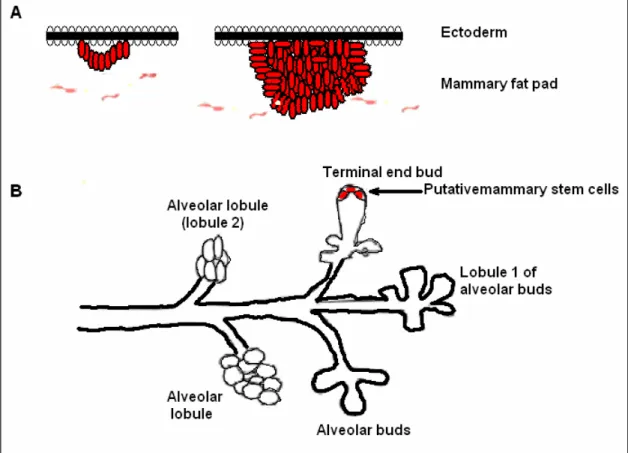
The mammary gland is a remarkable organ with respect to its development and functional differentiation. Unlike most mammalian organs, development of the mammary gland is primarily postpubertal. Mammary glands start to develop during the 4th week of gestation in mammals. Mammary epithelium is derived from epidermis, but
the initiation is dependent on the presence of a specialized mesenchyma, called fat pad. Signals from mammary fat pads underlying the epidermis direct epidermal cells to a mammary differentiation pathway and induce their migration into the mammary fat pad (Figure 1.6, part A) (Polyak, 2001).
Although, the genes governing the mammary gland development are not clearly identified, the homeobox genes are suggested as the possible candidates based on their roles in the development of other organs. Recent genetic and expression analyses
clarified the functions of homeobex genes at specific transition points in the mammary gland development (Lewis, 2000).
Figure 1.6. Mammary gland and its development (Adapted from Polyak, 2001).
Only few, poorly branched mammary ducts are formed during embryogenesis. The mammary gland remains in this rudimentary form until puberty. During puberty, hormones, particularly estrogen and progesterone, induce further elongation, branching and extension of the already existing ducts. This leads to the generation of lobules that contain a terminal duct splitting into alveoli. These lobules are relatively simple and not branched in nulliparous women (lobule 1 in Figure 1.6 part B). Fully mature gland (extensive branching, alvoelogenesis, and terminal differentiation) only occur during “full-term pregnancy” (Polyak, 2001).
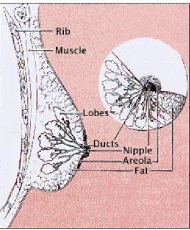
Figure 1.7. Main anatomic structures of breast (http://www.imaginis.com).
The smallest structure in breast is the tiny bulbs that can produce milk. These tiny bulbs form the lobules, and lobules forms the lobes. Ducts are the tiny tubes connecting the bulbs, lobules, and lobes. These ducts lead to the nipple in the center of a dark area of skin called areola.
The remaining part of the breast is filled with fat and vessels carrying the colorless fluid named “lymph”. Muscle tissue is present only under the breast and covers the ribs.
The main origin of the breast carcinoma is the epithelial tissue of the mammary gland, including the milk-producing lobules and the ducts that carry milk to the nipple. The stromal, vascular, or fatty components of the breast are not generally included in the transformation process, excluding some very rare cases. The progression profile somehow reflects the clinics of the disease. For example, inflammatory breast cancer (IBC) is an aggressive form of locally advanced breast cancer that affects approximately 5% of women with breast cancer (Kleer et al., 2000).
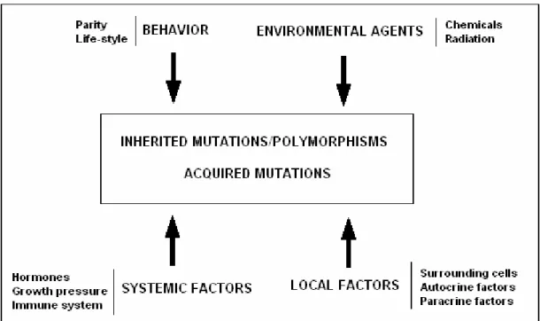
Breast cancer results from a combination of many factors including inherited mutations or polymorphisms of cancer susceptibility genes, environmental agents that influence the acquisition of somatic genetic changes and several other systemic and local factors (Figure 1.8) (Polyak, 2001). These factors may be grouped under sections of,
behavioral (i.e. parity, life-style), environmental (i.e. chemicals, radiation), systemic
(hormones, growth pressure, immune system), local factors (surrounding cells, autocrine factors, paracrine factors), and lastly genetic factors which is accepted as the major factor on the disease, since all the other factors may regulate and/or supplement the contribution of genetic factors.
Figure 1.8. Summary of factors influencing breast carcinogenesis (Adapted from Polyak, 2001).
The factors listed in Figure 1.8 actually act on the development of breast cancer in various combinations. For example, when we consider, parity, we must also mention the effect of hormones. So, the factors mentioned in Figure 1.8 is summarized below.
The frequency of breast cancer is clearly shown to be associated with the body-mass-index (bmi) of the patient. Although the relationship between the bmi and the development of breast cancer is complex, the underlying factor is supposed to be the elevated levels of estrogen due to the production in adipose tissue (DeVita et al., 2001).
The development of breast cancer in many women appear to be related to the exposure of female reproductive hormones. Early age at menarche, nulliparity, late age at first full term pregnancy, late age at menopause increase the risk of breast cancer due to the hormonal exposure levels (DeVita et al., 2001).
The natural history of breast cancer involves a sequential progression through defined clinical and pathologic stages starting with atypical hyperproliferation, progression to in situ then invasive carcinomas, and culminating in matestatic disease (Figure 1.9 and Table 1.3) (Polyak, 2001).
Figure1.9. A hypothetical multi stage model of breast carcinogenesis (Adapted from Polyak, 2001).
The stage at the time of diagnosis is very important in determining the treatment modalities and prognosis. So the staging of breast cancer is very important. Although many staging systems have been proposed, the most commonly used system is the one adopted by both the American Joint Committee (AJC) and the International Union Against Cancer (UICC). The staging system is a detailed TNM (tumor, nodes, metastasis) (Table 1.3).
Table 1.3. TNM Staging. Stage 0 Carcinoma in situ
Stage I Tumor 2 cm, axillary nodes not involved
Stage II Tumor between 2 and 5 cm and/or involved but mobile axillary lymph nodes
Stage III Tumor larger than 5 cm and/or fixed axillary lymph nodes; includes inflammatory breast cancer
1.2.2. Genetics of breast cancer
In breast cancer, the risk to close relatives of a case, averaged across all ages, is about two-fold (Ponder, 2001). 5-10% of the cases have a first- or second-degree relative with the disease. The remaining nearly 90% of cases are sporadic (non-inherited) (Figure 1.5) (Wooster, 2003).
The hereditary breast and ovarian cancer syndromes are shown to involve genetic alterations in various susceptibility genes such as BRCA1, BRCA2, p53, ATM, PTEN or
MSH2, MLH1, PMS1, MSH3, and MSH6 (Palevic, 2001). Two of these are regarded as
the major susceptibility genes, breast cancer susceptibility gene 1 (BRCA1) and breast cancer susceptibility gene 2 (BRCA2) (Venkitaraman, 2002). However, mutations in these genes account for only 2 to 3 percent of all breast cancers, which indicates the presence of other susceptibility genes (Wooster, 2003).
Recently, the structure and expression of CHEK2 was analyzed in breast cancer.
CHEK2 was found to be implicated in a significant proportion of sporadic breast
cancers, but unlikely to represent a susceptibility gene for a high proportion of hereditary breast cancer (Sullivan et al., 2002). In conclusion, CHEK2 1100DelC variant is a low penetrance, and low frequency predisposing allele (Offit et al., 2003). More recent experiments stated the association of this variant and prostate cancer risk (Meijers-Heijboers et al., 2003, and Dong et al., 2003).
1.2.2.1. Somatic mutations in breast cancer
Studies of sporadic breast cancers led scientists to understand the pathogenetic mechanisms underlying the development of breast cancer. Approximately 90% of all breast cancer cases are sporadic. The genes coding for growth factors and receptors, intracellular signaling molecules, regulators of cell cycle, genome maintenance mechanisms, adhesion molecules and proteases are the first targets of the somatic
- The tumor suppressor protein p53 plays a central role in regulating progression through cell cycle and the genome maintenance. p53 mutations have been detected in 15-45% of human breast cancer specimens in several studies.
- Cyclin proteins are regarded as the central regulators of cell cycle progression, which are also shown to be over expressed in breast cancer (Evan, 2001).
- The proto-oncogene bcl-2 and c-myc which suppress apoptosis over expressed in 30-45% of breast cancer cases (Evan, 2001).
- Frequent alterations of the FHIT locus in breast cancer, suggest its role in the pathogenesis of breast tumors. FHIT protein was shown to be directly involved in the control of cell growth and/or proliferation. (Ingvarsson, 2001).
1.2.2.2. Germline mutations in breast cancer
Clinical investigations of familial aggregation of breast cancer have identified several genetic syndromes with an autosomal dominant pattern of inheritance that features breast cancer (Tonin, 2000). Breast cancer cases due to germline mutations have several distinctive clinical features. For example, age-of-onset is relatively low than sporadic breast cancer, the prevalence of bilateral breast cancer is higher, and in the presence of associated tumors in affected individuals is noted in some families. Associated tumors may include ovarian, colon, prostate, and endometrial cancers and sarcomas. However, inherited breast cancer does not appear to be distinguished by histologic type, metastatic pattern, or survival characteristics (Vogelstein et al., 1998).
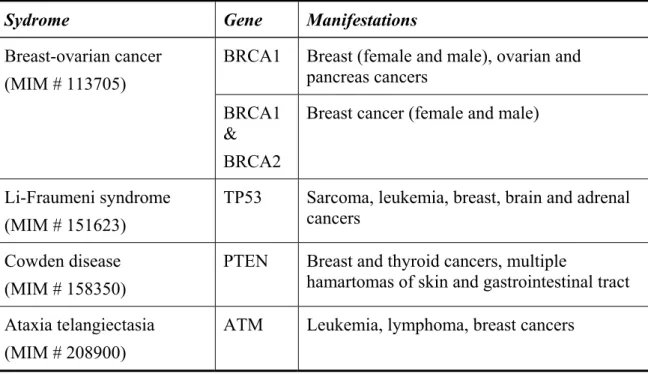
The syndromes in which genes are known or are suggested to cause inherited breast cancer and other cancers are shown in Table 1.4.
Table 1.4. Hereditary cancer syndromes that feature breast cancer (Tonin, 2000).
Sydrome Gene Manifestations
BRCA1 Breast (female and male), ovarian and pancreas cancers Breast-ovarian cancer (MIM # 113705) BRCA1 & BRCA2
Breast cancer (female and male)
Li-Fraumeni syndrome (MIM # 151623)
TP53 Sarcoma, leukemia, breast, brain and adrenal cancers
Cowden disease (MIM # 158350)
PTEN Breast and thyroid cancers, multiple
hamartomas of skin and gastrointestinal tract Ataxia telangiectasia
(MIM # 208900)
BRCA1 and BRCA2
The existence of the BRCA1 gene, which predispose to breast cancer, was demonstrated by linkage analysis in 1990 (Hall et al., 1990). Using polymorphic markers, which would distinguish the parental origins of alleles and are representative of different chromosomal regions, linkage was established to the long arm of chromosome 17 at region q21. Families with early age of onset (pre-menopausal) of breast cancer were more likely to be linked to the BRCA1 locus. Through an intense cloning effort, the identity of BRCA1 was discovered in 1994 (Miki et al., 1994, and, Brown MA, 1995). In the following year, a human BRCA1 gene knockout (Boyd, 1995) and the aberrant subcellular localization was identified (Chen et al., 1995).
In addition, linkage analyses provided sufficient evidence for the presence of another susceptibility gene (Wooster et al., 1994), which was identified about a year later (Wooster et al., 1995, and, Tavtigian et al., 1996). Germ-line mutations in BRCA1 and BRCA2 have been reported in at least two syndromes that feature breast-cancer: site-specific breast cancer and breast-ovarian cancer syndrome (Table 1.14). The striking feature common to families of both syndromes is the young age of onset of breast cancer (Tonin, 2000).
Population-based studies have reported lower risks of breast and ovarian cancer in mutation carriers. It has been suggested that other factors may modulate the risk in mutation carriers, and may account for the reduced penetrance. Recent studies have shown that lifestyle choices such as smoking may modulate the risk of breast cancer in mutation carriers. More than 100 mutations in each gene have been described to date, and the majority of the mutations is private and reported in only one family (Please refer to the Breast Information Core Data Base).
BRCA1 is comprised of 5.592 nucleotide pairs with 24 exons. BRCA2 is
comprised of 10,254 nucleotide pairs and 27 exons. The coding sequences of both genes are spread across large tracts of DNA, comprising more than 1,000,000 nucleotides. The large size and complexity of each gene, and the absence of “hot-spots” for mutations,
TP53
Li-Fraumeni syndrome (LFS), now known to be associated with germ line mutations in TP53, was first identified as a syndrome in 1969 in a description of four kindreds in which cousins or siblings had childhood soft-tissue sarcomas and other relatives had excessive cancer occurrence (Vogelstein et al., 1998). Underlying genetic defect in the Li-Fraumeni syndrome is a germline mutation in the TP53 gene (MIM# 191170) as first described by Malkin et al., in 1990. But now, there are nearly 250 independent germ-line TP53 mutations in numerous publications.
Li-fraumeni syndrome is associated with a variety of different tumor types occurring over a wide age range, including childhood. The definition of LFS originated from Li and Fraumeni’s work as a proband with a sarcoma aged under 45 years with a first-degree relative aged under 45 years with any cancer, plus an additional first- or second-degree relative in the same lineage with any cancer aged under 45 years or a sarcoma at any age (Li et al., 1988). Now, LFS is defined as a proband with any childhood tumor, or a sarcoma, brain tumor, or adrenocortical tumor aged under 45 years plus a first- or second-degree relative in the same lineage with a typical LFS tumor at any age, and an additional first- or second-degree relative in the same lineage with any cancer under the age of 60 years (Varley, 2003).
Bone and soft-tissue sarcomas, premenopausal breast carcinoma, brain tumors, adrenocortical carcinomas and leukemias are the first identified tumors of LFs. Subsequent studies reported wider range of tumors such as melanoma, Wilm’s tumor, and lung, gastric, and pancreatic carcinoma (Varley, 2003).
The cellular role of p53 is well characterized. p53 is a sequence specific DNA binding protein, that functions as a transcription factor. The sequence specific transcription factor activity appears to be essential for its role as a tumor suppressor (Picksley et al., 1994). The impact of p53 on multiple cellular functions such as gene transcription, DNA synthesis and repair, cell cycle arrest, senescence, and apoptosis is well documented (Hussain et al., 2001). The phosphorylation status of the protein is
ATM
ATM (ataxia telangiectasia mutated) is one of the key proteins involved in the cellular response to DNA damage. In the autosomal recessive disorder ataxia telangiectasia (A-T) ATM protein is defective. The heterozygous A-T gene carrier frequency in the population is ~1% and the disease incidence is ~1/40000. Affected individuals develop progressive cerebellar ataxia (loss of balance and coordination) such that most are wheelchair bound by their early teenage years. Telangiectasias are tortuous dilated blood vessels that develop in the eyes and sun-exposed skin. A-T is associated with a 30–40% lifetime risk of developing a malignancy, usually of lymphoid origin and occurring in childhood. And relevant studies showed that women with ATM mutation have an elevated risk of developing breast cancer. A-T individuals are also more susceptible to infections, and aspiration pneumonia is a common cause of death. Life expectancy is reduced, with a median age at death of ~30 years (Levitt et al., 2002).
PTEN
Cowden disease is best characterized by multiple hamartomatous lesions, especially of the skin, mucus membranes, colon, breast, and thyroid, and multiple facial trichilemmomas. Hamartomatous polyps of the colon also occur, and there are neoplasms of the thyroid and breast. Family-based analysis suggested an autosomal dominant mode of inheritance with high penetrance in both sexes, and a high frequency of breast cancer (up to 30%) in females. Linkage analysis of Cowden disease families revealed a locus on chromosome 10q22-23. PTEN was the strongest candidate gene that mapped to this interval on chromosome 10, and was previously shown to harbor somatic mutations in a number of tumor types, particularly breast cancer, that feature in Cowden disease. Therefore, a combination of linkage analysis and candidate gene approaches led to the discovery that individuals with Cowden disease harbored germline mutations in PTEN. Although the reported mutations are dispersed throughout the gene, there is a
1.3. RNASEL
RNASEL (MIM# 180435) encodes for the ubiquitously expressed ribonuclease L
(RNase L). The RNASEL gene maps to the hereditary prostate cancer (HPC) predisposition locus at 1q24-q25 (HPC1).
1.3.1. Prostate cancer
Prostate cancer (PC) is the second leading cause of cancer deaths in men >50 years of age and the most frequent visceral cancer in males (Silverman, 2003). Prostate cancer is a significant international public health problem, with a world-wide estimate of 239,000 deaths resulting from this disease annually, in the U.S. only (Xu et al., 2000).
The prostate is a walnut-sized gland of the male reproductive system located beneath the bladder and in front of the rectum that produces and stores the seminal fluid (Silverman, 2003). Precursor lesions known as prostate intraepithelial neoplasia (PIN) can progress after many years of overt carcinoma and finally to metastatic cancer (Figure 1.12) (Abate-Shen et al., 2002). The most common sites for metastasis are lymph nodes and bones (pelvis and axial skeleton) (Silverman, 2003).
Aging, hormonal, environmental, and genetic factors are all believed to play roles in the pathogenesis of prostate cancer.
This cancer type usually appears after the sixth decade, and so it is generally considered as a disease of aging. Prostate cancer is diagnosed in very few people younger than 50 years (<0.1% of all patients). The mean age of patients with this disorder is 72-74 years, and about 85% of patients are diagnosed after age of 65 years (Grönberg et al., 2003).
Prostate cancer is rare in males castrated before puberty and the tumor growth is inhibited by orciectomy or chemical hormone-ablation theraphy. Also, there is a large body of evidences indicating the role of “androgen signaling system” in the development
Environmental causes are found to be implicated in prostate cancer development by the geographic data on prostate cancer incidence and observations that relative risk of developing prostate cancer is associated with migrations between low and high incidence regions of the world (Siverman, 2003).
It has been recognized for some time that prostate cancer tends to cluster in families (Wang et al., 2002). Remarkably, men with three or more first degree relatives with prostate cancer have a 100-fold increased risk compared with men that have no family history of prostate cancer (Silverman, 2003). Segregation analysis suggests that this familial clustering can best be explained by at least one rare dominant susceptibility gene (Wang et al., 2002). And this dominant susceptibility gene must be rare, autosomal, highly penetrant for the hereditary prostate cancer with early onset (Silverman, 2003). However, there is also a considerable evidence on the presence of a complex genetic basis, involving multiple susceptibility genes and variable phenotypic expression (Simard et al., 2002). On the basis of linkage studies of families with high risk of PC, six PC-susceptibility loci were identified (Eeles et al., 1998, and, Wang et
al., 2002). 1. HPC1 (1q24-25), 2. HPCX (Xq27-q28), 3. PCAP (1q42), 4. CAPB (1p36), 5. HPC20 (20q13), and
6. HPC2 (17p11) (reviewed in Ostrander et al., 2000).
HCP1 was the first such prostate cancer locus, mapped in 1996 to chromosome
1q24-25 (Smith et al., 1996). Initial gene mapping studies placed RNASEL and several other genes in the critical HPC1 region in chromosome 1q25 (Carpten et al., 2000).
To overcome limitations due to genetic heterogeneity and a low frequency of mutations in any particular susceptibility gene, the International Consortium for Prostate Cancer Genetics (ICPCG) performed a joint analysis from 722 families. They have confirmed linkage of hereditary prostate cancer to the HPC1 locus (Xu, 2000, and, Xu et
al., 2001). A second important study was performed with 2410 individuals, including
662 men with prostate cancer compared several potential prostate cancer susceptibility loci (HPC1, PCAP, HPCX, and CAPB). They have demonstrated that only HPC1 commonly segregated within families with the most severe cases of prostate cancer (Goode et al., 2001).
The linkage of HPC1 to RNASEL suggests that RNase L directly or indirectly suppresses one or more steps is prostate tumorigenesis and/or metastasis (Figure 1.12).
Figure 1.10. Possible role of RNase L in prostate carcinogenesis (Adapted from Silverman, 2003).
1.3.2. RNase L
RNase L is a fascinating tightly regulated endoribonuclease of higher vertebrates that plays essential roles in mediating diverse types of cellular responses (Zhou, 1993). The activation of RNase L requires the production of unusual effector molecules, 2’,5’-linked oligoadenylates, p1-3A(2’p5’A)>=2 (2-5A) (Dong et al., 2001). 2-5As are
produced from ATP by 2-5A-synthetases (OAS enzymes). The genes coding for OAS enzymes are activated upon interferon treatment of mammalian cells (Dong et al., 1997). OAS enzymes were discovered in the mid-1970s by I.M.Kerr and colleagues. They are found to be activated by double stranded RNA (dsRNA). They convert ATP to PPi and a series of short 2’ to 5’ linked oligoadenylates, collectively referred to as 2-5As
(Figure 1.11) (Silverman, 2003).
IFN treatment of the cells activates the JAK-STAT pathway which also activates the expression of OAS genes (Stark et al., 1998). In humans, there are four related genes (OAS1, OAS2, OAS3, and OASL) encoding eight or more isoforms as a result of alternative splicing (Silverman, 2003).
Up to date, the only well-established biochemical function of 2-5A is the activation of RNase L (Zhou et al., 1997). The significance of the dsRNA requirement for 2-5A synthetase activity is that it is a common intermediate or by product of viral infections.
RNaseL protein consists of 741 amino acids. RNase L has been detected only in reptiles, birds and mammals. Only mouse and human RNase L sequences are available. The presence of RNase L was shown for many mammalian tissues.
C-terminal region contains the RNase domain and the kinase-like domain. N-terminal domain represses the ribonuclease domain in the C-N-terminal region in the absence of 2-5A. The N-terminal region of the protein is called the repressor part. It contains 9 ankyrin repeats. Anykrin repeats are common protein/protein interaction domains. The presence of two P-loop motifs (GTK) is the characteristic property of RNase L. Because the Lysine residues in these regions are the sites of 2-5A binding (Figure 1.12) (Silverman, 2003).
A kinase domain was assigned to the C-terminal region of the protein based on the sequence comparisons. But this assignment lacks experimental evidence. On the contrary, experimental evidence suggests that the kinase like motif is implicated in enzyme dimerization (Dong et al., 1999), which is the crucial step in enzyme activation (Figure 1.12) (Silverman, 2003).
Figure 1.13. Functional model for the activation of RNase L by 2-5A (Adapted from Silverman, 2003).
The binding of 2-5As leads to the formation of a potent dimeric endoribonuclease (Dong et al., 1995). 2-5A binding to the P-loops relieves binding of repressor domain. This conformational change ceases the inhibition by the ankyrin repeats on the dimerization and ribonuclease domains. Accessible dimerization domains enables the dimerization of the enzyme, which enables formation of active enzyme (Dong et al., 2001).
Cleavage sites for the RNase L enzyme are UpNp dinucleotide sequences (primarily UU and UA) (Silverman, 2003).
Considering the production of 2-5As and the 2-5A dependent activation of the enzyme, it may be concluded that the RNase L action is located in the vicinity of the dsRNA. This enables specificity to the system to degrade only the viral RNA. The experimental evidence is the preferential degradation of viral RNA in comparison to cellular RNA in EMCV-infected cells (Li et al., 1998). Along with PKR, RNase L constitutes the antiviral arm a group of mammalian stress response proteins (Williams, 1999).