Association of fragmented QRS complexes on ECG with
left ventricular diastolic function in hypertensive patients
Hipertansiyonlu hastalarda fragmente QRS komplekslerinin
sol ventrikül diyastolik fonksiyonu ile ilişkisi
Department of Cardiology, Gaziosmanpasa University Faculty of Medicine, Tokat; #Department of Internal Medicine, Gaziosmanpasa University Faculty of Medicine, Tokat
Hasan Kadı, M.D., Ayşe Kevser Demir, M.D.,# Köksal Ceyhan, M.D.,
İbrahim Halil Damar, M.D., Kayıhan Karaman, M.D., Çağrı Zorlu, M.D.,
Objectives: Diastolic dysfunction occurs as a result of in-terstitial fibrosis in hypertensive patients. Fragmented QRS (fQRS) on ECG signifies myocardial fibrosis in various clinical situations. We investigated whether fQRS on ECG is related to diastolic dysfunction in patients with hypertension.
Study design: The study population included 72 hyperten-sive patients with normal coronary angiogram. Fragmented QRS was defined as the presence of an additional R wave (R’), notching of the R or S wave or fragmentation in two contiguous leads corresponding to a major coronary artery. Echocardiography was performed to all patients to detect diastolic dysfunction. Diastolic dysfunction was regarded as non-severe if patients had normal diastolic function or grade 1 diastolic dysfunction or severe if they had grade ≥2 diastolic dysfunction.
Results: Thirty-two patients had fQRS on ECGs (fQRS [+] group) and there were 40 patients who did not have fQRS on their ECGs (fQRS [-] group). The two groups were simi-lar in terms of baseline characteristics. In patients with fQRS on the ECG, severe diastolic dysfunction was more preva-lent (59.4% vs. 7.5%, p<0.001). The duration of hypertension was longer in patients with fQRS on the ECG (p<0.001). The presence of fQRS on the ECG was an indicator for severe diastolic dysfunction (B=1.954; odds ratio=7; 95% confidence interval=1.4-35.4; p=0.018).
Conclusion: The presence of fQRS complexes on ECG pre-dicts more severe diastolic dysfunction in patients with hyper-tension.
Amaç: Hipertansiyonlu hastalarda diyastolik fonksiyon bo-zukluğu interstisiyel fibrozun bir sonucudur. EKG’de fragmen-te QRS (fQRS) farklı klinik durumlarda miyokart fibrozunu gösterir. Bu çalışmada, hipertensiyonlu hastalarda fQRS’nin ciddi diyastolik fonksiyon bozukluğuyla ilişkisi olup olmadığı araştırıldı.
Çalışma planı: Çalışmaya koroner anjiyografisi normal olan 72 hipertansiyonlu hasta alındı. Fragmente QRS, majör ko-roner arterin beslediği alanda en az iki ardışık derivasyonda ek bir R dalgasının varlığı (R’), R veya S dalgasının çentik-lenmesi ya da fragmentasyonu olarak tarif edildi. Diyastolik fonksiyon bozukluğunu belirlemek için tüm hastalara ekokar-diyografi yapıldı. Normal diyastolik işlevler veya evre 1 diyas-tolik işlev bozukluğu ağır olmayan, evre 2 veya üstü diyasdiyas-tolik işlev bozukluğu ise ağır diyastolik işlev bozukluğu olarak ka-bul edildi.
Bulgular: Hastaların 32’sinde EKG’de fQRS mevcut iken (fQRS [+] grup), 40 hastada yoktu (fQRS [–] grup). Her iki grup bazal özellikler bakımından benzerdi. fQRS (+) grup-ta ciddi diyastolik fonksiyon bozukluğu daha fazla bulundu (%59.4 ve %7.5, p<0.001). Hipertansiyonun süresi fQRS (+) grupta daha uzun idi (p<0.001). EKG’de fQRS’nin varlığının ciddi diyastolik fonksiyon bozukluğunun bir belirteci olduğu bulundu (B=1.954; odds oranı=7; %95 güven aralığı=1.4-35.4; p=0.018).
Sonuç: Hipertansiyonlu hastalarda EKG’de fQRS kompleks-lerinin varlığı ciddi diyastolik fonksiyon bozukluğu için bir be-lirteçtir.
Received:February 24, 2014 Accepted:August 19, 2014 Correspondence: Dr. Hasan Kadı. Balıkesir Üniversitesi Tıp Fakültesi,
Kardiyoloji Anabilim Dalı, Çağış Yerleşkesi, Balıkesir, Turkey. Tel: +90 266 - 612 14 61 e-mail: [email protected]
© 2015 Turkish Society of Cardiology
H
ypertension is one of the most common diseases in the world and the major cause of cardiac ar-rhythmias, left ventricular hypertrophy (LVH), coro-nary artery disease, stroke, renal failure, and systolic and diastolic heart failure. About half of hypertensive patients with heart failure symptoms have diastol-ic heart failure, a severe condition.[1] Causes of left ventricular (LV) diastolic dysfunction are impaired ventricular relaxation and decreased compliance in patients with hypertension. Although the pathophysi-ological mechanism of LV diastolic dysfunction in hypertensive patients is multifactorial, interstitial fi-brosis and accumulation of type I and III collagen in the myocardium play a significant role.[2] These alter-ations in hypertensive hearts account for the develop-ment of LV diastolic dysfunction.[3] On the other hand, it has been reported that degree of myocardial fibrosis is the most significant factor related to diastolic dys-function in patient with hypertension.[4]Fragmented QRS (fQRS) complexes are defined as changes in QRS morphology with different RSR’ patterns. Cardiac magnetic resonance imaging (CMR) and myocardial single-photon emission tomography (SPECT) studies have shown that presence of fQRS on ECG signifies myocardial fibrosis.[5,6] The presence of fQRS complexes on 12-lead ECG has been found to be associated with all-cause mortality and recurrent cardiac events.[7] Recently, Brenyo et al.[8] reported that the presence of fQRS complexes on ECG were an independent predictor for sudden cardiac death in a patient with idiopathic dilated cardiomyopathy.
The aim of this study was to investigate the rela-tionship between fQRS complexes on ECG and LV diastolic function in hypertensive patients. We hypoth-esized that fQRS on ECGs would be related to severe diastolic abnormalities in patients with hypertension.
PATIENTS AND METHODS Patients
Patients who underwent coronary angiography in our center between January 2011 and December 2012 were evaluated in this prospective case control study. Hypertensive patients who had normal coronary arter-ies were included in this study. Indications for coro-nary angiography were typical symptoms, positive exercise testing or myocardial perfusion imaging in the study group. Patients with coronary artery disease,
coronary anomalies, diabe-tes mellitus, other systemic illnesses (such as connec-tive tissue disease or renal failure), cardiomyopathy, moderate or severe valve disease, atrial fibrillation, typical left bundle block or right bundle block on the ECG (QRS duration >120
ms), incomplete right bundle block (QRS duration <120 ms and RSR’ patterns in V1-2 precordial leads), or poor quality imaging preventing analysis were ex-cluded. Patients who were unable to perform Valsalva maneuver were also excluded. Secondary forms of hypertension were not included in this study.
Diagnostic criteria of hypertension
Hypertension was defined according to the Adult Treatment Panel III (ATP-III)[9] criteria (blood pressure ≥130/85 mmHg or the use of antihypertensive drugs). ECG criteria for fQRS
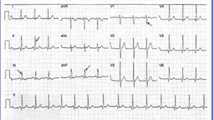
The resting baseline 12-lead ECG (filter range, 0.05 to 150 Hz; AC filter, 60 Hz; 25 mm/s; 10 mm/mV) was analyzed by two independent cardiologists blinded to the study. fQRS was defined as the presence of an ad-ditional R wave (R’), notching of the R or S wave, or the presence of fragmentation (more than one R’) in two contiguous leads corresponding to a major coro-nary artery.[10] Examples of 12-derivation ECG of two patients with and without fQRS in ECG are given in Fig. 1 and Fig. 2 respectively.
Echocardiographic examination
Prior to echocardiographic examination, the patients’ height, weight, systolic and diastolic blood pressure, and heart rate were measured after a period of at least 10 minutes’ rest. The echocardiographic examination was performed using the Vivid I system (GE Medical Systems, Andover, MA, USA). All the images were recorded on this system. At the end of the study, the recorded images were examined by two experienced cardiologists blinded to the study. On the recorded images, an average of 2 consecutive measurements was taken at the end of expiration. All the echocar-diographic measurements were taken according to the guidelines of the American Society of Echocardiog-raphy.[11]
Türk Kardiyol Dern Arş
150
Abbreviations: A Mitral late wave CMR Cardiac magnetic resonance E Mitral early wave EDT E deceleration time fQRS Fragmented QRS LV Left ventricular LVM LV mass
SPECT Single-photon emission tomography
M-mode measurements and calculations
Left ventricular volumes were estimated from end-diastolic and end-systolic dimensions by using the Teichholz formula[12] on the parasternal short-axis im-ages. Using the formula defined by Devereux et al.,[13] the LV mass (LVM) was calculated and indexed for body surface area. Increased LVM index was defined as LVM index ≥95 g/m2 for female and LVM index ≥115 g/m2 for male patients.[14]
Doppler measurements and calculations
Transmitral early (E) and late (A) velocities and their ratio (E/A) and the E deceleration time (EDT) were measured in the apical four-chamber view.
Myocardial velocities measurements and calculations
The myocardial velocities were measured at lateral and septal annulus levels on the apical four-chamber
images. Among the mitral annular tissue Doppler pa-rameters, peak diastolic early (e’), and late (a’) veloci-ties were measured. The pulmonary capillary wedge pressure was calculated using the E/e’ ratio as defined by Nagueh et al.[15]
Grading diastolic function
Grading of diastolic function was carried out according to Redfield et al.:[16]
1. Normal diastolic function: Subjects who had all
of the following Doppler findings were classified as having normal diastolic function: i) mitral E/A rate higher than 0.75 and lower than 1.5 and EDT higher than 140 msn, ii) change in E/A rate higher than 50% during peak Valsalva maneuver, and iii) E/e’ rate low-er than 9.
2. Grade 1 (impaired relaxation type) diastolic dysfunction: Patients with at least two of the
follow-ing Doppler criteria were considered as havfollow-ing Grade 1 (impaired relaxation) diastolic dysfunction: i) mitral E/A rate lower than 0.75 and EDT higher than 140 msn, ii) change in E/A rate lower than 50% during the peak Valsalva maneuver, and iii) E/e’ rate lower than 9.
3. Grade 2 (pseudo-normal type) diastolic dysfunc-tion: Patients with at least two of the following
Dop-pler criteria were considered as having Grade 2 (pseu-do-normal) diastolic dysfunction: i) mitral E/A rate higher than 0.75 and lower than 1.5 and EDT higher than 140 msn, ii) change in E/A rate higher than 50% during the peak Valsalva maneuver, and iii) E/e’ rate between 9 and 12.
4. Grade 3 (restrictive type) diastolic dysfunction:
Patients with at least two of the following Doppler criteria were considered as having Grade 3 (restric-tive) diastolic dysfunction: i) mitral E/A higher than 1.5 and EDT lower than 140 msn, ii) change in E/A rate higher than 50% (reversible Grade 3) or less than 50% (irreversible Grade 3) during the peak Valsalva maneuver, and iii) E/e’ rate more than 12.
5. Unclassified patients: Patients who had only one
of the above-mentioned Doppler criteria were consid-ered unclassified and excluded from the study.
Diastolic function was regarded as non-severe if patients had normal diastolic function or grade 1 dia-stolic dysfunction, and severe if they had grade ≥2 diastolic dysfunction.
Figure 1. 12-lead ECG from a hypertensive patient with Grade 2 diastolic dysfunction showing fragmented QRS complexes in leads II, III, aVF, and V1 (arrow heads).
Figure 2. 12-lead ECG from a hypertensive patient with Grade 1 diastolic dysfunction showing no fragmented fQRS on the ECG.
Türk Kardiyol Dern Arş
152
fQRS on their ECGs (fQRS [+] group), 40 patients did not have fQRS on their ECGs (fQRS [–] group). Both groups were compared in terms of demographic data, echocardiographic findings, and cardiovascu-lar drug use. The results are given in Tables 1-3. The groups were similar in terms of risk factors for coro-nary artery disease and cardiovascular drug use.
Whereas Grade 2 diastolic dysfunction (pseudo-normal type) was present in 3 patients (7.5%) with-out fQRS on their ECGs, it was present in 19 patients (59.4%) with fQRS on their ECGs. In patients with fQRS on their ECGs, Grade 2 diastolic dysfunction was statistically more prevalent (p<0.001). None of the patients had Grade 3 (restrictive) diastolic dys-function. The average duration of hypertension (year) was longer in the patient group with fQRS on their ECGs than in those without fQRS (15.72±4.3 versus 11.85±3.9), and the difference between the groups was significant (p=0.002). The LVM index (g/m2) was significantly higher in the patients with fQRS on their ECGs (131.6±14.9 vs. 95.7±13.3, p<0.001).
We classified those with normal diastolic func-tion and Grade 1 diastolic dysfuncfunc-tion as patients with non-severe diastolic dysfunction, and those with Grade 2 diastolic dysfunction as patients with severe diastolic dysfunction. To define factors that affected diastolic function, classified diastolic function (non-Statistical analyses
SPSS for Windows 18.0 (SPSS Inc. Chicago, Illinois, USA), a statistical package program, was used for sta-tistical analysis. The Kolmogorov-Smirnov test was used to evaluate whether the distribution of continu-ous variables was normal. Normally distributed, con-tinuous data are expressed as mean±standard devia-tion (SD). Categorical data are expressed as numbers with percentages. We used the Student’s t-test for nor-mally distributed continuous variables. Categorical data were compared using the Chi-square or Fisher’s exact test, where applicable. Severe diastolic dysfunc-tion was used as a dichotomous dependent variable, and multivariate logistic regression analysis was per-formed to identify independent variables. A p-value of <0.05 was considered statistically significant.
Informed consent was received from all patients. The study protocol was approved by the institutional ethics committee.
RESULTS
347 of the 2.082 consecutive patients who underwent a coronary angiography at our institution from January 2011 to December 2012 had normal coronary arteries. Seventy two patients were suitable for the study ac-cording to the above criteria. Thirty-two patients had
Table 1. Demographic findings in the fQRS (–) and fQRS (+) groups
Variable\Group fQRS (–) (n=40) fQRS (+) (n=32) p n % Mean±SD n % Mean±SD Age (year) 54.18±7.9 55±5.5 0.620 Male 11 27.5 11 34.4 0.529 Smoking 6 15 8 25 0.287 Hyperlipidemia 7 17.5 10 31.3 0.172
Hypertension duration (year) 11.85±3.9 15.72±4.3 <0.001
Body mass index (kg/ m2) 26.93±2.2 27.24±1.7 0.513
Body surface area (m2) 1.72±0.1 1.76±0.1 0.108
ACE-ARB 19 47.5 21 65.6 0.124
Beta blocker 14 35 12 37.5 0.826
Calcium canal blocker 6 15 10 31.3 0.099
STATIN 6 15 5 15.6 0.942
Increased LVM index 11 27.5 31 96.9 <0.001
fQRS: Fragmented QRS; SD: Standard deviation; ACE-ARB: Angiotensin converting enzyme inhibitor-Angiotensin receptor blocker; LVM: Left ventricular mass.
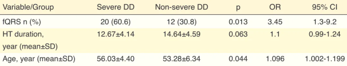
severe vs. severe) was accepted as a dichotomous dependent variable, and multivariate logistic regres-sion analysis was performed to search for indepen-dent variables. In the multivariate logistic regression analysis, age, hypertension duration, and fQRS on ECG were independent variables. Univariate analy-ses results of the patients in the severe and non-se-vere diastolic dysfunction groups are given in Table 4. Multivariate logistic regression analysis revealed that fQRS was an indicator of patients in the severe diastolic dysfunction (B=1.954; odds ratio=7; 95% confidence interval=1.4-35.4; p=0.018). Examples of
12-derivation ECG of two patients with and without fQRS in ECG are given in Figure 1 and 2 respectively.
DISCUSSION
The main finding of the present study is that LV diastolic dysfunction is more severe in hypertensive patients with fQRS on their ECGs compared to those without fQRS. Additionally, the presence of fQRS on ECG is shown to be an indicator of severe (Grade 2) diastolic dysfunction.
The risk of heart failure is higher in hypertensive Table 2. Echocardiographic findings in the fQRS (–) and fQRS (+) groups
Variable\Group fQRS (–) (n=40) fQRS (+) (n=32) p
Mean±SD Mean±SD
Mitral early wave (cm/s) 59.8±12.98 74.66±12.74 <0.001
Mitral late wave (cm/s) 90.98±12.7 80.13±11 <0.001
Mitral early wave/ Mitral late wave 0.66±0.13 0.946±018 <0.001
E wave deceleration time (ms) 234.6±27.6 195.75±23.08 <0.001
e’ (lateral), (cm/s) 9.2±1.3 8.4±1.2 0.006
e’ (septal), (cm/s) 7.38±1.93 6.53±1.74 0.056
E/e’ (average) 7.29±1.41 9.74±1.96 <0.001
Left ventricular mass index (g/m2) 95.7±13.3 131.6±14.9 <0.001
fQRS: Fragmented QRS, SD: Standard deviation, e’: Tissue Doppler mitral annular early wave.
Table 3. Left ventricular diastolic function findings in the fQRS (–) and fQRS (+) groups
Variable/Group fQRS (–) (n=40) fQRS (+) (n=32) p
n % n %
Normal 5 12.5 0 0 0.061*
Grade 1 diastolic dysfunction 32 80 13 40.6 <0.001
Grade 2 diastolic dysfunction 3 7.5 19 59.4 <0.001
*: Fisher’s exact test. fQRS: Fragmented QRS.
Table 4. Univariate analyses results of patients in the severe and non-severe diastolic dysfunction groups
Variable/Group Severe DD Non-severe DD p OR 95% CI
fQRS n (%) 20 (60.6) 12 (30.8) 0.013 3.45 1.3-9.2
HT duration, 12.67±4.14 14.64±4.59 0.063 1.1 0.99-1.24
year (mean±SD)
Türk Kardiyol Dern Arş
154
fQRS complexes are associated with interstitial myo-cardial fibrosis.[29] In the study,[29] researchers specu-lated that the reason of fQRS on ECG were produced zigzags of depolarization waves. The appearance of fQRS complexes on the ECGs of hypertensive pa-tients may be associated with this mechanism.
In the present study, advanced diastolic dysfunction in patients with fQRS can be explained by more dif-fuse and serious myocardial fibrosis. The significantly higher LVMI observed in patients with fQRS provides additional evidence for more advanced myocardial fi-brosis in these patients. The significantly longer dura-tion of hypertension in patients with fQRS also points to more diffuse and serious myocardial fibrosis. Ros-si[30] reported that myocardial collagen volume frac-tion, a measure of tissue collagen level, increased pro-gressively in hypertensive patients, and the severity of myocardial fibrosis increased with the increase in se-verity of hypertensive heart disease. It seems plausible to suggest that prolonged disease duration increases the severity of myocardial fibrosis, and consequently leads to further deterioration of LV diastolic function in patients with fQRS on their ECGs.
Limitations
Several limitations should be considered. The first limitation of the study is the small sample size because our study consists of selected cases. The absence of a qualitative measure of myocardial scarring, such as myocardial SPECT and cardiac magnetic resonance imaging, is another limitation. Further studies with larger numbers and a qualitative measure of myocar-dial scarring are needed to confirm these findings.
The most important finding of this study is that diastolic dysfunction is more severe in patients with fQRS on their ECGs than in those without fQRS. In addition, the presence of fQRS on the ECG is estab-lished as an independent risk factor for advanced dia-stolic dysfunction. Finally, it may be speculated that more severe diastolic abnormalities in hypertensive patients with fQRS can be explained by more diffuse and serious myocardial fibrosis in these patients. Conflict-of-interest issues regarding the authorship or article: None declared
REFERENCES
1. Bursi F, Weston SA, Redfield MM, Jacobsen SJ, Pakhomov
patients than normal individuals.[17] Left ventricular diastolic dysfunction is a common finding in hyper-tensive patients[18] and the major cause of heart failure in these patients. The mechanisms by which diastolic dysfunction develops in hypertensive patients are mul-tifactorial. A substantial body of evidence suggests that myocardial fibrosis and collagen deposition play an important role, and experimental and clinical stud-ies have shown that myocardial fibrosis is critically involved in development of diastolic dysfunction. In biopsy-proven studies, it was shown that myocar-dial collagen volume fraction frequently increased in hypertensive patients.[19,20] Moreover, one study revealed that antihypertensive treatment was related to regression of myocardial fibrosis.[21] In their study, Matsubara et al.[2] found that increased myocardial collagen concentration was associated with diastolic dysfunction in rats with unilateral renal ischemia. In another experimental study, Conrad et al.[22] reported that in spontaneous hypertensive rats, myocardial fibrosis accounted for the development of diastolic dysfunction and eventual diastolic heart failure by increasing myocardial stiffness. In other experimen-tal studies on hypertensive rats, these findings were confirmed.[23-25] These experimental findings have also been supported by clinical studies in patients with hypertension. Treatment with lisinopril, an angioten-sin converting enzyme inhibitor was associated with decreasing the concentration of myocardial collagen and improvement in diastolic function parameters in hypertensive patients.[26] In addition, it was demon-strated that collagen volume fraction assessed by endo-myocardial biopsy was related to parameters showing diastolic functions in patients with essential hyper-tension and treatment with losartan (an angiotensin receptor blocker) was associated with improvement of diastolic functions.[27] Thus, it appears that myocardial fibrosis is one of the main causes of impaired diastolic function in hypertensive patients.
Although endomyocardial biopsy is the gold stan-dard method in the diagnosis of myocardial fibrosis, it may cause major complications. CMR and SPECT are the most commonly used and accurate noninvasive methods for the detection of myocardial fibrosis. Us-ing these two methods, the presence of fQRS on ECG was shown to indicate myocardial fibrosis in studies of patients with idiopathic dilated cardiomyopathy,[28] coronary artery disease,[5] and collagen tissue disease. [6] Furthermore, an experimental study has shown that
14. Lang RM, Bierig M, Devereux RB, Flachskampf FA, Foster E, Pellikka PA, et al. Recommendations for chamber quantifi-cation. Eur J Echocardiogr 2006;7:79-108. CrossRef 15. Nagueh SF, Middleton KJ, Kopelen HA, Zoghbi WA, Qui-ñones MA. Doppler tissue imaging: a noninvasive technique for evaluation of left ventricular relaxation and estimation of filling pressures. J Am Coll Cardiol 1997;30:1527-33. CrossRef 16. Redfield MM, Jacobsen SJ, Burnett JC Jr, Mahoney DW, Bai- ley KR, Rodeheffer RJ. Burden of systolic and diastolic ven-tricular dysfunction in the community: appreciating the scope of the heart failure epidemic. JAMA 2003;289:194-202. CrossRef 17. Levy D, Larson MG, Vasan RS, Kannel WB, Ho KK. The
progression from hypertension to congestive heart failure. JAMA 1996;275:1557-62. CrossRef
18. Vasan RS, Benjamin EJ, Levy D. Prevalence, clinical features and prognosis of diastolic heart failure: an epidemiologic per-spective. J Am Coll Cardiol 1995;26:1565-74. CrossRef
19. Ciulla M, Paliotti R, Hess DB, Tjahja E, Campbell SE, Magri-ni F, et al. Echocardiographic patterns of myocardial fibro-sis in hypertensive patients: endomyocardial biopsy versus ultrasonic tissue characterization. J Am Soc Echocardiogr 1997;10:657-64. CrossRef
20. Pardo Mindán FJ, Panizo A. Alterations in the extracellu-lar matrix of the myocardium in essential hypertension. Eur Heart J 1993;14 Suppl J:12-4. 21. López B, Querejeta R, Varo N, González A, Larman M, Mar-tínez Ubago JL, et al. Usefulness of serum carboxy-terminal propeptide of procollagen type I in assessment of the cardio- reparative ability of antihypertensive treatment in hyperten-sive patients. Circulation 2001;104:286-91. CrossRef 22. Conrad CH, Brooks WW, Hayes JA, Sen S, Robinson KG, Bing OH. Myocardial fibrosis and stiffness with hypertrophy and heart failure in the spontaneously hypertensive rat. Circu-lation 1995;91:161-70. CrossRef
23. Kuwahara F, Kai H, Tokuda K, Takeya M, Takeshita A, Egashira K, et al. Hypertensive myocardial fibrosis and dia- stolic dysfunction: another model of inflammation? Hyperten-sion 2004;43:739-45. CrossRef
24. Brilla CG, Matsubara L, Weber KT. Advanced hypertensive heart disease in spontaneously hypertensive rats. Lisinopril-mediated regression of myocardial fibrosis. Hypertension 1996;28:269-75. CrossRef
25. Susic D, Fares H, Frohlich ED. Nebivolol prevents myocar- dial fibrosis and diastolic dysfunction in salt-loaded spontane-ously hypertensive rats. J Am Soc Hypertens 2012;6:316-23. 26. Brilla CG, Funck RC, Rupp H. Lisinopril-mediated regres-sion of myocardial fibrosis in patients with hypertensive heart disease. Circulation 2000;102:1388-93. CrossRef 27. Díez J, Querejeta R, López B, González A, Larman M, Mar-tínez Ubago JL. Losartan-dependent regression of myocardial fibrosis is associated with reduction of left ventricular chamber stiffness in hypertensive patients. Circulation 2002;105:2512-7. CrossRef S, Nkomo VT, et al. Systolic and diastolic heart failure in the community. JAMA 2006;296:2209-16. CrossRef
2. Matsubara LS, Matsubara BB, Okoshi MP, Cicogna AC, Janicki JS. Alterations in myocardial collagen content affect rat papillary muscle function. Am J Physiol Heart Circ Physi-ol 2000;279:H1534-9. 3. Brilla CG, Janicki JS, Weber KT. Impaired diastolic function and coronary reserve in genetic hypertension. Role of inter- stitial fibrosis and medial thickening of intramyocardial coro-nary arteries. Circ Res 1991;69:107-15. CrossRef 4. Ohsato K, Shimizu M, Sugihara N, Konishi K, Takeda R. His- topathological factors related to diastolic function in myocar-dial hypertrophy. Jpn Circ J 1992;56:325-33. CrossRef 5. Mahenthiran J, Khan BR, Sawada SG, Das MK. Fragmented QRS complexes not typical of a bundle branch block: a mark-er of greater myocardial perfusion tomography abnormalities in coronary artery disease. J Nucl Cardiol 2007;14:347-53. 6. Homsi M, Alsayed L, Safadi B, Mahenthiran J, Das MK.
Fragmented QRS complexes on 12-lead ECG: a marker of cardiac sarcoidosis as detected by gadolinium cardiac mag-netic resonance imaging. Ann Noninvasive Electrocardiol 2009;14:319-26. CrossRef 7. Das MK, Saha C, El Masry H, Peng J, Dandamudi G, Mahen- thiran J, et al. Fragmented QRS on a 12-lead ECG: a predic-tor of mortality and cardiac events in patients with coronary artery disease. Heart Rhythm 2007;4:1385-92. CrossRef 8. Brenyo A, Pietrasik G, Barsheshet A, Huang DT, Polonsky B, McNitt S, et al. QRS fragmentation and the risk of sud-den cardiac death in MADIT II. J Cardiovasc Electrophysiol 2012;23:1343-8. CrossRef 9. Expert Panel on Detection, Evaluation, and Treatment of High Blood Cholesterol in Adults. Executive Summary of The Third Report of The National Cholesterol Education Program (NCEP) Expert Panel on Detection, Evaluation, And Treat-ment of High Blood Cholesterol In Adults (Adult Treatment Panel III). JAMA 2001;285:2486-97. CrossRef
10. Das MK, Khan B, Jacob S, Kumar A, Mahenthiran J. Sig-nificance of a fragmented QRS complex versus a Q wave in patients with coronary artery disease. Circulation 2006;113:2495-501. CrossRef
11. Schiller NB, Shah PM, Crawford M, DeMaria A, Devereux R, Feigenbaum H, et al. Recommendations for quantitation of the left ventricle by two-dimensional echocardiography. American Society of Echocardiography Committee on Stan-dards, Subcommittee on Quantitation of Two-Dimensional Echocardiograms. J Am Soc Echocardiogr 1989;2:358-67. 12. Feigenbaum H. Echocardiographic evaluation of cardiac
chamber. In: Feigenbaum H, editor. Echocardiography. 5th ed. Philadelphia: Williams Wilkins; 1994. p. 134-80.
13. Devereux RB, Alonso DR, Lutas EM, Gottlieb GJ, Campo E, Sachs I, et al. Echocardiographic assessment of left ven-tricular hypertrophy: comparison to necropsy findings. Am J Cardiol 1986;57:450-8. CrossRef
Türk Kardiyol Dern Arş 156 30. Rossi MA. Pathologic fibrosis and connective tissue matrix in left ventricular hypertrophy due to chronic arterial hyperten-sion in humans. J Hypertens 1998;16:1031-41. CrossRef 28. Basaran Y, Tigen K, Karaahmet T, Isiklar I, Cevik C, Gurel E, et al. Fragmented QRS complexes are associated with cardiac fibrosis and significant intraventricular systolic dyssynchrony in nonischemic dilated cardiomyopathy patients with a nar-row QRS interval. Echocardiography 2011;28:62-8. CrossRef 29. Gardner PI, Ursell PC, Fenoglio JJ Jr, Wit AL. Electrophysio- logic and anatomic basis for fractionated electrograms record- ed from healed myocardial infarcts. Circulation 1985;72:596-611. CrossRef
Key words: Echocardiography; electrocardiography; heart failure,
diastolic; hypertension; ventricular dysfunction, left.
Anahtar sözcükler: Ekokardiyografi; elektrokardiyografi; kalp