706
http://journals.tubitak.gov.tr/medical/ © TÜBİTAK
doi:10.3906/sag-2102-47
Acute necrotizing encephalopathy of childhood: a single-center experience
Erhan AKSOY1,* , Ülkü ÖZTOPRAK1 , Halil ÇELİK1 , Fatih Mehmet Akif ÖZDEMİR1 , Mehpare ÖZKAN2 ,Hülya KAYALIOĞLU3 , Ayşegül DANIŞ4 , Özge KUCUR1 , Selman KESİCİ5 , Mutlu Uysal YAZICI6 ,
Ebru AZAPAĞASI6 , Yasemin TAŞCI YILDIZ7 , Nesrin CEYLAN1 , Saliha ŞENEL8 , Deniz YÜKSEL1
1Department of Pediatric Neurology, Faculty of Medicine, Dr Sami Ulus Maternity Child Health and Diseases Training and Research
Hospital, University of Health Sciences, Ankara, Turkey
2Department of Pediatric Neurology, VM Medical Park Pendik Hospital, İstanbul, Turkey
3Department of Pediatric Neurology, Faculty of Medicine, Muğla Sıtkı Koçman University, Muğla, Turkey
4Department of Pediatric Neurology, Faculty of Medicine, Abant İzzet Baysal University, Bolu, Turkey
5Department of Pediatric Intensive Care, Faculty of Medicine, Hacettepe University, Ankara, Turkey
6Department of Pediatric Intensive Care, Faculty of Medicine, Dr Sami Ulus Maternity Child Health and Diseases Training and
Research Hospital, University of Health Sciences, Ankara, Turkey
7Department of Pediatric Radiology, Faculty of Medicine, Dr Sami Ulus Maternity Child Health and Diseases Training and Research
Hospital, University of Health Sciences, Ankara, Turkey
8Department of Pediatrics, Faculty of Medicine, Dr Sami Ulus Maternity Child Health and Diseases Training and Research Hospital,
University of Health Sciences, Ankara, Turkey
* Correspondence: [email protected]
1. Introduction
Acute necrotizing encephalitis (ANE) is a rare but severe disease with neurological complications triggered by possible viral or bacterial infections, with multifocal brain lesions, especially in the bilateral thalamus. Mizuguchi et al. first described the form seen in childhood (acute necrotizing encephalitis of childhood; ANEC) in 1995 [1,2]. Although it has been reported with high rates from Japan so far, as a result of studies conducted worldwide,
ANE has been observed to be not specific to a particular race [1–4].
ANE often begins with upper respiratory infections (URIs), less frequently gastroenteritis, or acute otitis media [2–5]. Mostly influenza A, H1N1 (pdm09) and influenza B, along with parainfluenza, mycoplasma, chickenpox, human herpesvirus 6 and herpesvirus 7 (HHV-6 and HHV-7), enterovirus, reovirus, rotavirus, herpes simplex, coxsackie, measles, Rubella viruses, and diphtheria-Background/aim: Acute necrotizing encephalopathy is a rare type of acute encephalopathy characterized by multi-ocal brain lesions
and associated severe neurological findings and various organ dysfunctions may accompany it.
Materials and Methods: Patients with acute necrotizing encephalopathy of childhood diagnosed by pediatric neurology and pediatric
intensive care at Sami Ulus Maternity, Child Health and Diseases Training and Research Hospital between 2007 and 2020 were included in this study.
Results: Nine patients (six females, three males) with a mean age of 4.05 ± 1.94 years (age range 1–6.5) were included in this study. The
interval range between fever and encephalopathy in patients was 1–4 days. Influenza A (3H1N1, one untyped) was detected in four patients, influenza B in three patients, and no cause was found in two patients. Major clinical findings other than febrile encephalopathy in all patients were a hemodynamic shock in seven patients, seizures in six patients, vomiting in five patients, dystonia in three patients, and flaccid paralysis in the upper extremity in one patient. Despite all our treatment approaches, including plasmapheresis, moderate to severe neurological sequelae was observed in all of our patients, who survived even with significant radiological improvement. Three patients for whom we could not perform plasmapheresis died.
Conclusion: Our study revealed that thalamic involvement increased as the interval shortened, and brainstem involvement increased in
patients over four years of age. The presence of persistent vomiting accompanying encephalopathy during the parainfectious period and plasmapheresis treatment being a treatment option that could prevent mortality were cautionary for our study.
Key words: Febrile illness, encephalopathy, multifocal brain lesions, seizure(s), childhood
Received: 04.02.2021 Accepted/Published Online: 20.03.2021 Final Version: 30.04.2021 Research Article
tetanus-pertussis (DPT) vaccine are held responsible for the etiology. Recently, a case of SARS-coV2-related (COVID-19) ANEC has been reported [6]. Apart from these factors, genetic susceptibility (HLA DRB/HLA DQB) and environmental factors are other factors responsible for possible pathogenesis [2,5,7,8].
Despite the increasing number of studies in recent years, etiology and pathogenesis remain uncertain. However, the most common and accepted view regarding pathogenesis is the cytokine storm resulting from excessive cytokine release [5,8–11]. Proinflammatory cytokines are mostly triggered by viral infections. However, after some bacterial infections causing necrotizing encephalopathy, pediatric patients’ immune functions are temporarily impaired; therefore, several opportunistic viruses may cross the blood-brain barrier and cause an infection. According to cytokine storm hypothesis, cerebral involvement occurs secondary to blood-brain barrier dysfunction, endothelial damage and increased permeability due to several cytokines released, especially interleukin-6 (IL-6) and tumor necrosis factor-alpha (TNF-α). Various organ dysfunctions related to hypercytokinemia, such as shock and disseminated intravascular coagulation (DIC), liver dysfunction and acute renal failure, can be added to the clinical manifestation [5,8]. Although ANE is mostly seen as sporadic and nonrecurrent, fewer recurrent and familial (ANE1) cases have also been described [7]. Ran-binding protein 2 (RanBP2) is an essential protein for the energy metabolism of neuronal cells. The heterozygous missense mutation in the gene encoding RanBP2 (the most common c.1880C > T: p.Thr585met) creates a predisposition to ANE1 by causing energy breakdown. ANE1 is a rapidly progressive encephalopathy with incomplete penetrating OD inheritance. There are also ANE1 cases without RanBP2 mutation. Therefore, ANE1 has a complex heterogeneity [2,7,12].
In light of this information, this study has been discussed to share our center’s experiences regarding the diagnosis and treatment characteristics, clinical and radiological prognosis of our cases due to the rareness of ANEC.
2. Material and methods 2.1. Patients
Patients with ANEC diagnosed by pediatric neurology and pediatric intensive care (PICU) at Sami Ulus Maternity, Child Health and Diseases Training and Research Hospital between 2007 and 2020 were included in this study. The patients’ presentation symptoms, history of infection and vaccination, clinical, laboratory, neuroradiological findings, treatment and prognosis were retrospectively analyzed from medical records.
The patients were categorized according to the Neilson criteria. According to this categorization, cases
with clinical presentation in the form of febrile acute onset encephalopathy, absence of pleocytosis and protein elevation in cerebrospinal fluid (CSF), variable high values in liver function tests (LFT), and characteristic symmetrical multifocal magnetic resonance imaging (MRI) lesions, including bilateral thalamus involvement, were considered ANE. Patients with recurrent, familial, and ANE clinical symptoms without thalamic involvement on MRI were accepted as ANE1, a subtype of ANE [7,13]. Acute alteration in consciousness (range: somnolence-coma) and/or behavioral (e.g., confusion or irritability) changes were considered encephalopathy. The variability of consciousness level manifested itself from confusion to coma and was evaluated according to the Glasgow Coma Scale (GCS). Electroencephalography (EEG) was used in cases of encephalopathy or concomitant seizures. Especially acute disseminated encephalomyelitis (ADEM), which is mostly confused with ANEC in the differential diagnosis, along with cerebellitis, aseptic meningitis, viral or immune encephalitis, mitochondrial disease and other metabolic causes, Reye’ syndrome, malignancy, primer angiitis of the central nervous system, and autoimmune diseases were excluded by standard diagnostic tests (brain/ spinal MRI and/or CT, EEG), blood and CSF analyzes.
Yamamoto et al.’s ANE severity score (ANE-SS) was used to evaluate the treatment efficiency and prognosis of the patients. Accordingly, the presence of shock findings was scored as 3 points, being over four years old as 2 points, the presence of lesions in the brain stem on brain MRI as 2 points, platelet count less than 100 thousand, and CSF protein higher than 60 mg/dL were scored as 1 point each. This scoring was graded as low risk between 0 and 1 points, medium risk between 2 and 4 points, high risk between 5 and 9 points [14]. In the treatment, while intravenous (IV) pulse steroid was initiated in all patients, intravenous immunoglobulin (IVIG), plasmapheresis (PLEX), or their combination was added to some. Acute treatment onset time and duration, initial and control neuroimaging findings, clinical and radiological responses to treatment were evaluated.
2.2. Neuroradiological evaluation
Radiological protocols were applied on Philips Achieva 1.5 T magnetic resonance imaging (MRI) and/or Siemens Somatom 16-slice computed tomography (CT) device. A pediatric radiologist interpreted brain/spinal MRI scans of the children. The presence of pretreatment thalamus involvement, location, and distribution of multifocal lesions, limitation of enhancement and diffusion, presence of hemorrhage and/ or necrosis were compared with posttreatment findings. IBM SPSS (version 21.0, IBM Corp., Armonk, NY, USA) was used to perform data analysis. Data were summarized by number (percentage) and mean ± standard deviation
(or median and range). The groups were compared with each other. p ≤ 0.05 value was considered significant.
3. Results
Nine patients [six females (66.7%), three males (33.3%)] were included in this study. The mean age was 4.05 ± 1.94 (age range 1–6.5) years, and six patients were over four years old at the time of admission. Demographic, clinical, laboratory, radiological and treatment of the patients are shown in Table 1.
The mean interval time between fever and encephalopathy was 2 ± 1.94 (interval range 1–4) days. The cause of fever was URI in eight patients (88.8%) and both URI and gastroenteritis in one patient (11.1%) during the parainfectious period. Using nasopharyngeal swab reverse transcriptase polymerase chain reaction (RT-PCR) method, influenza A (3H1N1, one untyped) was detected in four patients (44.4%), influenza B in three patients (33.3%), and no cause was determined in two patients (22.2%). In two cases with influenza B and H1N1 (cases 4 and 5, respectively), mycoplasma IgM serology positivity was also detected in serum. However, no other agents were detected in blood culture, blood serology, blood and CSF viral PCR tests in any of the patients.
Except for febrile encephalopathy found in all patients, the main clinical findings were seizures (two status epilepticus, three generalized, one focal) in six patients (66.6%), and vomiting in five (55.5%). In addition to upper motor neuron examination findings in all patients, dystonia was present in three patients (33.3%) and bilateral flaccid paralysis in the upper extremity in one patient (11.1%). Signs of hemodynamic shock developed in seven patients (77.7%) at baseline or during clinical follow-up.
MRI findings are shown in Table 1 and illustrative examples in (Figures 1a–1j). The first brain MRI was performed on the first day of admission, and patients had a follow-up MRI following treatment after an average of 12 weeks (range 2–48 weeks). According to the MRI findings at the admission, bilateral thalamus involvement was found in eight patients (88.8%), brainstem in seven patients (77.7%), basal ganglia in six patients (66.6%), cerebellar in six patients (66.6%), and temporal involvement in five patients. While signal changes were less common in the hippocampus, internal capsule, external capsule, parietal, occipital, frontal, and white matter, cervical spinal cord involvement was observed in one patient (11.1%) (Figure 1h). Hemorrhage and/or necrosis were detected in five patients (55.5%), diffusion restriction in eight patients (88.8%) and enhancement in three patients (33.3%). Interval time between fever and encephalopathy was shorter in patients with thalamus lesions (p: 0.017). Besides, increased brain stem involvement was observed in patients older than four years old (p: 0.03).
The mean CSF protein of the patients was 84.7 ± 38.31 (29–142). In five (55.5%) patients, CSF protein level was above 60 mg/dL. No pleocytosis was found in the CSF in any of the patients. While hypertransanemia (from mild to markedly) was detected in seven patients (77.7%), the platelet value of two (22.2%) patients was below 100,000 (Table 1). The RanBP2 mutation could be studied in four patients, and no mutations were detected in any of them.
The patients’ median length of stay was three (range 0.5–28) weeks. Six of them (66.6%) were monitored as intubated on mechanical ventilation. In evaluating the severity of the disease, the average ANE-SS was 5.66 ± 1.94 (3–9), seven of them (77.8%) were in the high-risk group, and two (22.2%) were in the medium-risk group. The median immunotherapy initiation time was 1 (range: 1–5) days.
Five (55.5%) patients received IV pulse steroid, IVIG and PLEX treatment, two (22.2%) patients received IV pulse steroid and IVIG treatment, one (11.1%) patient received IV pulse steroid and PLEX treatment, and one (11.1%) patient received only IV pulse steroid treatment. When there was no clinical improvement in cases 3 and 5, PLEX was added to their treatment later. Simultaneously with immunotherapy, IV antibiotics, IV acyclovir, oral antiviral, and antiepileptic drug (AED) treatments for seizures were administered. A total of three patients in the ANE1 group died, with case no six on the 3rd day of the treatment, and cases 7 and 8 during their second attack occurred one year after the first attack. It was observed that PLEX treatment could not be applied to these three patients due to technical reasons. After acute attack treatment, all surviving patients were followed by a “slow” oral steroid taper over six weeks. The median duration of outpatient follow-up was 12 (range 0.5–144) weeks. According to the last examination, minor motor sequelae was found in two patients (22.2%) and moderate or severe motor sequelae in four patients (44.4%) (Table 1).
4. Discussion
ANEC is a critical and challenging pathology to manage in PICU. It is not uncommon, yet also underresearched. This study included nine patients with investigations for infectious sources, a clinical course based on laboratory and radiographic examinations, and a description of follow-up morbidity and mortality. Our findings showed that the majority of the patients had an identifiable viral infection, elevated liver function tests, and thalamic abnormalities, with high morbidity and even mortality if not treated by PLEX.
ANE characterized by acute encephalopathy and multifocal brain lesions is a severe clinical presence [5,7,8,14]. One study reported that ANEC was seen in 4% of 983 children with acute encephalopathy [9]. Although
Ta bl e 1. C ha rac ter ist ic o f s tud y p at ien ts. Ag e/ sex Pr es en tin g sy m pt om Inf ec tio us ag ent In ter va l a A bn or m al la bo ra to ry t es ts b, c Abn or m al sig ns o f cer eb ro s pin al fluid Radio log ic fin din gs (MRI) EEG Tr eat m ent O ut com e Fo llo w-u p (m on th)/ Re la ps (m on th) 1 3 y ea rs 1 m on th/F A ltera tio n in co ns cio usn es s Fe ve r URI Vom iti ng Sh ock Un kn ow n 3 H yp er tra na min as e (A ST : 139) (AL T: 98) N Tha la m us Bra in stem C er eb el lum Ba sa l ga ng lia En cep ha lo pa th y m ul tif oc al ep ilep tic ac tiv ity
IVMP (5), * PLEX (7) IVI
g (5) Mo to r im pa irm en t (m odera te) 12/-2 1 y ea r/F A ltera tio n in co ns cio usn es s Seizur es Fe ve r URI Vom iti ng Sh ock Infl uenza A 2 H yp er tra na min as e (A ST : 146) (AL T: 70) Pr ot ein: 78.5 m g/dL Tha la m us Bra in stem C er eb el lum Ba sa l ga ng lia En cep ha lo pa th y te m por oo cc ipit al ep ilep tic ac tiv ity (left)
IVMP (5), * PLEX (5) IVI
g (5) Mo to r im pa irm en t (mi ld) 1/-3 4 y ea rs 5 m on th /M A ltera tio n in co ns cio usn es s Seizur e Fe ve r URI Shock H1N1 3 Thr om bo cyt op eni a (P LT :49,000/) ND Ba sa l ga ng lia Bra in stem Tem po ra l En cep ha lo pa th y m ul tif oc al ep ilep tic ac tiv ity
IVMP (5), * PLEX (10) IVI
g (5) Mo to r im pa irm en t (m odera te) 3/-4 5 y ea rs 1 m on th/M A ltera tio n in co ns cio usn es s seizur es Fe ve r URI Mya lji Infl uenza B M yco pl asm a pn eum oni a 1 H yp er tra na min as e (mi ld) (A ST : 62) (AL T: 56) 2 leu ko cyt es/ mm 3 Pr ot ein: 71.7 m g/dL Tha la m us Bra in stem Tem po ra l En cep ha lo pa th y
IVMP (5). * PLEX ( 5) IVI
g (5) Mo to r im pa irm en t (mi ld) 1/-5 6 y ea rs 3 m on th/F A ltera tio n in co ns cio usn es s seizur es Fe ve r H1N1 Myco pl asm a pn eum oni a 1 H yp er tra na min as e (mi ld) (A ST : 54) (AL T: 41) Pr ot ein: 142 m g/dL Tha la m us Bra in stem C er vic al co rd (C4-C6) En cep ha lo pa th y
IVMP (5) * PLEX ( 7) IVI
g (5) Mo to r im pa irm en t (m odera te) 3/-6 4 y ea rs 7 m on th/M A ltera tio n in co ns cio usn es s sta tu s ep ilep ic us Fe ve r URI H1N1 2 No rm al ND Tha la m us C er eb el lum Ba sa l ga ng lia Tem po ra l En cep ha lo pa th y IVMP (5), * IVI g (5) Ex(3.d) -/-7 5 y ea rs 11 m on th/F A ltera tio n in co ns cio usn es s Fe ve r URI Vom iti ng Sh ock Un kn ow n 1 H yp er tra na min as e (A ST : 92) (AL T: 71) Pr ot ein: 99 m g/dL Tha la m us C er eb el lum Ba sa l ga ng lia Tem po ra l En cep ha lo pa th y IVMP (5), * Ex 12 (up t o se co nd ep iso de) 8 1 y ea rs 4 m on th/F A ltera tio n in co ns cio usn es s seizur es Fe ve r URI Vom iti ng Sh ock Infl uenza B 4 H yp er tra na min as e (A ST : 123) (AL T: 66) N Tha la m us C er eb el lum Bra in stem Ba sa l ga ng lia En cep ha lo pa th y IVMP (5), * IVI g (5) Ex 12 (up t o se co nd ep iso de)
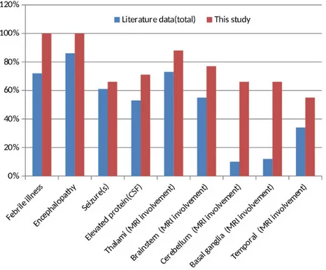
etiology is not fully elucidated, viral or bacterial infections trigger hypercytokinaemia, which is responsible for the disease’s pathogenesis [2,13,15–18]. The prodromal phase has a heterogeneous clinic in the form of the encephalopathy recovery phase. The shortness of interval in the prodromal period causes the cytokine storm to progress rapidly and aggressively. Therefore, on the first day, deep coma, which usually requires mechanical ventilation, and then upper motor neuron findings develop promptly [2,13,16,19]. Frequently, seizures, vomiting, cough and organ dysfunctions accompany the clinical picture. However, these reported findings are not specific to ANE and are seen in many diseases [5,14,16]. Comparison of our patients with febrile illness, seizure (s), encephalopathy, abnormal CSF, and radiological findings with ANE and ANE1 data in the literature (n: 123, total % rates) summary reported on a case basis shown in Table 2 and Figure 2 [2,16,20–25].
Radiological findings are essential in diagnosis due to the difficulties experienced in autopsy and its progressive deterioration. MRI is more sensitive than CT. Lesions mostly show a symmetrical multifocal distribution. T2 weighted and fluid-attenuated inversion recovery (flair) MRI mostly shows hyperintensity in the bilateral thalamus and brainstem. Bilateral thalamus involvement is one of the most defining features of this disease [5]. These lesions mostly show hypointensity and diffusion restriction in T1 weighted [26]. Thalamic involvement is also seen in Japanese encephalitis. However, symmetrical thalamic involvement and brain stem involvement rarely occur compared to ANE [25]. In brain MRI, we found that eight (88.8%) of our patients had bilateral thalamus involvement, seven (77.7%) had T2 and flair hyperintensity in the brainstem (T1 hypointensity in five and two, respectively), and eight patients (88, 8%) had multifocal involvement accompanied by restriction (Table 1). It is also stated that involvement is observed less frequently in the cervical spinal cord, cerebellum, medial temporal lobes (insular cortex) and other regions in the ANE. These radiological findings are consistent with autopsy findings [5,7,21]. As the disease progresses in ANE, the progression of radiological findings from edema to petechial hemorrhage and necrosis correlates with the severity of clinical findings [5,21]. Therefore, serial brain MRI is needed. A study conducted by Kubo et al. showed that lesions occurred in the brain before neurological symptoms were revealed using an advanced MRI device [10]. This condition is emphasized due to the cytokine storm that starts in the subclinical period, causing blood-brain barrier damage and an increase in vascular permeability. Also, MRI has an essential role in evaluating the effectiveness of treatment. The shrinkage of the lesions increases the survival rate as an indicator of treatment effectiveness. Results in ANE are
Ta bl e 1. (C on tin ue d) Ag e/ sex Pr es en tin g sy m pt om Inf ec tio us ag ent In ter va l a A bn or m al la bo ra to ry t es ts b, c Abn or m al sig ns o f cer eb ro s pin al fluid Radio log ic fin din gs (MRI) EEG Tr eat m ent O ut com e Fo llo w-u p (m on th)/ Re la ps (m on th) 9 6 y ea rs 5 m on th/F A ltera tio n in co ns cio usn es s sta tu s ep ilep tic us Fe ve r URI Shock Infl uenza B 1 H yp er tra na min as e Thr om bo cyt op eni a (A ST :140) (AL T: 73) (P LT : 95.000) 1 leu ko cyt es/ mm 3 Pr ot ein: 115.9 m g/dL Tha la m us C er eb el lum Bra in stem Tem po ra l En cep ha lo pa th y m ul tif oc al ep ilep tic ac tiv ity IVMP (5), * PLEX (5) Me nt al m ot or im pa irm en t (s ev er ity) 36/-M: m ale; F : f em ale; URI: u pp er r es pira to ry inf ec tio n; EEG: e le ct ro en cep ha log ra ph y; A ST : a sp ar ta te a min ot ra nsf era se; AL T: a la nin e a min ot ra nsf era se; P LT : p la te let; N: n or m al; ND: no t do cum en te d; IVMP : in tra ven ou s m et hy lp re dni so lo ne; IVI G : in tra ven ou s imm un e g lo bu lin; P LEX: p la sm ap her esi s; MRI: m ag net ic r es on an ce im ag in g. *: o ra l s ter oid m ain ten an ce t hera py a fter IVMP t re at m en t. ain ter va l (t im e b et w een f ev er a nd en cep ha lo pa th y); b o th er et io log ic al p os sib ili ties r ule d o ut (in cludin g inf ec tio us, m et ab olic, a ut oimm un e, a nd imm un e en cep ha lit is); cthe no rm al va lue f or AL T 8 t o 33 U/L, f or AL T 29 t o 33 IU/L, a nd P LT 150,000 t o 450,000/mm 3. M ild im pa irm en t: n ot a ffe ct in g t he q ua lit y o f lif e; m odera te o r s ev er e im pa irm en t: r eq uir in g s pe ci al t re at m en t o r e duc at io n. ANE c as es (5–8): c as e 5: si blin g de at h s to ry (w ith simi la r c linic al p ic tur e); ca ses 6 a nd 7: si blin g; c as es 7 a nd 8: p rio r ep iso des o f en cep ha lo pa th y f ol lo w in g f ev er .
generally not satisfactory and have a wide prognosis range from complete recovery (<10%) to persistent neurological deficits and death. The mortality rate is approximately 30% [2]. Being <1 year of age, the presence of coma findings accompanying brainstem lesions, hemorrhage and necrosis in MRI, various high levels in LFT and high protein values without pleocytosis in CSF examination, and thrombocytopenia are among the poor prognosis criteria [2,3,5,7,12,14]. In the study of Yamamoto et al. conducted with 18 patients, they showed that 66.7% of the patients were in the medium-risk group, and they had a high risk of mortality or severe neurological sequelae. Observing the recurrence increased the risk of neurological sequelae [5,14]. It was shown that patients with normal CSF protein and LFT values recovered more quickly [16]. Compared with the literature summary data, while febrile illness, encephalopathy, seizure(s), CSF elevated protein values and thalamus, brainstem, and temporal lobe involvement rates were similar in our patients, basal ganglia and cerebellum
involvement were observed to be higher in brain MRI (Figure 2). Also, hemorrhage and necrosis were detected in MRI findings in five (55.5%) patients, hypertransanemia in seven (77.7%) patients, and thrombocytopenia in two (22.2%) patients. While hypertransanemia was detected in 62% of the patients in a study, it was found at a rate close to our study [27]. Our study showed that two cases (cases 7 and 8) with recurrence died. At the same time, hemorrhage and necrosis were detected on brain MRIs in the frontal lobe of case 8 and in the thalamus of case 2. In the other three cases (1, 2, and 3) in which necrosis was detected in many brain regions, including the thalamus, it was observed that two of them had moderate and one had minor sequelae. As the ANE-SS values increased, it was seen that there was a trend towards moderate/ severity sequelae or mortality. Besides, our case 5, who had a history of a sibling’s death due to similar clinical complaints, had common multifocal lesions, including very rare cervical spinal cord (C4-C6) involvement. It was Figure 1. MRI images of our cases. a: in the coronal T2-weighted image of case 1, swelling and hyperintensity in the bilateral thalamic
and external capsules and hyperintensity in the left parietal lobe; b: bilateral restriction is seen in the apparent diffusion coefficient (ADC) of the same patient; c: in the ADC of case 2 bilateral thalami and periventricular white matter restriction; d–e: postcontrast T1-weighted images of the same patient reveal more pronounced contrast enhancement pattern in the right thalamus and Hemosiderin deposition is observed in the left thalamus in susceptibility weighted imaging (SWI); f–j: case 5. (f) axial T2-weighted images demonstrating bilateral brainstem, medial temporal lobe, external capsule, occipital involvement, (g) markedly restriction in lesions described in ADC, (h) axial T2-weighted cervical images shows C4-C6 hyperintensity, (ı–j) axial T2-weighted images demonstrating and the apparent diffusion coefficient (ADC) control MRI lesions were seen to regress almost completely after treatment.
Table 2. ANE/ANE1 with children cases ( literature summary and our data).
Study Clinical findings CSF Radiologic findings (MRI involvement region)
Cases number
(reference number)Febrile illness Encephalopathy Seizure(s) Elevated protein Thalamus Brainstem Cerebellum Basal ganglia Temporal
Levine et al., 2020 (summary of cases 2003-2019 ) (2) 56 75 49 55 58 49 9 8 38 Marco et al., 2010 (19) 2 3 3 2 3 0 0 1 0 Britton et al., 2017 (20) 4 4 1 ND 2 0 0 1 0 Mastoyianni et al., 2006 (15) 3 3 1 1 1 1 0 0 0 Koh et al., 2019 (21) 3 3 2 2 3 3 1 1 0 Dadak et al., 2020 (22) ND 1 1 ND 2 2 0 1 0 Lim et al., 2016 (23) 7 4 5 ND 7 1 ND ND ND Kim et al., 2004 (24) 13 13 13 5 14 12 2 3 4
Total of all cases
(123) 88 106 75 65 90 68 12 15 42
Our study (9) 9 9 6 5 8 7 6 6 5
CSF: cerebrospinal fluid; ND: not documented; MRI: magnetic resonance imaging.
observed that this patient had significant regression in control MRI lesions three months after IV pulse steroid + IVIG + PLEX treatments (Figures 1h–1j).
A quick evaluation of the anamnesis, examination, radiological, blood and CSF findings is critical in initiating immunotherapy as soon as possible, preferably within 24 h [17,19]. However, there is no consensus yet on standard treatment guidelines. It has been reported that acyclovir and appropriate antibiotic treatment, as well as early initiation of IV pulse steroid treatment as immunotherapy and administration of high-dose oseltamivir with steroid treatment, are most useful in intensive care conditions [28]. In addition to these, combined IVIG and PLEX treatment is recommended in selected cases [15,16,19]. Therefore, we applied PLEX treatment in six (66.6%) patients with severe clinical findings and diffuse radiological involvement in brain MRI (five combined with IV pulse steroid therapy and IVIG, one combined with IV pulse steroid). When there was no improvement in clinical findings in cases 3 and 5, it was observed that they had moderate sequelae in their follow-up, although PLEX was added to their treatment later. As mentioned before, excessive cytokine release, especially IL-6 and TNF-α, occurs in ANEC, just as in COVID-19 infection. We could not administer the anti-IL6 or anti-TNF α preparations currently available in our country due to the COVID-19 pandemic, considering the urgency of the treatment, since all of our cases were before the pandemic and the supply of these preparations required a prolonged effort at that time. However, instead of these preparations, we have been successfully applying PLEX treatment for the last 7–8 years to remove IL-6, TNF-α, and other proinflammatory cytokines from the circulation. As mentioned, our three (6–8) cases with ANE1 treated in previous periods when we could not technically apply PLEX died. This situation suggests that PLEX treatment, together with immunotherapy in the cytokine storm, is a very beneficial method for survival. Because our other case with ANE1 (case 5) has been followed up in an outpatient setting for 1.5 years, with significant resolution in follow-up MRIs and moderate motor impairment findings with PLEX applied differently from the treatment of these patients (Figures 1g and 1h). No RanBP2 mutation was detected in any of our ANE1 cases we could examine. Considering our patients who died, it was observed that ANE1 had a worse prognosis than sporadic ANE.
This study has several limitations. Concerning the number of patients, a large enough sample group was not obtained. Since this is a retrospective study, the statistical data’s homogenization has been difficult because of some deficiencies. Also, the follow-up periods were not long enough, and the follow-ups irregularly created a handicap
in evaluating the process from diagnosis to prognosis. However, the cohort analysis reflecting the disease’s clinical importance, diagnostic methods, and treatment strategies, especially PLEX efficiency, suggests that this study has a strong aspect. There are still few reports of in-depth analysis in the literature. Therefore, although our study’s sample size was limited, there was still certain significance.
In conclusion, it is not possible to conduct randomized controlled clinical studies because of its rare occurrence. The data obtained so far are based on the clinical experience of different centers. Therefore, it is necessary to increase the centers’ experience to ensure early diagnosis and treatment standardization and predict the prognosis. For this purpose, we think that our experiences will shed light on guiding other centers. In this context, according to our results, it was observed that thalamic involvement increased as the interval between fever and encephalopathy became shorter, and brainstem involvement increased in patients over four years of age. Besides, we think that resistant vomiting accompanying encephalopathy in the parainfectious period is a stimulus for ANE’s clinical presentation. Our other results were that, except for three cases in whom PLEX could not be performed and died, all of our surviving patients had neurological sequelae ranging from moderate to severe, despite the apparent radiological improvement observed within an average of three months from all treatment approaches, including PLEX. These findings show the severity of mortality and morbidity risks of ANEC, and PLEX treatment is a treatment option that can prevent mortality. Therefore, long-term outpatient clinical and, when necessary, radiological follow-up of our patients is critical in prognosis. However, more studies related to ANE are needed in terms of increasing the experience.
Acknowledgments
We would like to thank all children and their parents and also the medical staff at all participating institutions for their valuable assistance throughout this study.
Conflict of interest
None of the authors has any conflict of interest to disclose.
Financial disclosure
All authors do not have any financial or personal relationships with other persons or organizations that may inappropriately influence (bias) their work.
Ethics approval
The study was first approved by the University of Health Sciences, Dr. Sami Ulus Maternity and Children Training and Research Hospital (EC number: E-20/10-003).
References
1. Mizuguchi M, Abe J, Mikkaichi K, Noma S, Yoshida K et al. Acute necrotising encephalopathy of childhood: a new syndrome presenting with multifocal, symmetric brain lesions. Journal of Neurology, Neurosurgery & Psychiatry 1995; 58 (5): 555-561. doi: 10.1136/jnnp.58.5.555
2. Levine JM, Ahsan N, Ho E, Santoro JD. Genetic acute necrotizing encephalopathy associated with RANBP2: clinical and therapeutic implications in pediatrics. Multiple Sclerosis and Related Disorders 2020; 43: 102194. doi: 10.1016/j. msard.2020.102194
3. Ormitti F, Ventura E, Summa A, Picetti E, Crisi G. Acute necrotizing encephalopathy in a child during the 2009 influenza A (H1N1) pandemia: MR imaging in diagnosis and follow-up. American Journal of Neuroradiology 2010; 31 (3): 396-400. doi: 10.3174/ajnr.A2058
4. Lyon JB, Remigio C, Milligan T, Deline C. Acute necrotizing encephalopathy in a child with H1N1 influenza infection. Pediatric Radiology 2010; 40 (2): 200-205. doi: 10.1007/ s00247-009-1487-z
5. Wu X, Wu W, Pan W, Wu L, Liu K et al. Acute necrotizing encephalopathy: an underrecognized clinicoradiologic disorder. Mediators of Inflammation 2015; 2015: 792578. doi: 10.1155/2015/792578
6. Virhammar J, Kumlien E, Fällmar D, Frithiof R, Jackmann S et al. Acute necrotizing encephalopathy with SARS-CoV-2 RNA confirmed in cerebrospinal fluid. Neurology 2020; 95 (10): 445-449. doi: 10.1212/WNL.0000000000010250
7. Neilson DE. The interplay of infection and genetics in acute necrotizing encephalopathy. Current Opinion in Pediatrics 2010; 22 (6): 751-757. doi: 10.1097/MOP.0b013e3283402bfe 8. Ichiyama T, Nishikawa M, Yoshitomi T, Hayashi T, Furukawa
S. Tumor necrosis factor alfa, interleukin lβ, and interleukin 6 in cerebrospinal fluid from children with prolonged febrile seizures. Comparison with acute encephalitis/encephalopathy. Neurology 1998; 50 (2): 407-411. doi: 10.1212/wnl.50.2.407 9. Hoshino A, Saitoh M, Oka A, Okumura A, Kubota M et
al. Epidemiology of acute encephalopathy in Japan, with emphasis on the association of viruses and syndromes. Brain and Development 2012; 34 (5): 337-343. doi: 10.1016/j. braindev.2011.07.012
10. Kubo T, Sato K, Kobayashi D, Motegi A, Kobayashi O et al. A case of HHV-6 associated acute necrotizing encephalopathy with increase of CD56 bright NK cells. Scandinavian Journal of Infectious Diseases 2006; 38 (11-12): 1122-1125. doi: 10.1080/00365540600740520
11. Wang KY, Singer HS, Crain B, Gujar S, Lin DD. Hypoxic-ischemic encephalopathy mimicking acute necrotizing encephalopathy. Pediatric Neurology 2015; 52 1): 110-114. doi: 10.1016/j.pediatrneurol.2014.09.009
12. Singh RR, Sedani S, Lim M, Wassmer E, Absoud M. RANBP2 mutation and acute necrotizing encephalopathy: 2 cases and a literature review of the expanding clinico-radiological phenotype. European Journal of Paediatric Neurology 2015; 19 (2): 106-113. doi: 10.1016/j.ejpn.2014.11.010
13. Neilson D, Eiben R, Waniewski S, Hoppel C, Varnes M et al. Autosomal dominant acute necrotizing encephalopathy. Neurology 2003;61 (2): 226-230. doi: 10.1212/01. wnl.0000073544.28775.1a
14. Yamamoto H, Okumura A, Natsume J, Kojima S, Mizuguchi M. A severity score for acute necrotizing encephalopathy. Brain and Development 2015; 37 (3): 322-327. doi: 10.1016/j. braindev.2014.05.007
15. Okumura A, Mizuguchi M, Kidokoro H, Tanaka M, Abe S et al. Outcome of acute necrotizing encephalopathy in relation to treatment with corticosteroids and gammaglobulin. Brain and Development 2009; 31 (3): 221-227. doi: 10.1016/j. braindev.2008.03.005
16. Mastroyianni SD, Gionnis D, Voudris K, Skardoutsou A, Mizuguchi M. Acute necrotizing encephalopathy of childhood in non-Asian patients: report of three cases and literature review. Journal of Child Neurology 2006; 21 (10): 872-879. doi: 10.1177/08830738060210101401
17. Soriano-Ramos M, Navarro-Abia V, Enamorado NN, Camacho-Salas A, De Aragón AM et al. Steroids for familial acute necrotizing encephalopathy: a future investment? Clinical Neurology and Neurosurgery 2018; 174: 134-136. doi: 10.1016/j.clineuro.2018.09.014
18. Bloch C, Suter B, Fischmann A, Gensicke H, Rüegg S et al. Only a touch of the flu? The simultaneous manifestation of acute necrotizing encephalopathy in two consanguineous patients. Open Forum Infectious Diseases 2015; 2 (2): ofv013. doi: 10.1093/ofid/ofv013
19. Alsolami A, Shiley K. Successful treatment of influenza-associated acute necrotizing encephalitis in an adult using high-dose oseltamivir and methylprednisolone: case report and literature review. Open Forum Infectious Diseases 2017; 4 (3): ofx145. doi: 10.1093/ofid/ofx145
20. Marco EJ, Anderson JE, Neilson DE, Strober JB. Acute necrotizing encephalopathy in 3 brothers. Pediatrics 2010; 125 (3): e693-698. doi: 10.1542/peds.2009-1984
21. Britton PN, Dale RC, Blyth CC, Macartney K, Crawford NW et al. Influenza-associated encephalitis/encephalopathy identified by the Australian Childhood Encephalitis Study 2013–2015. The Pediatric Infectious Disease Journal 2017; 36 (11): 1021-1026. doi: 10.1097/INF.0000000000001650
22. Koh JC, Murugasu A, Krishnappa J, Thomas T. Favorable outcomes with early interleukin 6 receptor blockade in severe acute necrotizing encephalopathy of childhood. Pediatric Neurology 2019; 98: 80-84. doi: 10.1016/j. pediatrneurol.2019.04.009
23. Dadak M, Pul R, Lanfermann H, Hartmann H, Hehr U et al. Varying patterns of CNS imaging in influenza A encephalopathy in childhood. Clinical Neuroradiology 2020; 30 (2): 243-249. doi: 10.1007/s00062-018-0756-3
24. Lim HY, Ho VPY, Lim TCC, Thomas T, Chan DWS. Serial outcomes in acute necrotising encephalopathy of childhood: a medium and long term study. Brain and Development 2016; 38 (10): 928-936. doi: 10.1016/j.braindev.2016.05.002
25. Kim JH, Kim I-O, Lim MK, Park MS, Choi CG et al. Acute necrotizing encephalopathy in Korean infants and children: imaging findings and diverse clinical outcome. Korean Journal of Radiology 2004; 5 (3): 171-177. doi: 10.3348/kjr.2004.5.3.171 26. Carmo RLD, Alves Simão AK, Amaral LLFd, Inada BSY,
Silveira CF et al. Neuroimaging of emergent and reemergent infections. Radiographics 2019; 39 (6): 1649-1671. doi: 10.1148/rg.2019190020
27. Lee Y-J, Hwang S-K, Kwon S. Acute necrotizing encephalopathy in children: a long way to go. Journal of Korean Medical Science 2019; 34 (19): e143. doi: 10.3346/jkms.2019.34.e143
28. Okumura A, Mizuguchi M, Aiba H, Tanabe T, Tsuji T et al. Delirious behavior in children with acute necrotizing encephalopathy. Brain and Development 2009; 31 (8): 594-599. doi: 10.1016/j.braindev.2008.09.002