SCIENTIFIC ARTICLE
Reliability and diagnostic utility of radiographs in patients
with incomplete atypical femoral fractures
Ulas Akgun1&Umut Canbek1&Nevres Hurriyet Aydogan1
Received: 15 September 2018 / Revised: 13 March 2019 / Accepted: 21 March 2019 / Published online: 10 April 2019 # ISS 2019
Abstract
Objective The aim of this study was to evaluate the sensitivity and specificity of radiographs in identifying incomplete atypical femoral fractures and to determine interobserver and intra-observer reliability.
Materials and methods Anterior–posterior and lateral radiographs of 10 femurs with incomplete atypical femoral fractures confirmed using bone scintigraphy and magnetic resonance imaging, and 40 femurs without incomplete atypical femoral fractures confirmed using bone scintigraphy, were reviewed by 4 orthopedic surgeons and 4 radiology specialists. Sensitivity, specificity, accuracy, positive predictive value, and negative predictive value were calculated to determine a diagnosis of incomplete atypical femoral fractures. Interobserver reliability was measured using Fleiss’ kappa value, and intra-observer reliability was ascertained using Cohen’s kappa statistic.
Results Mean sensitivity, specificity, accuracy, and the positive and negative predictive values were found to be 89, 89, 89, 67, and 97% respectively, in diagnosing incomplete atypical femoral fractures using radiographs. Interobserver reliability was found to be at a good level (Fleiss’ kappa = 0.66, standard error = 0.03, 95% confidence interval = 0.61–0.71). Intra-observer Cohen’s kappa values ranged from 0.53 to 0.91.
Conclusion The use of radiography was satisfactory in identifying incomplete atypical femoral fractures, and the level of interobserver agreement was found to be good. As radiographs are associated with low positive predictive values, an advanced imaging method should be used when an increase in femoral cortical thickness is the only contributory factor to suspicion of an incomplete atypical femoral fracture.
Keywords Diagnostic accuracy . Femoral fractures . Insufficiency fractures . Reliability . Sensitivity and specificity
Introduction
Osteoporosis is a common disease in the elderly population. The increased risk of bone fractures causes significant morbidity and mortality in these patients, and it poses an economic burden on healthcare systems [1–3]. Bisphosphonates are the most com-monly used antiresorptive agents for osteoporosis treatment, and these drugs are potent inhibitors of osteoclast-mediated bone resorption [4,5]. The efficacy of bisphosphonates in reducing the risk of osteoporotic fractures has been demonstrated in extensive clinical studies and, generally, they have been found to be safe
[6]. However, after 2005, an increasing number of atypical subtrochanteric and femoral diaphysis fractures have been re-ported. The long-term use of bisphosphonates is thought to be the cause of these fractures [7–11].
The American Society of Bone and Mineral Research (ASBMR) formed a task force related to atypical femoral frac-tures (AFFs), and the first report about these fracfrac-tures was com-pleted in 2010 [12]. In 2013, the diagnostic criteria were reviewed, and the ASBMR working group defined complete AFFs as noncomminuted, transverse, or short oblique subtrochanteric or femoral shaft fractures that extend across the entire femoral shaft, often with the formation of a medial spike [13]. The periosteal or endosteal thickening of the lateral cortex, in the form ofBbeaking^ or Bflaring,^ is a major feature, whereas a generalized increase in the cortical thickness of the femoral diaphysis is a minor feature in the case definition of AFFs.
The long-term exposure of bisphosphonates leads to im-paired bone quality with the suppression of bone remodeling
* Ulas Akgun [email protected]
1
Faculty of Medicine, Department of Orthopaedics and Traumatology, Mugla Sitki Kocman University, 48000 Mentese, Mugla, Turkey
Check for
and accumulation of microdamage, particularly at sites of high bone turnover [14]. Incomplete AFFs (iAFFs) may also be encountered in the form of an insufficiency fracture at the lateral cortex of the subtrochanteric or diaphyseal region of the femur. In contrast to typical stress fractures, in which the compressive load of the femur passes to the medial surface, iAFFs occur along
the lateral cortex of the femur, where the exposure to tensile forces is high [15].
The radiological features of an iAFF may vary as focal cortical thickening, radiolucency as a dreaded black line, stri-ations or cavities [16]. Variations in the appearance of an iAFF can be interpreted differently in individual evaluations. Although several studies have evaluated the sensitivity,
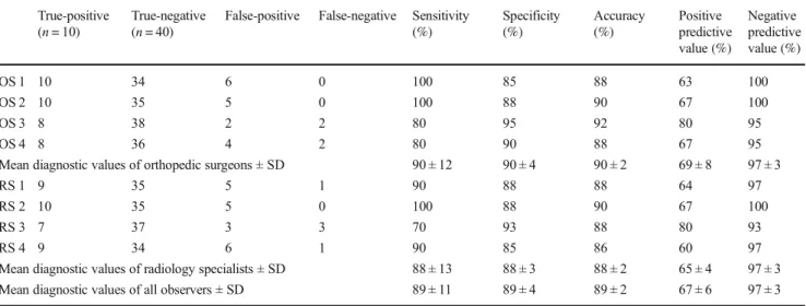
Fig. 1 a A woman aged 72 years presented with left thigh and knee pain, had been taking alendronate therapy for 8 years, and was diagnosed with an incomplete atypical femoral fracture in the left femur. Focal cortical thickening and a lucent line at the mid-diaphysis of the left femur (white arrow shows enlargement of thewhite box) is seen. Note that, without exception, all ob-servers evaluated this radiograph as an incomplete fracture in both reviews.b Increased isotope up-take is seen at the lateral cortex of the left mid-diaphyseal femur (black arrow) with technetium-99 m bone scintigraphy of the same patient.c Coronal T2 MRI shows incomplete fracture line (white arrow) and adjacent bone marrow edema ;
,.
c
l
\I
}
I I•
•
,;,
..
.
,
fl
•
•
I'
,
'
,~,
'
r.
•
-•
.
•
•
specificity, and reliability of diagnostic imaging procedures for complete AFFs, to the best of our knowledge, similar studies have not been performed in this regard for iAFFs [17,18]. The aim of this study was to evaluate the sensitivity and specificity of radiographs in identifying iAFFs and to determine interobserver and intra-observer reliability.
Materials and methods
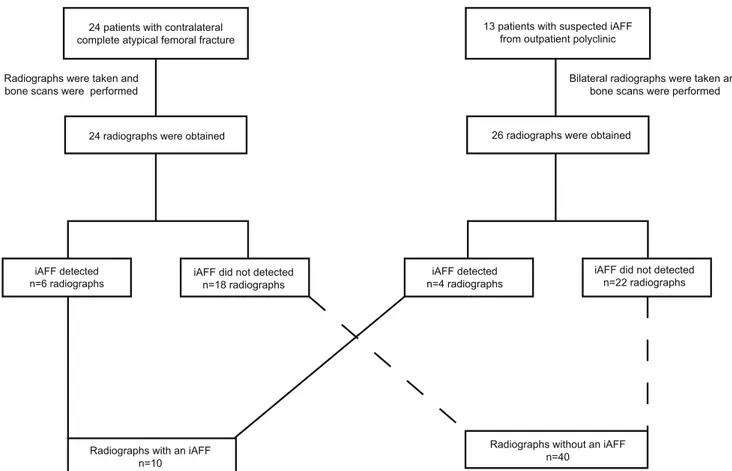
Approval for this study was granted by the Institutional Ethics Committee and informed consent waived for the retrospective review of medical records. A total of 24 consecutive patients presented to our clinic with complete AFFs between January 2014 and May 2018. The contralateral femur of the patients was examined, and iAFFs were identified in 6 of the patients. In addition, iAFFs were suspected in 13 patients with a history of long-term bisphosphonate use who presented at the outpa-tient polyclinic with knee or thigh pain. Bilateral femoral ra-diographs were obtained from these patients to conduct a com-parison with the asymptomatic side. An incomplete fracture was detected in 4 of the 13 patients, and none of them had bilateral involvement.
Evidence of an iAFF was confirmed in all cases using technetium-99 m bone scintigraphy, along with radiography. Presence of focally increased isotope uptake at the suspected
area was accepted as an iAFF. Magnetic resonance imaging (MRI) was also performed for 7 of the 10 patients in whom iAFF was detected and bone marrow edema was seen in all these cases (Fig.1). MRI could not be performed for 3 pa-tients; 2 of them had a cardiac implant, and 1 of them did not consent to undergo the MRI owing to claustrophobia. Lack of evidence of focally increased isotope uptake on the bone scans was accepted as a negative diagnosis, and a further MRI ex-amination was not performed for these cases.
High-resolution digital images of the anterior–posterior and lateral radiographs of 50 femurs of 37 patients (10 femurs with iAFFs and 40 femurs without iAFFs) were included in this study. The flow diagram of the radiographs used in the study is shown in Fig.2.
All 37 patients were women aged >50 years old (mean 67.1 ± 6.9 years), and all of them had been receiving bisphos-phonate treatment for >5 years (mean 7.5 ± 1.4 years). The characteristics of patients with and without iAFFs are given in Table1.
None of the images included any clinical information re-lated to the patients. The radiographs were numbered using a random sequence and evaluated separately by four indepen-dent orthopedic surgeons and four radiology specialists with clinical experience ranging from 10 to 22 years. Before the evaluation, the observers were informed about the radiological characteristics of AFFs related to bisphosphonate treatment,
24 radiographs were obtained
iAFF detected n=6 radiographs
iAFF did not detected n=18 radiographs Radiographs were taken and
bone scans were performed
Radiographs with an iAFF n=10
24 patients with contralateral complete atypical femoral fracture
13 patients with suspected iAFF from outpatient polyclinic
Bilateral radiographs were taken and bone scans were performed
26 radiographs were obtained
iAFF detected n=4 radiographs
iAFF did not detected n=22 radiographs
Radiographs without an iAFF n=40
Fig. 2 The flow diagram of radiographs used in this study.iAFF incomplete atypical femoral fracture
'
'
and they were shown images of iAFFs from the literature [9, 13,15]. When performing the assessments, the observers were blinded to the age of the patients, duration of bisphosphonate use, pain, and other personal clinical information. The ob-servers were asked to evaluate the presence or absence of iAFFs according to the 2013 ASBMR criteria (Table2) and to mark the location of an identified iAFF with a digital mark-er. The evaluations of the observers were recorded on standard data collection forms, and the data obtained from the forms were recorded electronically by two different researchers to prevent errors. Two weeks after the first evaluation, the same radiographs presented in a different sequence were evaluated once again by the observers.
Sensitivity, specificity, accuracy, and the positive and negative predictive values in the determination of iAFFs were calculated for each observer. Interobserver and intra-observer reliability were measured using Fleiss’ kappa and Cohen’s kappa respec-tively. The kappa values were evaluated as <0 poor agreement, 0.01–0.20 agreement at an insignificant level, 0.21–0.40 weak agreement, 0.41–0.60 moderate agreement, 0.61–0.80 good agreement, and 0.81–1.00 very good agreement [19]. The data obtained in this study were analyzed using IBM SPSS Statistics for Windows, version 22 (IBM, Armonk, NY, USA).
Results
Mean sensitivity 89% (range = 70–100%), specificity 89% (range = 85–95%), accuracy 89% (range = 86–92%), positive predictive value 67% (range = 60–80%), and negative predic-tive value 97% (range = 93–100%) were reported for the
identification of iAFFs using radiography. The orthopedic sur-geons and radiology specialists demonstrated similar diagnos-tic accuracy in detecting iAFFs. The diagnosdiagnos-tic statisdiagnos-tical values of each observer are shown in Table3.
Overall interobserver reliability in identifying iAFFs was found to be at a good level (Fleiss’ kappa = 0.66, standard error = 0.03, 95% confidence interval = 0.61–0.71). The inter-observer reliability within orthopedic surgeons and radiology specialists showed similar results (Fleiss’ kappa = 0.67, stan-dard error = 0.06, 95% confidence interval = 0.55–0.78, and Fleiss’ kappa = 0.63, standard error = 0.06, 95% confidence interval = 0.51–0.74 respectively). The intra-observer Cohen’s kappa values ranged from 0.53 to 0.91 (Table4).
Discussion
In recent years, the frequency of AFFs, which are thought to be due to the prolonged use of bisphosphonates, is increasing [20]. The ASBMR formed a task force related to AFFs and defined the major and minor characteristics of these fractures [12,13]. The characteristics of iAFFs are described as lateral cortex in-volvement only, periosteal or endosteal thickening of the lateral cortex, the presence of unilateral or bilateral prodromal symp-toms, and generalized cortical thickening in the femur diaphysis. The radiographic appearance of iAFFs varies as they are characterized by a wide range of features. Focal elevations that resemble bumps on the lateral cortex of the femur are fre-quently the only sign of an iAFF on early radiographs. Sometimes, a transverse radiolucent line in aBmound^-shaped focal elevation, defined as the Bdreaded black line,^ is
Table 2 American Society of Bone and Mineral Research Task Force 2013 revised case definitions for atypical femoral fractures [13]
Major features Minor features
The fracture is associated with minimal or no trauma, as in a fall from a standing height or less
Generalized increase in the cortical thickness of the femoral diaphysis The fracture line originates at the lateral cortex and is substantially transverse in
its orientation, although it may become oblique as it progresses medially across the femur
Unilateral or bilateral prodromal symptoms such as dull or aching pain in the groin or thigh
Complete fractures extend through both cortices and may be associated with a medial spike; incomplete fractures involve only the lateral cortex
Bilateral incomplete or complete femoral diaphysis fractures The fracture is noncomminuted or minimally comminuted Delayed fracture healing
Localized periosteal or endosteal thickening of the lateral cortex is present at the fracture site (Bbeaking^ or Bflaring^)
Table 1 Characteristics of patients with and without an incomplete atypical femoral fracture (iAFF)
iAFF (n = 10) Non-iAFF (n = 27) p* value
Age ± SD (years) (range) 69.3 ± 4.3 (62–76) 68.9 ± 7.7 (57–82) 0.853
Female gender (%) 10 (100%) 40 (100%) N/A
Duration of BP treatment ± SD, years (range) 7.8 ± 0.9 (7–10) 7.7 ± 1.7 (5–12) 0.674 BP bisphosphonate, SD standard deviation, N/A not applicable, as all the patients were women
apparent [9,16]. Although several studies have evaluated the diagnostic accuracy and interobserver reliability of radio-graphs for complete AFF, to the best of our knowledge, sim-ilar studies have not been performed in this regard on iAFFs [17,18]. Therefore, we aimed to evaluate the diagnostic accu-racy, interobserver reliability, and intra-observer reliability of radiography in diagnosing iAFFs.
We used radiographs of 10 iAFFs and 40 radiographs of femurs without iAFFs in a similar age group for this study. It can be difficult to diagnose an iAFF in patients with extreme lateral femoral bowing or generalized cortical thickening of the femur. Accordingly, advanced imaging methods with high sensitivity, such as bone scintigraphy or MRI, were used to confirm a diagnosis of iAFFs in the study [21].
In global medical diagnostic testing, a positive predictive value >90%, sensitivity >85%, and specificity >85% are usual-ly considered to beBhigh^ [22,23]. Sensitivity, specificity, and
accuracy were high (89%), the positive predictive value was low (67%), and the negative predictive value was high (97%) in the current study in diagnosing iAFFs using radiography. These results indicate that the radiographs exhibit a satisfactory level of effectiveness in the screening of the iAFFs.
Although theBfalse-negative^ assessment rate among ob-servers was low (Fig.3), numerousBfalse-positive^ interpre-tations of iAFFs were made during the analysis, as reflected by the low mean positive predictive value. When we reviewed theBfalse-positive^ cases, we observed that some observers marked diffuse cortical thickenings (Fig.4) as iAFFs.
We think that there is a lack of clarity in the definition of diffuse cortical thickening according to the ASBMR criteria, and uncertainty about the diagnostic importance of cortical thickening. Moreover, the presence of diffuse cortical thick-ening in AFFs is also controversial. Niimi et al. found no significant difference with respect to femoral cortical thicken-ing between osteoporotic patients with long-term bisphospho-nate use and those not receiving bisphosphobisphospho-nate treatment [24]. In addition, Koeppen et al. compared femoral cortical thickenings in patients aged >55 years with ordinary femoral fractures and those with AFF, and they found no significant difference; therefore, they recommended that this criterion should be revised [25]. Within the ASBMR criteria, there are clinical criteria, such as prodromal syndrome, in addition to the radiological criteria. Another reason for a low positive predictive value may be that the observers had no clinical information about the patients. In our opinion, if an iAFF is suspected both clinically and radiologically, it would be rea-sonable to use an advanced imaging modality.
When the interobserver reliability was evaluated, the kappa values were found to be at a good level of agreement. In a more detailed examination of the intra-observer kappa values, the agreement was found to be at a moderate level in 1 orthopedic
Table 4 Intra-observer reliabilities of each observer for the determination of iAFFs
Cohen’s kappa SE Minimum– maximum (95% CI) p value OS 1 0.91 0.06 (0.79–1.00) <0.001 OS 2 0.53 0.13 (0.34–0.82) <0.001 OS 3 0.65 0.14 (0.37–0.93) <0.001 OS 4 0.88 0.08 (0.73–1.00) <0.001 RS 1 0.67 0.12 (0.44–0.91) <0.001 RS 2 0.75 0.11 (0.54–0.95) <0.001 RS 3 0.73 0.13 (0.48–0.98) <0.001 RS 4 0.91 0.07 (0.77–1.00) <0.001
CI confidence interval, OS orthopedic surgeon, RS radiology specialist, SE standard error
Table 3 Sensitivity, specificity, positive predictive value, and negative predictive value of the radiographs for each observer True-positive
(n = 10)
True-negative (n = 40)
False-positive False-negative Sensitivity (%) Specificity (%) Accuracy (%) Positive predictive value (%) Negative predictive value (%) OS 1 10 34 6 0 100 85 88 63 100 OS 2 10 35 5 0 100 88 90 67 100 OS 3 8 38 2 2 80 95 92 80 95 OS 4 8 36 4 2 80 90 88 67 95
Mean diagnostic values of orthopedic surgeons ± SD 90 ± 12 90 ± 4 90 ± 2 69 ± 8 97 ± 3
RS 1 9 35 5 1 90 88 88 64 97
RS 2 10 35 5 0 100 88 90 67 100
RS 3 7 37 3 3 70 93 88 80 93
RS 4 9 34 6 1 90 85 86 60 97
Mean diagnostic values of radiology specialists ± SD 88 ± 13 88 ± 3 88 ± 2 65 ± 4 97 ± 3
Mean diagnostic values of all observers ± SD 89 ± 11 89 ± 4 89 ± 2 67 ± 6 97 ± 3
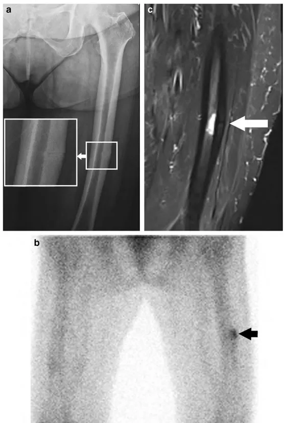
Fig. 4 a A woman aged 75 years presented with hip and back pain, and had been taking alendronate therapy for 9 years. The radiographic image was rated false-positive for the diagnosis of an iAFF by several observers. Increased cortical thickness along the subtrochanteric region of the prox-imal femur (white arrow) is seen on radiography. Two orthopedic sur-geons (OS1 and OS2) and two radiology specialists (RS2 and RS4) marked the subtrochanteric area as an iAFF in the first evaluation. In
the second review, the same orthopedic surgeons and one radiology spe-cialist (RS2) interpreted the same area as an incomplete atypical fracture. b Bone scintigraphy examination was performed owing to the increased cortical thickness of the right femur and a suspected iAFF. No increased isotope uptake in the two femurs was observed and the patient was eval-uated as not having an incomplete fracture
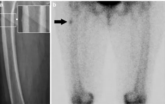
Fig. 3 a A woman aged 64 years presented with right hip pain, and had been taking alendronate therapy for 8 years. The radiographic image was rated false-negative for the diagnosis of an iAFF by several observers. The radiographic image of the right femur shows small periosteal focal cortical thickening (white arrow shows enlargement of the white box) at the subtrochanteric region. Two orthopedic surgeons (OS3 and OS4) and three radiology specialists (RS1, RS3, and RS4) interpreted this
radiograph as normal in the first evaluation. In the second evaluation, no incomplete fracture was detected by three orthopedic surgeons (OS2, OS3, and OS4) and two radiology specialists (RS1 and RS2).b Increased isotope uptake is seen at the lateral cortex of the right subtrochanteric region (black arrow) with technetium-99 m bone scintigraphy of the same patient. Unfortunately, MRI could not be performed owing to the patient suffering from claustrophobia
surgeon, at a good level in 1 orthopedic surgeon and 3 radiology specialists, and at a very good level in 2 orthopedic surgeons and 1 radiology specialist. Based on these results, it can be interpreted that the observers made similar judgements for the same cases.
Adams et al. evaluated the sensitivity, specificity, and reli-ability of the radiological characteristics of complete AFFs as defined by the ASBMR criteria. According to that study, the two most sensitive fracture characteristics in the differentia-tion of AFFs were a transverse fracture pattern in the lateral cortex and a lack of comminution (94 and 93% respectively), and the determination of generalized cortical thickening ex-hibited the lowest sensitivity (49%) [17]. Moreover, interob-server reliability for the identification of generalized cortical thickening was evaluated asBweak^ (kappa = 0.16).
The current study had both limitations and strengths. We think that the use of advanced imaging methods for the con-firmation of the presence or absence of iAFFs is more objec-tive than the determination of AFFs by a committee decision using only the radiographs. The limitations of this study are that no power analysis was performed before the study and the number of radiographs with iAFFs was small. In addition, the observers only evaluated the radiographs of one femur for each patient, whereas in clinical practice, a comparison with the contralateral side assists in the diagnosis of an . If the observers in the current study had been able to compare the images with those obtained for the contralateral side, their findings on diffuse cortical thickening might have been con-siderably enhanced, leading to a lower false-positive rate and higher mean positive predictive value.
In conclusion, the use of radiography was satisfactory in identifying iAFFs, and the level of interobserver agreement was found to be good. As radiographs are associated with low positive predictive values, an advanced imaging method should be used when an increase in femoral cortical thickness is the only contributory factor to a suspected iAFF.
Compliance with ethical standards
Conflicts of interest The authors declare that they have no conflicts of interest.
Ethical approval All of the procedures performed in this study involving human participants were in accordance with the ethical standards of the institutional and/or national research committee and with the 1964 Declaration of Helsinki and its later amendments or comparable ethical standards.
Informed consent The need to obtain informed consent was waived as this research comprised a retrospective review of the medical records.
References
1. Burge R, Dawson-Hughes B, Solomon DH, Wong JB, King A, Tosteson A. Incidence and economic burden of
osteoporosis-related fractures in the United States, 2005-2025. J Bone Miner Res. 2007;22(3):465–75.
2. Fischer S, Kapinos KA, Mulcahy A, Pinto L, Hayden O, Barron R. Estimating the long-term functional burden of osteoporosis-related fractures. Osteoporos Int. 2017;28(10):2843–51.
3. Hopkins RB, Tarride JE, Leslie WD, et al. Estimating the excess costs for patients with incident fractures, prevalent fractures, and nonfracture osteoporosis. Osteoporos Int. 2013;24(2):581–93. 4. Cummings SR, Cosman F, Lewiecki EM, et al. Goal-directed
treat-ment for osteoporosis: a progress report from the ASBMR-NOF working group on goal-directed treatment for osteoporosis. J Bone Miner Res. 2017;32(1):3–10.
5. Silverman S, Christiansen C. Individualizing osteoporosis therapy. Osteoporos Int. 2012;23(3):797–809.
6. Pazianas M, Abrahamsen B. Safety of bisphosphonates. Bone. 2011;49(1):103–10.
7. Abrahamsen B, Eiken P, Eastell R. Subtrochanteric and diaphyseal femur fractures in patients treated with alendronate: a register-based national cohort study. J Bone Miner Res. 2009;24(6):1095–102. 8. Clout A, Narayanasamy N, Harris I. Trends in the incidence of
atypical femoral fractures and bisphosphonate therapy. J Orthop Surg. 2016;24(1):36–40.
9. Feldman F. Atypical diaphyseal femoral fractures—new aspects. Skeletal Radiol. 2012;41(1):75–81.
10. Goh SK, Yang KY, Koh JS, et al. Subtrochanteric insufficiency fractures in patients on alendronate therapy: a caution. J Bone Joint Surg Br. 2007;89(3):349–53.
11. Odvina CV, Zerwekh JE, Rao DS, Maalouf N, Gottschalk FA, Pak CY. Severely suppressed bone turnover: a potential complication of alendronate therapy. J Clin Endocrinol Metab. 2005;90(3):1294– 301.
12. Shane E, Burr D, Ebeling PR, et al. Atypical subtrochanteric and diaphyseal femoral fractures: report of a task force of the American Society for Bone and Mineral Research. J Bone Miner Res. 2010;25(11):2267–94.
13. Shane E, Burr D, Abrahamsen B, et al. Atypical subtrochanteric and diaphyseal femoral fractures: second report of a task force of the American Society for Bone and Mineral Research. J Bone Miner Res. 2014;29(1):1–23.
14. Ng AC, Png MA, Chua DT, Koh JS, Howe TS. Review: epidemi-ology and pathophysiepidemi-ology of atypical femur fractures. Curr Osteoporos Rep. 2014;12(1):65–73.
15. Porrino JA Jr, Kohl CA, Taljanovic M, Rogers LF. Diagnosis of proximal femoral insufficiency fractures in patients receiving bis-phosphonate therapy. AJR Am J Roentgenol. 2010;194(4):1061–4. 16. Dell R, Greene D. A proposal for an atypical femur fracture treat-ment and prevention clinical practice guideline. Osteoporos Int. 2018;29(6):1277–83.
17. Adams AL, Xue F, Chantra JQ, et al. Sensitivity and specificity of radiographic characteristics in atypical femoral fractures. Osteoporos Int. 2017;28(1):413–7.
18. Rosenberg ZS, La Rocca Vieira R, Chan SS, et al. Bisphosphonate-related complete atypical subtrochanteric femoral fractures: diag-nostic utility of radiography. AJR Am J Roentgenol. 2011;197(4): 954–60.
19. Landis JR, Koch GG. The measurement of observer agreement for categorical data. Biometrics. 1977;33(1):159–74.
20. Starr J, Tay YKD, Shane E. Current understanding of epidemiolo-gy, pathophysioloepidemiolo-gy, and management of atypical femur fractures. Curr Osteoporos Rep. 2018;16(4):519–29.
21. Matcuk GR Jr, Mahanty SR, Skalski MR, Patel DB, White EA, Gottsegen CJ. Stress fractures: pathophysiology, clinical presenta-tion, imaging features, and treatment options. Emerg Radiol. 2016;23(4):365–75.
22. Arroll B, Schechter MT, Sheps SB. The assessment of diagnostic tests: a comparison of medical literature in 1982 and 1985. J Gen Intern Med. 1988;3(5):443–7.
23. Sheps SB, Schechter MT. The assessment of diagnostic tests. A survey of current medical research. JAMA. 1984;252(17):2418–22. 24. Niimi R, Kono T, Nishihara A, et al. Cortical thickness of the femur and long-term bisphosphonate use. J Bone Miner Res. 2015;30(2): 225–31.
25. Koeppen VA, Schilcher J, Aspenberg P. Atypical fractures do not have a thicker cortex. Osteoporos Int. 2012;23(12):2893–6.
Publisher’s note Springer Nature remains neutral with regard to jurisdic-tional claims in published maps and institujurisdic-tional affiliations.
![Table 2 American Society of Bone and Mineral Research Task Force 2013 revised case definitions for atypical femoral fractures [ 13 ]](https://thumb-eu.123doks.com/thumbv2/9libnet/3849186.35062/4.892.261.821.91.187/american-society-mineral-research-revised-definitions-atypical-fractures.webp)