INTRODUCTION
Prostate cancer is the most common cancer in men and the second most common reason of cancer-related deaths.1Depending on the rise of routine measurements of prostate specific antigen (PSA), the incidence of prostate cancer and radical prostatectomy (RP) is being increased.2 In this respect, the correct and detailed evaluation of RP specimens became very important for predicting prognosis.3,4 Many histological parameters such as Gleason score, extraprostatic extension, involvement of seminal vesicles, status of lymph nodes, and surgical margins can be directive for prognosis in addition to preoperative PSA levels.5
In this study, the aim was to investigate the relationship between biochemical recurrence and preoperative PSA
level, Gleason score, tumor volume, surgical margin status, extraprostatic extension and histopathological features in prostate cancer cases treated with RP.
METHODOLOGY
A total number of 117 cases diagnosed with prostatic adenocarcinoma and treated by RP in Izmir Ataturk Training and Research Hospital Urology Clinic from 2001 to 2013 were included in this study. Patients were grouped as < 60 years and ≥ 60 years of age.6All of the specimens were evaluated with same procedure as all surgical margins were inked and all of the specimens were submitted totally. All the hematoxylen-eosin slides were reviewed, and staged according to 2010 AJCC staging system. In cases with high grade prostatic intraepithelial neoplasia (HGPIN), the percentage of HGPIN was measured and classified as <5% as focal, 5-20% as moderate, and >20% as extensive. Intraluminal mucin and foamy cytoplasm was estimated as positive even seen in a single gland.
The relations between all of the histopathological parameters (Gleason score, extraprostatic extension, perineural invasion, vascular invasion, ganglion involvement, tumor positive surgical margins, necrosis, involvement of seminal vesicules, intraluminal mucin, collagenous micronodules and foamy cytoplasm) and pathological stage and recurrence were studied in addition to the preoperative PSA levels, biochemical
ORIGINAL ARTICLE
The Histopathological Parameters Affecting Biochemical
Recurrence in Radical Prostatectomies
Yelda Dere1, Aysegul Aksoy Altinboga2, Kaan Bal3, Aylin Calli4, Murat Ermete4and Aysegul Akder Sari4
ABSTRACT
Objective: To determine the relationship between biochemical recurrence and other histopathological factors in prostate
cancer.
Study Design: Analytical study.
Place and Duration of Study: Pathology and Urology Departments, Izmir Ataturk Training and Research Hospital,
between 2001 - 2013.
Methodology: 117 cases diagnosed with prostatic adenocarcinoma and treated by radical prostatectomy were reviewed
retrospectively for histopathological features; whereas, other prognostic findings were noted. PSA levels and many other histopathological parameters were assessed in order to put forth their effect on biochemical recurrence.
Results: PSA level (p<0.001), tumor volume (p<0.001), Gleason score (p<0.001), extraprostatic extension (p<0.001),
perineural invasion (p<0.001), ganglion involvement (p=0.040), vascular invasion (p<0.001), positive surgical margins (p<0.001), presence of tertiary pattern (p=0.004) and the involvement of the seminal vesicles (p<0.001) were found to be statistically related to the pathological stage. Age, perineural invasion, high grade tertiary pattern, intraluminal mucin, collagenous micronodules and foamy cytoplasmic changes were unrelated to recurrence.
Conclusion: Histopathological features can be helpful in predicting prognosis in prostatic adenocarcinomas. However
some of the histopathological factors such as intraluminal mucin and foamy cytoplasmic changes may not reflect high recurrence.
Key Words: Prostatic adenocarcinoma. Gleason score. PSA. Prostate.
1 Department of Pathology, Mugla Sitki Kocman University,
Faculty of Medicine, Mugla, Turkey.
2 Department of Pathology, Ankara Ataturk Training and Research
Hospital, Ankara, Turkey.
3 Department of Urology / Pathology4, Izmir Katip Celebi
University Ataturk Training and Research Hospital, Izmir, Turkey.
Correspondence: Dr. Yelda Dere, Assistant Professor, Department of Pathology, Mugla Sitki Kocman University, Faculty of Medicine, Mugla, Turkey.
E-mail: [email protected]
recurrence and follow up time up to recurrence. Biochemical recurrence was accepted as postoperative PSA level >0.2 ng/ml.
The Kolmogorov-Smirnov test and Q-Q Plot were used to verify the normality of the distribution of continuous variables. Since none of the continuous variables is normally distributed, descriptive statistics are reported as the median (min-max and IQR) and categorical variables were given as frequencies (percentages). Statistical analysis of clinical data between two groups
consisted of Mann-Whitney U-test for non-parametric data, whereas the Chi-square/Fisher's exact tests were used for categorical variables. In addition, Cox regression was used for the analyses of the risk predictors of recurrence status (present or absent). Analyses were performed with IBM SPSS Statistics for Windows, Version 22.0 (IBM Corp. Released 2013. Armonk, NY: IBM Corp.) software and two-tailed p-value less than and equal to 0.05 (p ≤0.05) was considered statistically significant.
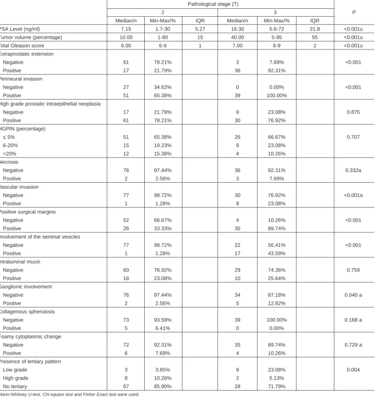
Table I: Characteristics of the tumors and the relation with pathological stage.
Pathological stage (T)
2 3 P
Median/n Min-Max/% IQR Median/n Min-Max/% IQR
PSA Level (ng/ml) 7.15 1.7-30 5.27 16.30 5.6-72 21.8 <0.001u
Tumor volume (percentage) 10.00 1-80 15 40.00 5-95 55 <0.001u
Total Gleason score 6.00 6-9 1 7.00 6-9 2 <0.001u
Extraprostatic extension Negative 61 78.21% 3 7.69% <0.001 Positive 17 21.79% 36 92.31% Perineural invasion Negative 27 34.62% 0 0.00% <0.001 Positive 51 65.38% 39 100.00%
High grade prostatic intraepithelial neoplasia
Negative 17 21.79% 9 23.08% 0.875 Positive 61 78.21% 30 76.92% HGPIN (percentage) ≤ 5% 51 65.38% 26 66.67% 0.707 6-20% 15 19.23% 9 23.08% >20% 12 15.38% 4 10.26% Necrosis Negative 76 97.44% 36 92.31% 0.332a Positive 2 2.56% 3 7.69% Vascular invasion Negative 77 98.72% 30 76.92% <0.001a Positive 1 1.28% 9 23.08%
Positive surgical margins
Negative 52 66.67% 4 10.26% <0.001
Positive 26 33.33% 35 89.74%
Involvement of the seminal vesicles
Negative 77 98.72% 22 56.41% <0.001 Positive 1 1.28% 17 43.59% Intraluminal mucin Negative 60 76.92% 29 74.36% 0.759 Positive 18 23.08% 10 25.64% Ganglionic involvement Negative 76 97.44% 34 87.18% 0.040 a Positive 2 2.56% 5 12.82% Collagenous spherulosis Negative 73 93.59% 39 100.00% 0.168 a Positive 5 6.41% 0 0.00%
Foamy cytoplasmic change
Negative 72 92.31% 35 89.74% 0.729 a
Positive 6 7.69% 4 10.26%
Presence of tertiary pattern
Low grade 3 3.85% 9 23.08% 0.004
High grade 8 10.26% 2 5.13%
No tertiary 67 85.90% 28 71.79%
RESULTS
The median age of 117 cases was 67 (46-81) years and the median preoperative PSA level was 8.2 ng/ml (range: 1.7-72). The median volume of tumor was 15% (range: 1-95).
The distribution of total Gleason score (GS) and pathological stage were as follows: 56 cases were GS 6 (47.86%), 42 cases were GS 7 (35.90%), 6 cases were GS 8 (5.13%), 13 cases were GS 9 (11.11%). Seventy eight cases were T2 (66.67%) and 39 were T3 (33.33%). High grade tertiary pattern was observed in 10 cases (8.55%). High grade prostatic intraepithelial noeplasia (HGPIN) was observed in 91 cases (77.80%), and the percentage of it was distributed as 77 cases ≤ 5% (65.81%), 24 were 6-20% (20.51%) and 16 were >20% (13.68%).
Among all prognostic factors; PSA level (p<0.001), tumor volume (p<0.001), Gleason score (p<0.001), extraprostatic extension (p<0.001), perineural invasion (p<0.001), ganglion involvement (p=0.040), vascular invasion (p<0.001), positive surgical margins (p<0.001), presence of tertiary pattern (p=0.004), and the involvement of the seminal vesicles (p<0.001) were found to be statistically related to the pathological stage. Other characteristic features of the tumors and the relationship between pathological stage and prognostic factors were summarized in Table I.
A total number of 85 cases were available for follow-up and the median follow-up time was 13.43 (1-72) months. Biochemical recurrence was detected in 18 (15.4%) patients. Median time for recurrence was found as 19 (1-72) months.
HGPIN, extraprostatic extension, perineural invasion, tumor positive surgical margins, vascular invasion, necrosis and involvement of seminal vesicles was detected in 65 (76.5%), 42 (49.4%), 65 (76.5%), 45 (52.9%), 5 (5.9%), 4 (4.7%) and 9 (10.6%) of 85 cases,
respectively. In histological evaluation, ganglion involve-ment was found in 2 (2.4%) of cases while intraluminal mucin, collagenous micronodules and foamy cytoplasm were determined in 22 (25.9%), 3 (3.5%) and 8 (9.4%) of followed up cases, respectively.
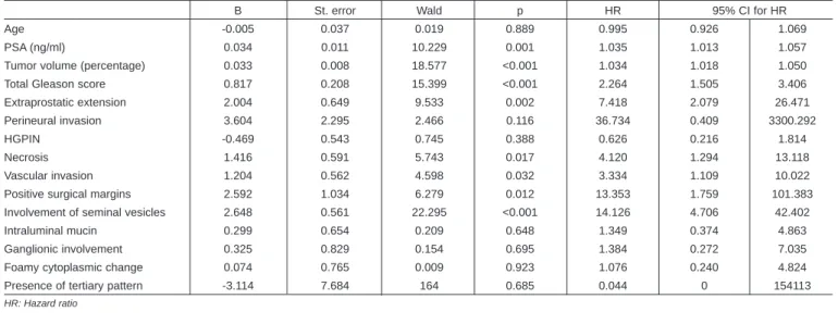
Among all the factors; preoperative PSA level (p=0.001), tumor volume (p<0.001), total GS (p<0.001), extraprostatic extension (p=0.002), necrosis (p=0.017), lymphovascular invasion (p=0.032), positive surgical margins (p=0.012), the involvement of the seminal vesicles (p<0.001) was found as statistically related to recurrence. The factors found to be related to recurrence were summarized in Table II. Since collagenous spherulosis was only positive for three patients, it was not analyzed with Cox regression.
High grade tertiary pattern was observed in 10 cases among all the patients with the most Gleason score of 3+3=6 (n=9). Also one case with a GS of 4+3=7 had a high grade tertiary pattern. Among these ten patients, 8 were available for follow-up and none of them had recurrence. The presence of high grade tertiary pattern was not statistically related to recurrence (p=0.685). Additional histopathological factors such as collagenous micronodules, intraluminal mucin and foamy cytoplasm were also found as unrelated to biochemical recurrence.
DISCUSSION
Being the most common type of cancer in men, prostate cancer and its prognostic factors have been the subject of many researches. Gleason score is a grading system used for determining optimal treatment and predicting prognosis, which is based on the sum of the most common primary and secondary histological patterns, in addition to the pathological stage and tumor positive surgical margins.3,7
One of the major controversies in pathological evaluation of radical prostatectomy specimens is the measurement of tumor volume. Tumor volume is one of the major
Table II: Factors related to recurrence using Cox regression.
B St. error Wald p HR 95% CI for HR
Age -0.005 0.037 0.019 0.889 0.995 0.926 1.069
PSA (ng/ml) 0.034 0.011 10.229 0.001 1.035 1.013 1.057
Tumor volume (percentage) 0.033 0.008 18.577 <0.001 1.034 1.018 1.050
Total Gleason score 0.817 0.208 15.399 <0.001 2.264 1.505 3.406
Extraprostatic extension 2.004 0.649 9.533 0.002 7.418 2.079 26.471
Perineural invasion 3.604 2.295 2.466 0.116 36.734 0.409 3300.292
HGPIN -0.469 0.543 0.745 0.388 0.626 0.216 1.814
Necrosis 1.416 0.591 5.743 0.017 4.120 1.294 13.118
Vascular invasion 1.204 0.562 4.598 0.032 3.334 1.109 10.022
Positive surgical margins 2.592 1.034 6.279 0.012 13.353 1.759 101.383
Involvement of seminal vesicles 2.648 0.561 22.295 <0.001 14.126 4.706 42.402
Intraluminal mucin 0.299 0.654 0.209 0.648 1.349 0.374 4.863
Ganglionic involvement 0.325 0.829 0.154 0.695 1.384 0.272 7.035
Foamy cytoplasmic change 0.074 0.765 0.009 0.923 1.076 0.240 4.824
Presence of tertiary pattern -3.114 7.684 164 0.685 0.044 0 154113
factors effecting biochemical recurrence because small tumors (<5% of all prostate volume), which can totally be cured, are the most important forms of prostatic adenocarcinoma.3
Apart from visual estimate of tumor percentage in which tumor areas were measured directly under the microscope, many different stereobiological methods are used for detecting tumor volume because no optimal method was agreed upon. This is one of the main reasons of the restricted use of tumor volume in predicting prognosis.3,8 In this study, tumor volume is measured by the eyeball estimate of the percentage of prostatic tissue involved by tumor, and was found to be a prognostic factor for predicting biochemical recurrence (p=0.00).
On the other hand, it is still controversial whether age is a major prognostic factor or not.9 Some studies found younger patients had a better outcome than older patients, after radical prostatectomy.9 In this study, we regrouped the cases into < 60 and ≥60 years and no significant difference was found between these groups (p=0.889).
Many prognostic parameters can be determined by careful evaluation of RP specimens. Among all these parameters, vascular invasion (lymphatic or venous) has no role in pathological staging. Isolated effect of vascular invasion on prognosis remains a mystery because of being observed generally in advanced tumors.10In this study, vascular invasion was mostly observed in advanced (T3) tumors and showed significant relation-ship with biochemical recurrence (p=0.032). Similar to this, perineural invasion was detected in all advanced tumors (100%) but not found to be significantly related to biochemical recurrence (p=0.116).
In addition to the known classical prognostic histopatho-logical factors such as Gleason score, extraprostatic extension, vascular invasion, perineural invasion, positive surgical margins, we also investigated the prognostic role of the presence of intraluminal mucin, collagenous micronodules and tumoral cells with foamy cytoplasm.
Intraluminal mucin can be seen in routine hematoxylen-eosin slides as amorphous basophilic secretions in the lumina of carcinomatous glands and rarely recognized in benign prostatic hyperplasias.8 Collagenous nodules are paucicellular eosinophilic fibrous micro-scopic nodules invaginated into aciner lumina and specific for carcinoma which can incidentally be found in 2-13% of prostatic adenocarcinomas.11 Some tumor cells contain vacuolated or abundant foamy cytoplasm. These tumors are known as foamy gland carcinomas
and graded by Gleason system.12-14 The three
histological features mentioned above are known to be associated with prostatic adenocarcinoma; however, their effect on prognosis remains a mystery.9,15 In this
study, these histological features were found to be unrelated to biochemical recurrence and pathological stage; however, foamy cytoplasm was detected more commonly in advanced (T3) cases. But intraluminal mucin was partially equal in T2 (n=15) and T3 (n=13) with a slight dominancy for early stage group. No significant relationship was found between these histological features and recurrence. Depending on the small number of cases with these histological features, it is not possible to make any accurate comments regarding the prognostic role of these histological findings. Ganglion involvement is one of the main factors effecting extraprostatic extension. Sakamoto studied upon this factor but could not prove the actual effect on extraprostatic extension.16 In our study, ganglion involvement showed significant relation to pathological stage but recurrence was not statistically related to ganglionic involvement.
In the recent years, Gleason grading system was revised and the presence of a high grade tertiary component seen in <5% of tumor was found to correlate with pathological stage and progression.17,18 In our study, in contrast with the literature, patients with same Gleason score but having a higher tertiary pattern (n=10) showed no significant difference for recurrence according to the presence of high grade tertiary pattern (p=0.983). This may also be related to the limited number of cases with high grade tertiary pattern and may be overcomed by including more patients with tertiary pattern.
Intraluminal mucin, collagenous micronodules and foamy cytoplasm were found to be unrelated to prognosis. However, it is important to know that these features were commonly noted in advanced tumors in the English literature. These features were studied only in a few studies showing no exact relation to prognosis.9,13-15 Depending on the small number of cases showing these features, studies with larger case series are needed to put forth the real effect of intraluminal mucin, collagenous micronodules and foamy cytoplasm on recurrence.
CONCLUSION
The findings support the observation that reporting histopathological features such as Gleason score, tumor volume, extraprostatic extension, tumor positive surgical margins, lymphovascular invasion, necrosis, involvement of seminal vesicles, can be helpful in predicting recurrence in prostatic adenocarcinomas.
REFERENCES
1. Siegel RL, Miller KD, Jemal A. Cancer Statistics, 2015.
CA Cancer J Clin 2015; 65:5-29.
2. Buhmeida A, Pyrhönen S, Laato M, Collan Y. Prognostic factors in prostate cancer. Diag Path 2006; 1:4
3. Montironi R, Mazzucchelli M, Scarpelli M, Lopez-Beltran A, Mıkuz G. Prostate carcinoma I: prognostic factors in radical prostatectomy specimens and pelvic lymph nodes. Bju Int 2005; 97:485-91.
4. Epstein JI, Srigley J, Grignon D, Humphrey P. Recommendations for the reporting of prostate carcinoma. Hum Pathol 2007;
38:1305-9.
5. Vickers AJ, Cronin AM, Björk T, Manjer J, Nilsson PM, Dahlin A,
et al. Prostate specific antigen concentration at age 60 and
death or metastasis from prostate cancer: Case-control study.
BMJ 2010; 341:c4521.
6. Sakr WA, Wheeler TM, Blute M, Bodo M, Calle-Rodrigue R, Henson DE et al. Staging and reporting of prostate cancer-sampling of the radical prostatectomy specimen. Cancer 1996;
78:366-8.
7. Eichelberger LE, Koch MO, Daggy JK, Ulbright TM, Eble JN, Cheng L. Predicting tumor volume in radical prostatectomy specimens from patients with prostate cancer. Am J Clin Pathol 2003; 120:386-91.
8. Noiwan S, Rattanarapee S. Mucin production in prostatic adenocarcinoma: A retrospective study of 190 radical prostatectomy and/or core biopsy specimens in department of pathology, Siriraj Hospital, Mahidol University, Thailand. J Med
Assoc Thai 2011; 94:224-30.
9. Freedland SJ, Presti JC, Kane CJ, Aronson WJ, Terris MK, Dorey F, et al. Do younger men have better biochemical outcomes after radical prostatectomy? Urology 2004; 63: 518-22. 10. Ito K, Nakashima J, Mukai M, Asakura H, Ohigashi T, Saito S,
et al. Prognostic implication of microvascular invasion in
bio-chemical failure in patients treated with radical prostatectomy.
Urol Int 2003; 70:297-302.
11. Baisden BL, Kahane H, Epstein JI. Perineural invasion, mucinous fibroplasia, and glomerulations: diagnostic features of limited cancer on prostate needle biopsy. Am J Surg Pathol 1999; 23:918-924.
12. Dema A, Taban S, Lazar E, Borda A, Lazureanu C, Herman D
et al. Pseudobenign prostate carcinomas: Causes of
false-negative biopsy results. Rom J Morphol Embryol 2011; 52: 963-74.
13. Warrick JI, Humphrey PA. Foamy gland carcinoma of the prostate in needle biopsy: Incidence, gleason grade, and comparative a-Methylacyl-CoA Racemase vs. ERG Expression.
Am J Surg Pathol 2013; 37: 1709-14.
14. Berman DM, Yang J, Epstein JI. Foamy gland high-grade prostatic intraepithelial neoplasia. Am J Surg Pathol 2000; 24:140-4. 15. Arangelovich V, Tretiakova M, SenGupta E, Krausz T, Yang XJ.
Pathogenesis and significance of collagenous micronodules of the prostate. Appl Immunohisto M M 2003; 11:15-9.
16. Sakamoto N, Hasegawa Y, Koga H, Kotoh S, Kuroiwa K, Naito S. Presence of ganglia within the prostatic capsule: Ganglion involvement in prostatic cancer. Prostate 1999; 40:167-71. 17. Mosse CA, Magi-Galluzzi C, Tsuzuki T, Epstein JI. The
prognostic significance of tertiary gleason pattern 5 in radical prostatectomy specimens. Am J Surg Pathol 2004; 28: 394-8. 18. Vis AN, Schroder FH, Van der Kwast TH. The actual value of the surgical margin status as a predictor of disease progression in men with early prostate cancer. Eur Urol 2006;
50:258-65.