Contents lists available atScienceDirect
Bioorganic Chemistry
journal homepage:www.elsevier.com/locate/bioorg
Synthesis and evaluation of the antitumor activity of Calix[4]arene
L
-proline
derivatives
Mehmet Oguz
a,b, Alev Gul
a, Serdar Karakurt
c,⁎, Mustafa Yilmaz
a,⁎ aSelcuk University, Department of Chemistry, 42075 Konya, TurkeybDepartment of Advanced Material and Nanotechnology, Selcuk University, 42075 Konya, Turkey cSelcuk University, Department of Biochemistry, Konya 42075, Turkey
A R T I C L E I N F O Keywords: Calixarene L-Proline Cytotoxicity Apoptosis Anticancer agent A B S T R A C T
The unique conformational properties, functionality, low toxicity, and low cost make calixarene-based com-pounds a valuable candidate against cancer. The aim of the present study is the synthesis of the upper rim and lower rim-functionalizedL-proline-based calix[4]arene derivatives and evaluation of their cytotoxic potential for
human cancerous cells as well as to determine the death mechanism. Synthesized calix[4]arene (3, 8a, 8b 13a, and 13b) derivatives were characterized by different spectroscopic techniques such as1HNMR,13CNMR, and
FTIR. In vitro effects of compounds 3, 8a, 8b, 13a and 13b were tested on human cancerous cells (HEPG2, PC-3, A-549, and DLD-1) as well as human healthy epithelium cell (PNT1A). Results show that compounds 3, 8a, 8b and 13b have cytotoxic potential on human colorectal carcinoma cells (DLD-1) with IC50values of 43 µM,
45.2 µM, 64.57 µM, and 29.35 µM respectively. Apoptosis ratios of cell death were investigated with flow cyt-ometer using 7-AAD and AnnexV as markers. Cytotoxic potential of 8a was found to be higher due to in-creased apoptosis, when compared with healthy cells the apoptotic cell death was significantly (p < 0.0001) increased up to 1.7-fold and 2.4-fold in DLD-1 and A549 cells, respectively. In conclusion, theseL-proline derived
calix[4]arenes with their selective cytotoxic potential on human cancerous cells may be a potential candidate for the treatment of human CRC and lung cancer.
1. Introduction
Cancer is the major cause of death in the world, and especially in developing countries, the mortality rate has been increased sig-nificantly[1–3]. According to the world health organization, about 9.6 million new cases have been reported in 2018 and it is expected to rise by about 70% over the next two decades[4]. Increased morbidity and mortality rates of cancer lead investigators to find new and active strategies against cancer. Although there are many synthetic drugs used for the treatment of the several types of cancers, the severe side effects and low cytotoxicity makes them ineffective for the cure[5–8]. Indeed, one of the biggest challenges for pharmaceutical industries is to explore new anticancer agents that are safe and effective[9–12].
One of the important third generation supramolecular compounds is calixarene possesses ring and flexible conformation, which make them suitable for adding functional groups including amino acids, amide, amine, alcohol, ester, aldehyde and alkyl derivatives[13–16]. Phenolic rings of calixarenes interact with each other by methylene bridges from the o-position of the phenolic structures to the hydroxyl groups, which
causes the formation of a hydrophobic gap capable of complexing with various guest molecules [17–22]. Beside these gaps, the molecules carried by the lower and upper rim of calixarene may also interact with the proteins and nucleic acids, which modulates the activity of many enzymes, the proliferation of cancerous cells and metabolic pathways [23–26]. The exceptional structural properties make calixarene a va-luable candidate against cancer[27–29]. Recent studies proved that calixarenes are capable of use in pharmaceutical applications as an anti-cancer drug, the drug carrier, antiviral and antithrombotic activities [30–33]. Some calixarene derivatives have been reported for its effec-tive activity against bacteria, fungi, antitumor, antimicrobials, can-cerous cells, and enveloped viruses, but also against thrombosis or fi-brosis diseases[34–36]. Ding et al., prepared calix[4]arene derivatives functionalized 2-dimethylaminoethyl groups on its hydrophilic face vis-à-vis the larger and somewhat more polar N-(2-dimethylamino)ethyl acetamide groups and tested their anti-tumor activities. They observed effectiveness of inhibiting the growth of several human cancer cell lines, as well as drug-resistant cancer cells[37]. Alkyl acid and alkyl ester groups from lower rim position containing calix[4]arenes were
https://doi.org/10.1016/j.bioorg.2019.103207
Received 19 February 2019; Received in revised form 10 July 2019; Accepted 15 August 2019
⁎Corresponding authors.
E-mail addresses:[email protected](S. Karakurt),[email protected](M. Yilmaz).
Available online 19 August 2019
0045-2068/ © 2019 Elsevier Inc. All rights reserved.
sion is upregulation of matrix metalloproteinases (MMPs) that catalysis the degradation of ECM and releasing of proline into the matrix. As a result of proline metabolism (P5C cycle), glutamate and ornithine are produced, which further converted to α-ketoglutarate (α-KG) entering the tricarboxylic acid (TCA) cycle and arginine entering the urea cycle, respectively[44,45]. Fu et al. synthesized calix[4]arene modified with L- orD-proline and used them against human cervical cancer, the second cause of death in the female in the world. They observed that the chiral proline modified calix[4]arene prevented L1 pentamer formation of HPV[46].
In regards of these studies, we designed and synthesized upper and lower rim-functionalizedL-proline-based calix[4]arene derivatives, and investigated their cytotoxic potential for various human cancerous cells and determined the mechanism of cell death.
2. Materials and methods 2.1. Instruments/materials
All materials and reagents used in the experiments were supplied from various commercial sources and used without further purification. NMR measurements obtained from Varian (400 MHz), D2O and
DMSO‑d6 for calix[4]arene derivatives were used as deuterated
sol-vents. Infrared spectra were measured using a Bruker spectrometer transform-infrared (FT-IR) spectrometer. HR-MS spectra were per-formed by Synapt G1 High Definition Mass Spectrometer. Elisa plate reader (Thermo multiscan-90), Flow cytometer (BD FACS Aria III). 2.2. Synthesis
The different derivatives of calixarenes presented in Scheme 1–3 (1a, 1b, 4a, 4b, 5a, 5b, 6a and 6b, 7a, 9a, 9b, 10a, 10b, 11a, 11b)
stirring for 72 h at room temperature. The reaction was completed when water-insoluble material was not detected. The liquid was re-moved and drained. The remaining solid was washed with acetone and the solution was filtered. The precipitate was washed with acetone several time and then recrystallized from ethanol/water (1:1) to yield a white solid (7.4 g; yield, 82%). IR: 1612 cm−1COO−.1HNMR (D
2O): δ,
1.40–1.82 (broad m, 12H CHCH2), 1.82–2.20 (broad m, 4H, CHCH2),
2.75–2.98 (broad m, 4H, CH2N), 3.18–3.35 (broad m, 4H, CH2N),
3.54–3.67 (broad t, 4H, NCH), 3.67–3.82 (broads, 8H, ArCH2N),
3.83–3.88 (broads, 8H, ArCH2Ar), 7.03 (s, 8H, ArH). 13C NMR (D2O):
δ, 20.9, 29.9 (2 × CH2), 28.7 (ArCH2Ar), 52.4, 55.9 (2 × CH2N), 66.1
(CHN), 121.4, 127.8, 129.5, 149.4[3], 171.8 (C]O). 2.4. General procedure for the synthesis of compounds 7a and 7b
Compound 6a and 6b (0.95 g, 1 mmol) dissolved in dry THF (100 mL) in two separate flaks and the solution ofL-proline methyl ester hydrochloride (0.99 g 6 mmol) added dropwise containing pyridine (1 mL) and stirred for 48 h at room temperature. The reaction was monitored by TLC. When the reaction finished excess of the solvent was removed off in vacuo. The residue was extracted with chloroform/water several times. Organic layer was separated and dried with MgSO4. After
filtration of magnesium sulfate, the solvent was evaporated. 7a: Yield 74%;1H NMR (400 MHz, DMSO): δ (ppm) 1.07 (s, 36H, But), 1.83–1.97
(m, 8H, CH2(proline)), 2.13–2.22 (m, 8H, CH2(proline)) 3.24–3.35 (m,
8H, CH2(proline)), 3.69–3.78 (m, 16H, (12H, OCH3and 4H, ArCH2Ar)),
4.48–4.62 (m, 4H, ArCH2Ar), 4.93 (s, 4H, OCH2), 5.08 (s, 4H, OCH2),
5.20–5.29 (m, 4H, CH(proline)), 6.75 (s, 8H, ArH).
7b: Yield 68%;1H NMR (400 MHz, DMSO) δ (ppm): 1.73–1.89 (m,
8H, CH2(proline)), 2.03–2.19 (m, 8H, CH2(proline)) 3.23–3.38 (m, 8H,
CH2(proline)), 3.59 (s, 12H, OCH3), 3.68 (s 4H, ArCH2Ar), 4.23–4.31
(m, 8H, OCH2), 4.61 (d, 4H, J = 15.4 Hz, ArCH2Ar), 4.93 (d, 4H, CH
(proline)), 6.44–6.58 (m, 4H, ArH), 6.71–6.83 (m, 8H, ArH).
Scheme 1. The schematic route for the synthesis of water-solubleL-proline based calix[4]arene derivative. Reagents and conditions: (i) AlCl3, Toluene; (ii)L-proline,
Scheme 2. The schematic route for the synthesis ofL-proline based calix[4]arene derivatives. Reaction conditions; (i) BrCH2COOCH3, K2CO3, Acetone; (ii) KOH,
ethanol/water; (iii) SOCl2, THF; (iv) pyridine;L-Proline methyl ester hydrochloride, THF; v) KOH, ethanol/water.
Scheme 3. The schematic route for the synthesis ofL-proline based calix[4]arene derivatives. Reaction conditions; (i) BrCH2COOCH3, K2CO3, Acetone; (ii) KOH,
4H, ArCH2Ar)), 3.96 (d, 4H, J = 14.6 Hz, ArCH2Ar), 4.38 (s, 4H,
OCH2), 4.82 (s, 4H, OCH2), 4.94–5.10 (m, 4H, CH(proline)), 7.17 (s,
8H, ArH).13C NMR (100 MHz, DMSO): δ (ppm) 172.7, 168.1, 152.2,
147.5, 134.7, 125.3, 4.6, 59.0, 45.6, 34.54, 31.4, 30.8, 29.0, 24.8. 8b: Yield 80%. IR: 1729 cm−1 C]O, 1658 cm−1 NeC]O. 1H NMR
(400 MHz, DMSO) δ (ppm): 1.86–2.49 (m, 16H, CH2 (proline)),
2.91–3.73 (m, 8H, CH2 (proline) ve 4H ArCH2Ar), 3.97 (d, 4H,
J = 14.1 Hz, ArCH2Ar), 4.55–4.73 (m, 8H, OCH2), 4.93–5.31 (m, 4H,
CH(proline)), 6.86–7.14 (m, 4H, ArH), 7.28 (m, 8H, ArH).13C NMR
(100 MHz, DMSO): δ (ppm) 174.0, 168.1, 156.5, 135.18, 128.9, 122.4, 72.2, 59.1, 45.6, 31.6, 29.0, 25.0. The HR-MS data of 8a and 8b (1267.6733 and 1043.4077, respectively) supported a molecular for-mula 8a: C72H92N4O16and 8b: C56H60N4O16(Figs. S7 and S11).
2.6. Synthesis of compounds 12a and 12b
Compound 11a and 11b (0.95 g, 1 mmol) were dissolved in dry THF (50 mL) in two separate flasks and the solution ofL-proline methyl ester hydrochloride (0.66 g 4 mmol) was added dropwise in the pre-sence of pyridine (0.5 mL). The mixture was allowed to stir for 30 h at room temperature. The reaction was monitored by TLC. After formation of products, excess of the solvent was removed off at low pressure. The residue was extracted with chloroform/water several times. Organic layer was separated and dried with MgSO4. After filtration the solvent
was evaporated, and residue was dried at room temperature, 12a: Yield 75%.1H NMR (400 MHz, DMSO): δ (ppm) 1.05 (s, 18H, But), 1.16 (s,
18H, But), 1.85–2.11 (m, 8H, CH2CH2(proline)), 3.38 (m, 4H,
CH2(proline)), 3.65 (s, 6H, OCH3), 3.68 (bs, 4H, ArCH2Ar), 4.38–4.46
(m, 4H, ArCH2Ar), 4.49–4.63 (m, 4H, OCH2), 4.76–4.83 (m, 2H, CH
(proline)), 7.05 (s, 4H, ArH), 7.10 (s, 4H, ArH). 12b: Yield 65%.1H
NMR (400 MHz, DMSO): δ (ppm) 1.71–1.96 (m, 8H, CH2CH2(proline)),
3.13 (b, 4H, CH2(proline)), 3.41–3.67 (m, 10H, (OCH3ve ArCH2Ar)),
4.19–4.31 (m, 4H, ArCH2Ar), 4.76–4.82 (m, 4H, OCH2), 5.06–5.17 (m,
2H, CH(proline)), 6.41–7.08 (m, 12H, ArH). 2.7. Synthesis of compound 13a and 13b
Compound 12a and 12b (0.95 g) were dissolved in ethanol (50 mL) in two separate flasks containing aqueous KOH solution. The reaction mixture was then stirred and heated at reflux for 8 h. Solvent was re-moved under reduced pressure, the residue was precipitated with 0.1 M HCl. The solid material was then filtered and washed till neutralization with water several time. The target compounds 13a, b was obtained as white solid. 13a: Yield 85%.1H NMR (400 MHz, DMSO): δ (ppm) 1.07
(s, 18H, But), 1.13 (s, 18H, But), 1.80–2.27 (m, 8H, CH2CH2(proline)),
3.33 (m, 4H, CH2(proline)), 3.62–3.87 (m, 4H, ArCH2Ar), 4.36 (d, 4H,
J = 12.05 Hz, ArCH2Ar), 4.53–4.72 (m, 4H, OCH2), 4.86–4.91 (m, 2H,
CH(proline)), 7.12 (s, 4H, ArH), 7.18 (s, 4H, ArH).13C NMR (100 MHz,
CDCl3): δ (ppm) 173.6, 167.1, 152.0, 150.6, 147.0, 141.1, 133.5, 128.8, 127.4, 125.6, 74.19, 59.25, 45.93, 34.35, 33.99, 31.86, 31.36, 30.9, 29.1, 24.9, 22.3. 13b: Yield 80. 1H NMR (400 MHz, DMSO): δ
(ppm) 1.71–1.93 (m, 8H, CH2CH2(proline)), 3.17–3.24 (m, 4H,
CH2(proline)), 3.48–3.52 (m, 4H, ArCH2Ar), 3.96–4.11 (m, 4H,
ArCH2Ar), 4.25–4.43 (m, 4H, OCH2), 4.78–4.91 (m, 4H, 2H, CH
(Missouri, ABD). The cells were incubated with DMEM, Ham’s F-12, RPMI 1640 and EMEM mediums, respectively and supplemented with 10% FBS (fetal bovine serum), 1% L-glutamine and 1% penicillin/ streptomycin at 37 °C in a 5% CO2 atmosphere and 95% humidity.
Following the confluence, the cells were transferred to 96-well plates and 6-well plates for cytotoxicity studies and apoptosis/comet assay studies, respectively.
2.9. Determination of IC50values for chemical treatment
The cells were counted with the BioRad TC20 automatic cell counter and death/live ratio was calculated by using 0.04% “Trypan Blue”. 1 × 10⁴of the cells were incubated for 24 hr. then treated with various
concentration of L-proline-based calix[4]arenes (3, 8a, 8b.13a, and 13b) ranging between 0 and 200 µM. The viability of the cells and cytotoxicity of the compounds were determined spectrophotometrically by using Alamar Blue reagent as an indicator (Invitrogen, Thermo Fischer Scientific, Waltham, MA, USA)[52]. The IC50value was
cal-culated from the sigmoidal plot of cell inhibition and statistical analyses were done by GraphPad Prism 5.0.
2.10. Apoptosis assays
2.5 × 10⁵of the cells from PNT1A, DLD-1, and A549 cell lines were
transferred into 6-well plates and incubated at 37 °C, 5% CO2for 24 hr.
Then, PNT1A, DLD-1, and A549 cells were treated with equivalent concentration as IC50 values of compound 3, 8a, 8b.13a and 13b.
Following 48 hr incubation, the cells were incubated with Annexin V and 7-AAD and analyzed by flow cytometer (BD FACS Aria III, software: FACS Diva software).
3. Results and discussion
The target of this work is to prepare an effective antitumor agent using calix[4]arene by usingL-proline amino acid (Fig. 1). In this re-gard, compound 3 was designed and readily prepared using simple reaction conditions (Scheme 1). First of all, p-tert-butylcalix[4]arene (1) as the precursor was treated with Lewis acid (AlCl3) in the presence of
phenol to remove upper rim tert-butyl groups to obtain calix[4]arene (2). Then, the calix[4]arene (2) was reacted withL-proline (Mannich reaction) in the presence of glacial acetic acid and formaldehyde to synthesize compound 3 as L-proline functionalized calix[4]arene (3) reported in published work[46,51].
Compound 8a, 8b, 13a, and 13b were synthesized using an acid chloride method. The L-proline-substituted calix[4]arene derivatives 8(a,b) and 13(a,b) were obtained in five steps (Scheme 2and3). The parent compounds 1, 4, 5 and 6 were synthesized according to litera-ture[47–49]. Calix[4]arene tetraester derivative (4) was synthesized in one step from p-tert-butylcalix[4]arene (1) with a yield of 79% by re-acting with methyl bromoacetate and K2CO3in acetone under reflux
conditions. Base hydrolysis of tetraester derivative (4) in ethanol pro-duced acid derivative (5) in 85% yield. In the next step, carboxylic acid derivative (5) was converted into acid chloride (6) via thionyl chloride in THF at inert conditions. Compound 7 (a.b) was synthesized using an
acid chloride derived from calixarene (6) andL-proline methyl ester hydrochloride in the presence of pyridine in THF. Finally, compound 7(a.b) was hydrolyzed with KOH in ethanol to furnish final product 8(a,b). The chemical structures of all derivatives were completely characterized by1H,13C NMR, and IR spectroscopic techniques.
In functional group analysis of 3, presence of C]O (COO−)
stretching band at 1612 cm−1 confirmed the derivatization of
calix-arene withL-proline (Fig. S1). In1H NMR spectrum (Fig. S2), the ap-pearance of different peaks of methyl protons ofL-proline from 1.63 to 3.38 ppm further confirmed the purity of the synthesized compound. In addition protons of ArCH2Ar at 3.86 and 3.98 ArCH2Ar, the presence of
singlet aromatic protons at 7.07 (s, 8H, ArH) were confirmed the structure of compound 3 (Fig. S2).
1HNMR,13CNMR, and FTIR spectroscopy are used to elucidate the
structure of compound 8 (a,b) and 13 (a, b). In FTIR spectrum of compounds 8 (a, b), presence of stretching bands at 1732 and 1729 cm−1 for C]O, 1650 and 1658 cm−1for NeC]O defines the
presence ofL-proline functionality on calix[4]arene (Figs. S4 and S8). Presence of the signal at 1.16 ppm for singlet t-butyl group and the peaks from 1.96 to 3.27 ppm forL-proline methylene, at 4.38 ppm show the presence of OCH2protons of ester linkage and finally singlet
aro-matic signal (8Hs) at 7.17 ppm for aroaro-matic protons explain the com-plete structure of 8a (Fig. S5). The synthesis of 8b was confirmed by the appearance of proline protons at from 1.86 to 3.73 ppm and the pre-sence of aromatic signals at 7.81 ppm and at 7.28 ppm for aromatic rings (Fig. S9). The 13C NMR spectra of 8a and 8b showed the
ap-pearance of the carbon signals at 173.7 and at 174.0 ppm belong to C] O groups. In addition, carbon signals at 168.1 confirmed the presence of NeC]O groups (Figs. S6 and S10).
The formation of 13a and 13b was confirmed by the appearance of the characteristic carbonyl bands at about 1732 cm−1and 1738 cm−1,
amide bands at 1650 cm−1and 1654 cm−1in the FTIR spectra (Figs. S12
and S16). The structure of 13a was also confirmed by the appearance of proline methyl protons from 1.80 to 3.33 ppm in the1H NMR spectra. In
addition, the appearance of p-tert butyl protons at 1.07 (18H) and 1.13 (18H), aromatic protons at 7.12 (4H) and 7.18 (4H) confirmed the structure of 13a (Fig. S13). The 13C NMR spectra of 13a showed the
appearance of the carbon signal at 173.6 ppm belong to C]O group and signal at δ167.1 belong to NeC]O (Fig. S14). The appearance of proline methyl protons at from 1.71 to 3.24 in the1H NMR spectrum, the
pre-sence of carbon signals at 173.6 ppm and 168.2 ppm (C]O and NeC] O) in13C NMR spectrum confirmed the structure of compound 13b (Figs.
S17 and S18). The spectral data were in agreement with the desired structures. Besides, the HR-MS spectra results also supported the struc-tures of the synthesized compounds (Figs. S7, S11, S15 and S19). 3.1. Effects ofL-proline-based Calix[4]arene derivatives on viability and
proliferation of human cancerous and healthy cells
In order to determine the cytotoxic effects of compound 3, 8a, 8b, 13a, and 13b on proliferation and viability of human DLD-1, PC-3,
HEPG2, A549 and PNT1A cells, the cells were treated with various concentration of the compounds. Different functional group on calix[4] arene behave differently on the viability of the cells. They have selec-tively and dose-dependently inhibited proliferation and viability of the cancerous cells as well as healthy epithelium cell (Table 1).
As shown inTable 1, when the compounds were compared, com-pound 13b was found to be the highest cytotoxic potential on human CRC cell (DLD-1) and human hepatocarcinoma cell (HEPG2) with IC50
values as 29.35 µM and 64.45 µM, respectively (Fig. S23). On the other hand, when the healthy epithelium cell (PNT1A) was considered, al-though 13b has lower IC50value on DLD-1 than PNT1A, the best choice
seems compound 3 which has IC50value as 43 µM for DLD-1, but no
cytotoxic potential on human epithelial cells (> 200 µM) (Fig. 2). The molecular mechanism of this effect lies in the genetic difference be-tween two cells. The BRAF, KRAS, p53, and SMAD genes that act as key enzymes in the proliferation of DLD-1 cells are mutated and their ex-pressions are much lower than in healthy PNT1A cells. Therefore, it is not a good candidate for the treatment of other cancerous cells. The pH of cancerous cells is around 6.0–6.5 and at that pH theL-proline groups (pI = 6.3) in the upper rim of the compound 3 form zwitterion at this pH, and makes it soluble in water. Besides, compound 13a was also found a potent inhibitor for human lung carcinoma cell (A549) and prostate cells (PC-3) with the IC50 values as 15.7 µM and 23.38 µM,
respectively (Fig. S22). When compared with other compounds, com-pound 13a seems the perfect candidate against the proliferation of A549 since it has the lowest ratio between A549 vs PNT1A (Healthy cell) as 0.39. In addition, when the compounds 8a and 8b were com-pared to healthy epithelium cells, the best toxicity has been seen in the DLD-1 cells with 45.2 µM and 64.5 µM IC50 values, respectively (Figs. S20 and S21).
To clarify the molecular mechanism of cell death, the cells were subject to flow cytometric analysis with 7-AAD and Annexin-V dyes. When compared the compound 8a, 8b, 13a, and 13b, it was shown that compound 3 does not progress the cells through the apoptosis (p > 0.05). On the other hand, Compound 8a significantly inhibited the viability of DLD-1 and A549 cells via apoptosis. Apoptotic cell death was increased as 1.93-fold and 2.02-fold in DLD-1 and A549 cells, re-spectively (Fig. S20). Furthermore, compound 8b and 13a were also increased the apoptotic cell death of DLD-1 cells as 4.3-fold and 15.3-fold, respectively (Figs. S21 and S22).
Activation of death receptors signaling and Bcl-2 (pro-apoptotic Fig. 1. The structure ofL-proline based calix[4]arene derivatives (3, 8a, 8b, 13a, and 13b).
Table 1
Calculated IC50values (µM) forL-proline-based calix[4]arene derivatives.
PNT1A A549 PC-3 DLD-1 HEPG2
3 > 200 > 200 94.50 43.00 147.0
8a 66.10 49.50 55.90 45.20 105.5
8b 105.6 80.95 91.61 64.57 107.7
13a 40.06 15.70 23.38 59.55 73.94
gene) pathways may increase the early apoptosis which clarifies the extrinsic or intrinsic pathways of apoptosis. On the other hand, the intrinsic and extrinsic pathways were merged in the late phase of apoptosis that is characterized by DNA fragmentation. In both stages of apoptosis, morphological and phenotypical changes occur[53].
When the cells were treated with 8a, 8b and 3, 1.9%, 4.3% and 0.9% of the DLD-1 cells were death due to early apoptosis and 7.9%, 8.8% and 4.8% of the cells were death due to late apoptosis, respec-tively. Besides, 8a, 13a and 3 increased early apoptosis of A549 cells as
18.3%, 9.3%, and 5.3%, and increased late apoptosis as 12.9%, 9.5% and 7.8%, respectively (Fig. 3).
4. Conclusion
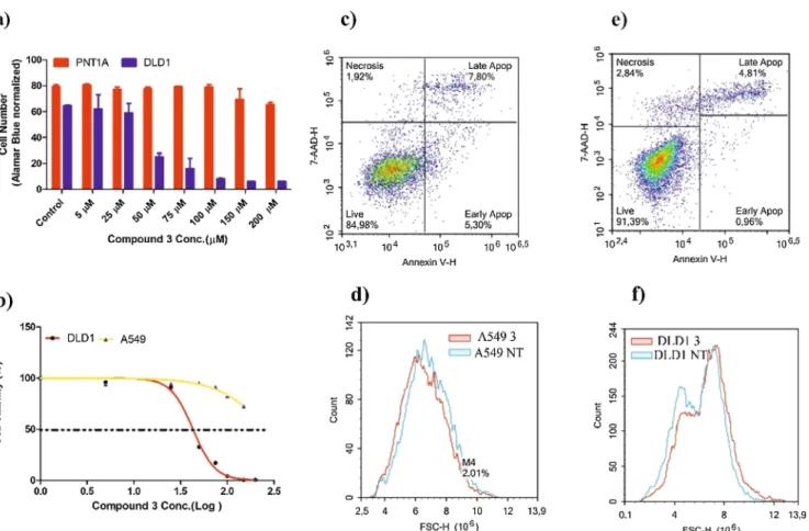
In conclusion, calix[4]arene molecule was successfully functiona-lized withL-proline at upper and lower rims. The lower rimL-proline derivative (8a) has been shown the highest toxicity against human CRC (DLD-1) and lung carcinoma (A549) cells. Compound 8a leads Fig. 2. Effects of compound 3 on viability, proliferation, and apoptosis of the A549, DLD-1, and PNT1A cells. The cells were treated with compounds (0–200 µM) and incubated 48 h at 37 °C, 5% CO2. (a) Alamar Blue assay was used to investigate viability and cytotoxicity. Viability of PNT1A and DLD-1 cells after treatment with
different concentrations (0–200 µM) of compound 3 for 48 h. (b) IC50value of compound 3 on A549 and DLD-1 cells; (c) and (e) A549 and DLD-1 cells treated with
compound 3; (d) and (f) comparison of histogram plots of compound 3 treated cells with non-treated cells of A549 and DLD-1, respectively. Overlapping plots of apoptotic cells of NT (Non-treated cells) and Compound 3 (43 µM) treated A549 and DLD-1 cells. The results represent mean ± SD of three independent experiments (n = 4).
Fig. 3. Effect of compound 3, 8a and 13a on A549 and DLD-1 cell abpoptosis. Apoptosis was examined by flow cytometry using Annexin V and 7-AAD staining. The scatterplots illustrate the effects of Compound 3, 8a (S16), 8b (S17), and 13a (S18). The gate was obtained from annexin-V negative and 7-AAD negative cells. The lower left quadrant represents the early apoptosis (annexin V+, 7-AAD−), the upper left quadrant represents the late apoptosis (annexin V+, 7-AAD+), the lower
right quadrant represents the alive cells (annexin V−, 7-AAD−), and the upper right quadrant represents the necrosis (annexin V−, 7-AAD+). The bars represent the
apoptotic cell death especially the early stage of apoptosis. Besides this, other compounds also showed proliferation of various human can-cerous cells including, liver, prostate, lung and colorectal carcinomas in a dose-dependent manner. The present study opens a way to develop selective and cost-effective inhibitors as new antitumor agents. Declaration of Competing Interest
The authors declare no conflict of interest. Acknowledgements
We would like to thank the Scientific and Technological Research Council of Turkey, Turkey (TUBITAK-Grant Number 116Z173) and the Research Foundation of Selcuk University (SUBAP-Grant Number: 17201066 and 17201064) for financial support of this work and is a part of Mehmet Oguz Ph.D. thesis and Alev Gul master thesis. Appendix A. Supplementary material
Supplementary data to this article can be found online athttps:// doi.org/10.1016/j.bioorg.2019.103207.
References
[1] E. Salvati, F. Stellacci, S. Krol, Nanosensors for early cancer detection and for therapeutic drug monitoring, Nanomedicine 10 (23) (2015) 3495–3512. [2] R.L. Siegel, K.D. Miller, A. Jemal, Cancer statistics, 2015, CA: Cancer J. Clin. 65 (1)
(2015) 5–29.
[3] Y.-E. Choi, J.-W. Kwak, J.W. Park, Nanotechnology for early cancer detection, Sensors 10 (1) (2010) 428–455.
[4] F. Bray, J. Ferlay, I. Soerjomataram, R.L. Siegel, L.A. Torre, A. Jemal, Global cancer statistics 2018: GLOBOCAN estimates of incidence and mortality worldwide for 36 cancers in 185 countries, CA: Cancer J. Clin. 68 (6) (2018) 394–424.
[5] K. Kalogerakos, C. Sofoudis, N. Baltayiannis, Early breast cancer: a review, Cancer Therapy 6 (2) (2008).
[6] M. Perfezou, A. Turner, A. Merkoçi, Cancer detection using nanoparticle-based sensors, Chem. Soc. Rev. 41 (7) (2012) 2606–2622.
[7] F.R. Hirsch, W.A. Franklin, A.F. Gazdar, P.A. Bunn, Early detection of lung cancer: clinical perspectives of recent advances in biology and radiology, Clin. Cancer Res. 7 (1) (2001) 5–22.
[8] M.M. Naseer, M. Ahmed, S. Hameed, Functionalized calix[4]arenes as potential therapeutic agents, Chem. Biol. Drug Des. 89 (2) (2017) 243–256.
[9] S.B. Nimse, T. Kim, Biological applications of functionalized calixarenes, Chem. Soc. Rev. 42 (1) (2013) 366–386.
[10] F.N. Pur, Calixdrugs: calixarene-based clusters of established therapeutic drug agents, Mol. Diversit. 20 (3) (2016) 781–787.
[11] F. Perret, A.W. Coleman, Biochemistry of anionic calix[n]arenes, Chem. Commun. 47 (26) (2011) 7303–7319.
[12] G.M.L. Consoli, G. Granata, G. Fragassi, M. Grossi, M. Sallese, C. Geraci, Design and synthesis of a multivalent fluorescent folate–calix [4] arene conjugate: cancer cell penetration and intracellular localization, Org. Biomol. Chem. 13 (11) (2015) 3298–3307.
[13] S. Karakurt, T.F. Kellici, T. Mavromoustakos, A.G. Tzakos, M. Yilmaz, Calixarenes in lipase biocatalysis and cancer therapy, Curr. Org. Chem. 20 (10) (2016) 1043–1057. [14] E. Akceylan, A. Uyanik, S. Eymur, O. Sahin, M. Yilmaz, Calixarene-proline
func-tionalized iron oxide magnetite nanoparticles (Calix-Pro-MN): an efficient recycl-able organocatalyst for asymmetric aldol reaction in water, Appl. Catal. A: Gen. 499 (2015) 205–212.
[15] A. Uyanik, M. Bayrakci, S. Eymur, M. Yilmaz, Upper rim-functionalized calix [4] arene-based l-proline as organocatalyst for direct asymmetric aldol reactions in water and organic media, Tetrahedron 70 (49) (2014) 9307–9313.
[16] S. Eymur, E. Akceylan, O. Sahin, A. Uyanik, M. Yilmaz, Direct enantioselective aldol reactions catalyzed by calix [4] arene-based L-proline derivatives in the water, Tetrahedron 70 (30) (2014) 4471–4477.
[17] S.D. Brown, J.A. Plumb, B.F. Johnston, N.J. Wheate, Folding of dinuclear platinum anticancer complexes within the cavity of para-sulphonatocalix [4] arene, Inorg. Chim. Acta 393 (2012) 182–186.
[18] M. Oguz, A.A. Bhatti, S. Karakurt, M. Aktas, M. Yilmaz, New water soluble Hg2+ selective fluorescent calix [4] arenes: synthesis and application in living cells imaging, Spectrochim. Acta Part A: Mol. Biomol. Spectrosc. 171 (2017) 340–345. [19] P. Neri, J.L. Sessler, M.-X. Wang, Calixarenes and Beyond, Springer, 2016. [20] R.V. Rodik, V.I. Boyko, V.I. Kalchenko, Calixarenes in bio-medical researches, Curr.
Med. Chem. 16 (13) (2009) 1630–1655.
[21] M. Shinde, R. Khurana, N. Barooah, A. Bhasikuttan, J. Mohanty, Metal ion-induced supramolecular p K a tuning and fluorescence regeneration of ap-sulfonatocalix-arene encapsulated neutral red dye, Org. Biomol. Chem. 15 (18) (2017) 3975–3984. [22] S. Sayin, E. Akoz, M. Yilmaz, Enhanced catalysis and enantioselective resolution of
racemic naproxen methyl ester by lipase encapsulated within iron oxide nano-particles coated with calix [8] arene valeric acid complexes, Org. Biomol. Chem. 12 (34) (2014) 6634–6642.
[23] E. Ozyilmaz, M. Bayrakci, M. Yilmaz, Improvement of catalytic activity of Candida rugosa lipase in the presence of calix [4] arene bearing iminodicarboxylic/phos-phonic acid complexes modified iron oxide nanoparticles, Bioorg. Chem. 65 (2016) 1–8.
[24] A. Yousaf, S.A. Hamid, N.M. Bunnori, A. Ishola, Applications of calixarenes in cancer chemotherapy: facts and perspectives, Drug Des., Develop. Therapy 9 (2015) 2831.
[25] P.L. Padnya, E.A. Andreyko, O.A. Mostovaya, I.K. Rizvanov, I.I. Stoikov, The synthesis of new amphiphilic p-tert-butylthiacalix [4] arenes containing peptide fragments and their interaction with DNA, Org. Biomol. Chem. 13 (21) (2015) 5894–5904.
[26] M. Durmaz, M. Yilmaz, A. Sirit, Synthesis of chiral calix [4] arenes bearing ami-nonaphthol moieties and their use in the enantiomeric recognition of carboxylic acids, Org. Biomol. Chem. 9 (2) (2011) 571–580.
[27] T. Läppchen, R.P. Dings, R. Rossin, J.F. Simon, T.J. Visser, M. Bakker, P. Walhe, T. Van Mourik, K. Donato, J.R. Van Beijnum, Novel analogs of antitumor agent calixarene 0118: synthesis, cytotoxicity, click labeling with 2-[18 F] fluor-oethylazide, and in vivo evaluation, Eur. J. Med. Chem. 89 (2015) 279–295. [28] V. Bagnacani, F. Sansone, G. Donofrio, L. Baldini, A. Casnati, R. Ungaro,
Macrocyclic nonviral vectors: high cell transfection efficiency and low toxicity in a lower rim guanidinium calix [4] arene, Org. Lett. 10 (18) (2008) 3953–3956. [29] D. Santos, J. Medeiros-Silva, S. Cegonho, E. Alves, F. Ramilo-Gomes, A.O. Santos,
S. Silvestre, C. Cruz, Cell proliferation effects of calix [4] arene derivatives, Tetrahedron 71 (40) (2015) 7593–7599.
[30] Â. de Fátima, S.A. Fernandes, A.A. Sabino, Calixarenes as new platforms for drug design, Curr. Drug Discov. Technol. 6 (2) (2009) 151–170.
[31] V. Kuete, S.B. Tankeo, M.E. Saeed, B. Wiench, P. Tane, T. Efferth, Cytotoxicity and modes of action of five Cameroonian medicinal plants against multi-factorial drug resistance of tumor cells, J. Ethnopharmacol. 153 (1) (2014) 207–219. [32] V. Saluja, B.S. Sekhon, Calixarenes and cucurbiturils: pharmaceutial and biomedical
applications, J. Pharm. Educat. Res. 4 (1) (2013) 16.
[33] L. Barbera, G. Gattuso, F.H. Kohnke, A. Notti, S. Pappalardo, M.F. Parisi, I. Pisagatti, S. Patanè, N. Micali, V. Villari, Self-assembly of amphiphilic anionic calix [4] arenes and encapsulation of poorly soluble naproxen and flurbiprofen, Org. Biomol. Chem. 13 (23) (2015) 6468–6473.
[34] H. Zhou, D.-A. Wang, L. Baldini, E. Ennis, R. Jain, A. Carie, S.M. Sebti, A.D. Hamilton, Structure–activity studies on a library of potent calix [4] arene-based PDGF antagonists that inhibit PDGF-stimulated PDGFR tyrosine phosphor-ylation, Org. Biomol. Chem. 4 (12) (2006) 2376–2386.
[35] P. Sreedevi, J.B. Nair, P. Preethalayam, B.S. Jeeja, C.H. Suresh, K.K. Maiti, R.L. Varma, Calix [4] arene based redox sensitive molecular probe for SERS guided recognition of labile iron pool in tumor cells, Anal. Chem. (2018).
[36] X.M. Chen, Y. Chen, Q. Yu, B.H. Gu, Y. Liu, Supramolecular assemblies with near-infrared emission mediated in two stages by Cucurbituril and amphiphilic calix-arene for lysosome-targeted cell imaging, Angew. Chem. Int. Ed. 57 (38) (2018) 12519–12523.
[37] R.P. Dings, J.I. Levine, S.G. Brown, L. Astorgues-Xerri, J.R. MacDonald, T.R. Hoye, E. Raymond, K.H. Mayo, Polycationic calixarene PTX013, a potent cytotoxic agent against tumors and drug resistant cancer, Invest. New Drugs 31 (5) (2013) 1142–1150.
[38] P. Rouge, V.S. Pires, F. Gaboriau, A. Dassonville-Klimpt, J. Guillon, S.D. Nascimento, J.-M. Leger, G. Lescoat, P. Sonnet, Antiproliferative effect on HepaRG cell cultures of new calix [4] arenes, J. Enzyme Inhib. Med. Chem. 25 (2) (2010) 216–227.
[39] L. Latxague, F. Gaboriau, O. Chassande, J.-M. Leger, V. Pires, P. Rouge, A. Dassonville-Klimpt, S. Fardeau, C. Jarry, G. Lescoat, Antiproliferative effect on HepaRG cell cultures of new calix [4] arenes Part II, J. Enzyme Inhibition Med. Chem. 26 (2) (2011) 204–215.
[40] L. An, L.-L. Han, Y.-G. Zheng, X.-N. Peng, Y.-S. Xue, X.-K. Gu, J. Sun, C.-G. Yan, Synthesis, X-ray crystal structure and anti-tumor activity of calix [n] arene poly-hydroxyamine derivatives, Eur. J. Med. Chem. 123 (2016) 21–30.
[41] E. Adams, Metabolism of proline and of hydroxyproline, International Review of Connective Tissue Research, Elsevier, 1970, pp. 1–91.
[42] J.M. Phang, The regulatory functions of proline and pyrroline-5-carboxylic acid, Current Topics in Cellular Regulation, Elsevier, 1985, pp. 91–132.
[43] J.M. Phang, Proline metabolism in cell regulation and cancer biology: recent ad-vances and hypotheses, Antioxidants Redox Signal. (2017).
[44] J.M. Phang, W. Liu, O. Zabirnyk, Proline metabolism and microenvironmental stress, Annu. Rev. Nutr. 30 (2010) 441–463.
[45] S.N. Dixit, J.M. Seyer, A.H. Kang, Covalent structure of collagen: amino-acid se-quence of chymotryptic peptides from the carboxyl-terminal region of α2-CB3 of chick-skin collagen, Eur. J. Biochem. 81 (3) (1977) 599–607.
[46] D.Y. Fu, T. Lu, Y.X. Liu, F. Li, M.I. Ogden, Y. Wang, Y. Wu, M. Mocerino, Enantioselective Inhibition of human papillomavirus L1 pentamer formation by chiral-proline modified Calix [4] arenes: targeting the protein interface, ChemistrySelect 1 (19) (2016) 6243–6249.
[47] E.M. Collins, M.A. McKervey, E. Madigan, M.B. Moran, M. Owens, G. Ferguson, S.J. Harris, Chemically modified calix [4] arenes. Regioselective synthesis of 1, 3-(distal) derivatives and related compounds. X-Ray crystal structure of a diphenol-dinitrile, J. Chem. Soc., Perkin Trans. 1 (12) (1991) 3137–3142.
[48] F. Arnaud-Neu, E.M. Collins, M. Deasy, G. Ferguson, S.J. Harris, B. Kaitner, A.J. Lough, M.A. McKervey, E. Marques, Synthesis, X-ray crystal structures, and cation-binding properties of alkyl calixaryl esters and ketones, a new family of