Cukurova Medical Journal
Rib Tuberculosis Treatment By Steel Greft which has
Fractured after a Thoracic Trauma: 10-year Follow up Case
Kostal Tüberkülozunun Çelik Tel ile Tedavisi ve
Toraks Travması Sonucu Greft
Fraktürü ;10 Yıllık Takip
Murat Öncel1, Güven Sadi Sunam1
1Selçuk Üniversitesi Selçuklu Tıp Fakültesi Göğüs Cerrahi Bölümü. KONYA
Çukurova Üniversitesi Tıp Fakültesi Dergisi (Cukurova Medical Journal) 2013; 38 (2):295-298.
ABSTRACT
Rib tuberculosis is an uncommon form of osteoarticular tuberculosis. The physical and radiological examination can often mimic other conditions ,including primary and metastatic chest wall tumors . A 60 year-old male patient had a mass in the chest wall. Surgery aiming diagnosis and treatment based on physical and radiological findings were performed. Histopathological examination of the mass specimen revealed tuberculosis. The chest wall defect was supported with a stell greft after the six month . The patient had fallen from stairs and hit his chest six year after . The case had a significant fracture in the steel graft . This is unusual manifestation of chest wall grefts . This, chest wall, showing a softer steel materials should be used in grafts that would break a long-term follow-up is presented.
Key Words: Rib tuberculosis, steel greft fracture.
ÖZET
Kaburga tüberküloz osteoartiküler tüberkülozun nadir bir formudur. Fiziksel ve radyolojik muayene sıklıkla primer ve metastatik göğüs duvarı tümörlerini taklit edebilir. 60 yaşındaki erkek hasta, göğüs duvarında kitle ile başvurdu. Fiziksel ve radyolojik bulgulara dayanarak Cerrahisi hedefleyen bir girişim planlandı ve kitle çıkartıldı . Histopatolojik incelemede tüberküloz saptandı. Göğüs duvarı defekti altı ay sonra bir çelik greft ile desteklenmiştir. Hasta merdivenlerden düştü ve altı yıl sonra greft olan bölgeye bir darbe aldı.Yapılan tetkiklerde çelik grefttte kırılma tesbit edildi. Bu göğüs duvarı greftlerinde alışılmadık bir durumdur. Toraks duvarı ve özellikle kaburga greftlerinde kırılma ve diğer komplikasyonları önlemek açısından daha yumuşak malzemeler kullanılmalıdır. Tarihi değeri olması ve uzun dönem takiplerinin olması nedeniyle ilginç bir vaka olarak sunduk.
Anahtar Kelimeler: Kosta tüberkülozu,çelik tel fraktürü.
INTRODUCTION
Costa osteomyelitis, which is among benign formations, is a rare disease, and trauma, hematogen spread, lung, pleura tuberculosis and fungi infections play important roles in its etiology1. Surgical resection sometimes provides
both diagnosis and treatment. Rib involvement within skeleton tuberculosis is very rare; however, it is the second widespread disease next to metastatic lesions among the destructive lesions of the ribs2.
Olgu Sunumu / Case Report
Öncel ve Sunam Cukurova Medical Journal
In this report, an interesting 10-year follow-up of a patient who applied with a mass complain in chest wall and who received a rib tuberculosis diagnosis.
Case
1st Hospitalization: The case presented with a chest pain, a complain of breath shortness, a mass enlargement on the right chest growing for a month in October 1993. History of the patient indicated that he was chronic obstructive
pulmonary disease( COPD). There was a hard mass of 5 x 7 cm size located at the fifth right sternocostal. Results of laboratory tests including whole blood cell count, blood chemistry and urinalysis were all within the reference limits. There was not any anomaly in the lung graph .
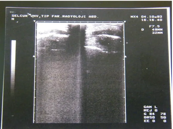
Thorax ultrasonography (USG) revealed a cystic mass lesion of 42 x 32 mm with a thick membrane, which creates an extrinsic pressure on parietal pleura, extending intrathoracically and contouring the end of the fifth right rib ( Figure 1) .
Figure 1. Preoperative USG showed rib abcess
Surgery aiming diagnosis and treatment based on these finding was planned, and the front end of the 5th rib and the mass were excised without resecting the pleura. Histopathological examination of the mass specimen revealed chronic granulamatous inflammation.
Monthly follow-ups indicated that the patient suffered from a flail chest at the removed thoracic region, he was re-hospitalized due to continuing respiratory difficulties despite the medical treatment on may 1994.
2nd Hospitalization: Physical examination revealed a chest wall defect of 5x7x2 cm sizes localized on the anterior wall of right hemithorax. The existing defect was supported with a stainless-steel graft and a mersylon mesh in the operation. The problem of flail chest recovered
during the post operation period 3rd
Hospitalization: June 1999. The patient had fallen from stairs and hit his chest a month before and applied again to the hospital with complaints of pains at the former operation site, resulting in an 296
Cilt/Volume 38 Yıl/Year 2013 Steel Graft Repair in Rib Tberculosıs: A 10-Year Follow up Case
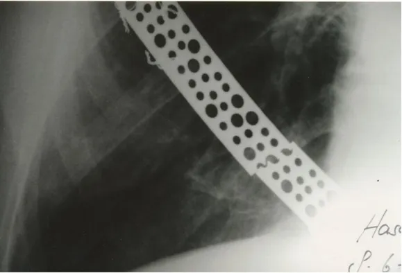
increase of the shortness of breath. Chest X-ray of the patient revealed a significant fracture in the steel graft (Figure 2). The patient was re-operated,
the graft region was incised and the steel graft was fixed with stainless steel wires. Flail chest recovered during the post operation period.
Figure 2. Chest-X ray shows a fracture in the steel graft
DISCUSSION
Rib tuberculosis is a rare disease. In a series of 5,000 skeleton tuberculosis, it was reported at 1.7%3. Asnis et al. in 1994, 24,361 cases of tuberculosis. In this series, musculoskeletal tuberculosis accounted as 1.8%. There were 400 cases of rib tuberculosis recorded in the world literature in 1997 (4). Rib tuberculosis ofen occurs up to 18 months after enfenction and <50% of patients have active pulmonary disease4. Usually one rib is affected, with the 5th rib being the most frequent. It sometimes starts simultaneously, multiple in different ribs.
The most frequent symptom observed in patients is the painful local tumor, which lasts approximately a month. It may then be fistulized
and drained by itself. Thus, the diagnosis of rib tuberculosis may be given in delay or surgically2-6.
Minimal rib destruction may be present in lung graphs. However, anamnesis of previous tuberculosis, USG, bone scintigraphy, C.T and percutaneous biopsy may be used for a definite diagnosis.
We employed USG in the diagnosis of our case, and upon the indication of abscess and ostemyelitis by USG, we operated the patient without considering further investigations.
In the treatment of rib tuberculosis, medical tuberculosis treatment should be started if it is possible to diagnose in advance and follow the case for a period 1 to 3 months. If no recovery is observed, surgical resection should be planned2,5,6.
Öncel ve Sunam Cukurova Medical Journal
Although single rib resurrections do not cause a problem, grafting may be required in multiple grafting7. While single rib anterior resection was performed in our case in contrary to the literature, an increase was observed in the paradoxical respiration, which required a treatment. Normally, different rates of paradoxical respiration is present at the lower and lateral ribs in cases with COPD. This finding observed in such patients is called “hoover sign”. We therefore concluded that since our case had already experienced a paradoxical respiration associated with COPD, removal of the rib caused the increase of this respiratory difficulties as well as paradoxical respiration.
Selection of prosthetic material is a complex issue since there are numerous materials used for this purpose. For example ,metallic instruments ,methyl methacrylate sandwich, silicone, Teflon, or acrylic materials have been utilized. Selection of the material is left to the discretion of the surgeon
7-8
. In non-tumor cases, biodegradable ribs may be used both in the reconstruction of chest wall and after thoracotomies. The flexibility and durability have been found similar with healthy ribs8. However, since biodegradable ribs are absorbed by the body within maximum three years. Therefore, we did not consider biodegradable ribs in our case. We preferred to repair the steel graft instead of replacement in our case with a view to avoid from removing the implanted graft and replacing it since this will create both material costs and management and infection problems.
In conclusion, in the light of the experience obtained in our case, we recommend using softer
graft materials that are more adaptable to the anatomy in cases where a chest wall reconstruction is to be made and graft durability is involved. Our cases showed interesting for the broken the steel implant material,follow up long term and especially the thoracic surgeon shouldn’t use like this material .They should use a titanium contain material instead of .
REFERENCES
1. Fonkalsrud EW. Chest wall abnormalities .In: Banue AA, editor. Glenn’s Thoracic and Cardiovascular Surgery. USA:Appleton & Lange. 1991; 515.
2. Khalil A, Breton C, Tassart M, Korzec J, Bigot J,Carette M. Utulity of CT scan for diagnosis of chest wall tuberculosis. Eur Radiol. 1999; 9:1638-42. 3. Carstensen E. Surgical treatment of chest wall
tuberculosis. Chirurg. 1980; 51:163-5.
4. Asnis DS, Niegowska A. Tuberculosis of the rib. Clinical Infectious Disease. 1997; 24:1018-9. 5. Chang JH, Kim SK, Lee WY. Diagnostic issues in
tuberculosis of the ribs with a review of 12 surgically proven cases. Respirology. 1999;4: 249-53.
6. Faure E, Souilamas R, Riquet M, Chehab A, Le Pimpec-Barthes F, Manac’h D, Debesse B. Cold abscess of the chest wall: a surgical entity?. Ann Thorac Surg. 1998; 66:1174-78.
7. Deschamps C, Tirnaksiz BM, Darbandi R, Trastek VF, Allen MS, Miller DL, Arnold PG, Pairolero PC. Early and long- term results of prosthetic chest wall reconstruction. J Thorac Cardiovasc Surg 1999; 117: 588-92.
8. Hirata T, Fukuse T, Mizuno H, Hitomi S, Wada H. Clinical application of biodegradable rib connecting pins in thoracotomy. Thorac Cardiovasc Surg. 1999; 47: 183-7.
Yazışma Adresi / Address for Correspondence:
Dr. Murat Öncel
Selçuk Üniversitesi Selçuklu Tıp Fakültesi Göğüs Cerrahi Bölümüı KONYA e-mail: [email protected] geliş tarihi/received :18.07.2012 kabul tarihi/accepted:16.11.2012 298