Okajimas Folia Anat. Jpn., 76(5): 255-262, December, 1999
The Variations of the Subclavian Artery and Its Branches
ByAhmet H. YUCEL, Emine KIZILKANAT and CengizO. OZDEMIR
Department of Anatomy, Faculty of Medicine, Cukurova University, 01330 Balcali, Adana Turkey- Received for Publication, June
19,1999-Key Words: Subclavian artery, Vertebral artery, Arterial variation
Summary: This study reports important variations in branches of the subclavian artery in a singular cadaver. The origin of the left vertebral artery was from the aortic arch. On the right side, no thyrocervical trunk was found. The two branches which normally originate from the thyrocervical trunk had a different origin. The transverse cervical artery arose directly from the subclavian artery and suprascapular artery originated from the internal thoracic artery. This variation provides a short route for posterior scapular anastomoses. An awareness of this rare variation is important because this area is used for diagnostic and surgical procedures.
The subclavian artery, the main artery of the
upper extremity, also gives off the branches which
supply the neck region. The right subclavian arises
from the brachiocephalic trunk, the left from the
aortic arch. Because of this, the first part of the
right and left subclavian arteries differs both in the
origin and length. The branches of the subclavian
artery are vertebral artery, internal thoracic artery,
thyrocervical trunk, costocervical trunk and dorsal
scapular artery. On the left, all branches except the
dorsal scapular arise from the first part; on the
right, the costocervical trunk usually springs from
the second part").
The vertebral artery is the first and largesi
branch of the subclavian artery. Its extracranial
part arises from the superoposterior
aspect of the
subclavian, usually enters the foramen of sixth
cer-vical transverse processes,
rarely the seventh,
curves medially behind the lateral mass of the atlas
and then enters the cranium via the foramen mag-
num. Its intracranial part joins its fellow to form the
basillar artery at the lower pontine border.
In this study, the variations of the branches oi
the subclavian arteries in the cadaver of an eighty
year-old man were described. On the left side, the
vertebral artery arose directly from the aortic arch
but on the right side, it was as usual. However on
the right side, there was no thyrocervical trunk and
the transverse cervical artery arose directly from
the subclavian artery. It was also observed the righi
suprascapular artery began from the internal
tho-racic artery.
The variations of the subclavian artery and its
branches have a great importance both in blood
vessels surgery and in angiographic investigations.
Subjects
This work is based on a dissection carried out in
the Department
of Anatomy in the Faculty of
Medicine of the cukurova University in 1996-199'7
academic year. A dissection was made of neck
re-gion of 80-year-old male cadaver. After seeing
var-iations, the dissection was completed by following
the course of the subclavian arteries and their
branches.
Findings
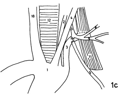
In our case, the left vertebral artery originated
from the superior aspect of the aortic arch between
the left subclavian artery and the left common
ca-rotid artery instead of the left subclavian artery. It
first ascended behind the carotid sheath for about
9.5 cm and then passed through the foramen of fifth
cervical vertebra. Thyrocervical trunk arising from
the superior aspect of the first part of the left
sub-clavian artery gave off a common trunk . The
trans-verse cervical artery and the suprascapular artery
originated from this trunk near the medial border
of the scalenus anterior muscle and crossed this
muscle anteriorly (Fig. 1.a, b, c).
256 A.H. Yiicel et aL
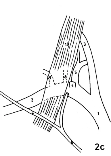
On the right side, the vertebral artery arose as
usual, but there was no thyrocervical trunk. Two
branches originated from the second part of the
subclavian artery at a distance of 1 cm: The first
branch, the costocervical trunk, situated medially
and it divided into the deep cervical artery as
su-perior branch and the susu-perior intercostal artery as
inferior branch (Fig. 2.a, b, c). The lateral branch,
the transverse cervical artery, passed deep to the
scalenus anterior muscle and inclined laterally
to-ward the posterior cervical triangle.
The main finding is that the suprascapular artery
arose from the internal thoracic artery instead of
the thyrocervical trunk; it first crossed anterior to
the scalenus anterior muscle run parallel to the
clavicle behind this bone and then turn backward
passing deep to the inferior belly of omohyohid
muscle.
Discussion
The subclavian artery and its branches have
many variations in their origin, course, level of
as-cending and termination in the neck2i3). The right
subclavian artery may directly arise from the aortic
arch or both subclavian arteries may originate from
the common trunk arising from the aortic arch.
The variations in the origin and the course of
extracranial part of the vertebral artery are also
common").
The vertebral artery may originate
from the aortic arch or the left external carotid
artery. Atypical artery positions, turtuosity and
duplication of the vertebral artery are frequent
variations in this artery4'5'''10). The origin of the
vertebral artery directly from the aorta usually
oc-curs on the left and the rate of its incidence has
been reported as 2.5% and 4.5%1).
The frequency of the left vertebral artery arising
directly from the aortic arch and following a course
between the left common carotid artery and the left
subclavian artery is from 1% to 5%11.12).
The vertebral artery enters the sixth cervical
transverse foramen at the rate of 90%1'9). This
ar-tery sometimes enters to the fifth cervical
trans-verse foramen. According to Lippert and Pabst"),
the frequency of this variation is 5%. von Eich-
horn")has
suggested that the blood flow factors
and ageing may cause the variations of vertebral or
basillar artery positions. In our case, the leftverte-
bral artery arose from the aortic arch and entered
the fifth cervical transverse foramen.
The suprascapular artery and the transvers cer-
vical artery are normally the branches of thyrocer-
vical trunk. There is no standard pattern for the
branching of the transverse cervical artery. Some of
its branches may arise separately or as common trunk. These types of branching show an incidence of 50%. In our case, no thyrocervical trunk origi- nating from the subclavian artery on the right side was observed. Two arteries which are normally the branches of the thyrocervical trunk had the unusual origin: a) The transverse cervical artery was a direct branch of the right subclavian artery and b) the su-prascapular artery arose from the internal thoracic artery. The frequency of the suprascapular artery arising from the internal thoracic artery has been established at 4.1%14). This variation of the supra- scapular artery provides a short route to the poste- rior scapular anastomoses supplying the upper ex-tremity via the internal thoracic artery originated directly from the suprascapular artery. Thus this short route has significant importance because it gives a collateral support to the upper extremity when obstruction or ligation occurs not only in the third part but also in the first part of the subclavian artery. The importance of this case is a cluster of variations of the branches of the subclavian artery was found in one anatomical specimens.
The origin and the course of subclavian artery and its branches, which also supply the brain be-sides the upper extremity, must be precisely deter-mined for accurate diagnostic interpretation as well as the performance of interventional or surgical procedures such as the construction of a subclavio-vertebral bypass, balloon dilatation of subclavian artery stenosis, treatment of aortic coarctation by plasty with the subclavian artery and artery dissec-
tion4'15-19).
The variations of the subclavian artery are explained by embryologic development. The early limb bud receives blood via intersegmental arteries which contribute to a primitive capillary plexus. In
the upper limb bud the lateral branch of the sev-enth intersegmental artery usually persists as the subclavian artery. Because of multiple and plexi- form sources of this artery, variations such as di-vergence in the mode and proximodistal level of
branching, aberrant vessels connecting other prin-cipal vessels are fairly common. The vertebral and internal thoracic artery develop from longitudinal arteries of intersegmented anastomoses. Caudal shifting of the aorta may cause the longitudinal torsion and bending of the proximal parts of seg-mental arteries by resulting the abnormal con-nections between the longitudinal arteries and the subclavian artery2). The anomalies found in the subclavian artery may also cause the pathologic conditions. Rodrigez et al. 2°) reported a case with dysphagia lusoria caused by an abnormal right subclavian artery associated with aortic coarctation. Therefore, the variations as well as physiological
The Variation of the Subclavian Artery 257
factors should be considered as causes of certain diseases related to the vessels.
References
1) Grant JCB. An Atlas of Anatomy. 6th ed. The Williams and Wilkins Co Baltimore 1972; pp. 432-447.
2) Williams PL, Bannister LH, Berry MM, Collins P, Dyson M, Dussek JE and Ferguson MWJ. Gray's Anatomy. 38th
ed. Churchill Livingstone, London 1995; pp. 1529-1536. 3) Daseler EH and Anson BJ. Surgical anatomy of the
clavian artery and its branches. Surgery Obstetrics and
Gynecology 1959; 108:149-174.
4) Stefanov S. Angiography of certain pathologic forms of the extracranial portion of the vertebral artery. Folia Med
(Plovdiv) 1965; 7(4):247-251.
5) Schmidt H and Pfingst E. Duplication of the vertebral artery. Fortschr Geb Rontgenstr Nuklearmed 1973; 118(6):636-640.
6) Distelmaier P and Wappenschmidt J. Atypical termination
of the vertebral artery and of the inferior posterior ebellar artery. ROFO Fortschr Geb ROntgenstr
armed 1976; 124(3):253-256.
7) Huber G and Piepgras U. Origin of the left vertebral artery from the left external carotid artery. ROFO Fortschr Geb
Rontgenstr Nuklearmed 1976; 125(1):63-66.
8) Barth H. Pathomorphological studies on the effect of generative changes in the area of the lateral cervical spine
and the course of the vertebral artery. Zentralbl Neurochir
1985; 46(2):119-125.
9) Tschabitcher M, Fuss FK, Matula C and Klimpel S. Course of the arteria vertebralis in its segment VI from the origin
to its entry into the foramen processus transversi. Acta
Anat (Basel) 1991; 140(4):373-377.
10) Hauke P. Turtuosity of the carotid and vertebral arteries:
angiographic and clinical findings. Otsch Med Wochenschr
1973; 98(50):2381-2384.
11) Lippert H and Pabst R. Arterial variations in man. fication and frequency. J.F. Bergmann Verlag Miinchen
1985.
12) Vorster W, du Plooy PT and Meiring JH. Abnormal origin of internal thoracic and vertebral arteries. Clinical
omy 1998; 11:33-37.
13) von Eichhorn M. Causes of variations in the pathway of the basilar and vertebral arteries. Gegenbaurs Morphol Jahrb
1991; 136(1):127-134.
14) Bergman RA, Thompson SA, Afifi AK and Saadeh FA. Compendium of Human Anatomic Variation. Urban and
Schwarzenberg, Baltimore 1988.
15) Habel J. Radiologic presentation of the vertebral arteries and the right carotid artery by means of direct
clavicular injection of the subclavian artery. J Beige Radiol
1967; 50(3):171-179.
16) Araneda I, Gomez 0, Arretz C, Leon L, Eimbcke F and Zilleruelo R. Treatment of aortic coarctation in children by plasty with the subclavian artery. Rev Chil Pediatr 1986; 57(5):401-404.
17) Burger K and Luther B. Surgical treatment of extracranial
occlusive processes of the vertebral artery by contraction of a subclavio-vertebral bypass. Zentralbl Chir 1989; 114(3):
181-189.
18) Thomassen L and Krakenes J. Balloon dilatation of clavian artery stenosis and brachiocephalic trunk stenosis.
Tidsskr Nor Laegeforen 1989; 109(10):1063-1068. 19) Frauchiger B and Bernays DR. Spontaneous dissection and
stenosis of the vertebral artery. Schweiz Med Wochenschr
1991; 121 (35):1243-1248.
20) Rodrigez Cuartero A, Rodrigez Cuartero F and Pelaez Redondo J. Dysphagia lusoria, caused by an abnormal right
subclavian artery associated with aortic coarctation. Rev
Esp Enferm Apar Dig 1979; 55(2):215-222.
258 A.H. Yficel et aL Plate I
la
The Variation of the Subclavian Artery 259
Explanation of Figures
Plate I
Fig. 1.a. The branches arising from the left side of aortic arch. b) The left vertebral artery originated from aortic arch. The left common carotid artery was pulled up to demonstrate the vertebral artery. c) The schematic representation of the left side. 1:
Aortic arch; 2: Vertebral artery; 3: Subclavian artery; 4: Common carotid artery; 5: Internal thoracic artery; 6: Thyrocervical
trunk; 7: Inferior thyroid artery; 8: Transverse cervical artery; 9: Suprascapular artery; 10: Brachiocephalic trunk; 11: Scalenus anterior muscle; 12: Trachea.
260 A.H. Yficel et al. Plate II
2a
The Variation of the Subclavian Artery 261
2c
Plate II
Fig. 2.a. The branches arising from the right side of aortic arch. b) The scalenus anterior muscle was dissected and retracted to demonstrate the right subclavian artery and its branches. c) The schematic representation of the right side. 1: Brachiocephalic trunk; 2: Subclavian artery; 3: Common carotid artery; 4: Inferior thyroid artery; 5: Vertebral artery; 6: Internal thoracic artery; 7: Suprascapular artery; 8: Costocervical trunk; 9: Transvers cervical artery; 10: Scalenus anterior muscle.