E-mail: [email protected] doi:10.3906/sag-0902-16
Imaging liver transplantation candidates using
gadolinium enhanced MDCT angiography
Aim:To report our experience with gadolinium enhanced computed tomography angiography (Gd-CTA) for end-stage liver disease patients who are candidates for liver transplantation as an alternative to iodinated contrast enhanced CTA and gadolinium enhanced magnetic resonance (MR) angiography.
Materials and methods:Seventeen patients with end-stage liver disease were evaluated with Gd-CTA. The patients were 10 men (58.8%) and 7 women (41.1%) (age range: 37-59 years). Multidetector CT (MDCT) was performed using 16-detector-row CT scanners. 3D images of the celiac artery, superior mesenteric artery, portal vein, and inferior vena cava (IVC) were obtained for thorough assessment using multiple plane reconstruction and maximum-intensity projection. Adequate images were defined by complete opacification of the celiac, superior mesenteric, hepatic propria, bilateral hepatic arteries, main portal vein and bilateral intrahepatic portal branches, and IVC.
Results:All 17 Gd-CTAs were rated as diagnostic. Adequate CT arteriography (celiac, common hepatic, hepatic propia, and bilateral hepatic artery) and portography were obtained in all the patients. Engorged gastric and coronary varices were detected in 5 patients (29.4%). Severe splenorenal shunt was found in 2 patients (11.7%). Thrombus of the portal vein was observed in 3 patients (17.6%). The radiological findings and operational findings were not discordant in any of the operated patients. Conclusion:The degree of enhancement and image quality achieved using these gadolinium-enhanced MDCT angiographies appear adequate for angiographic evaluation of liver transplantation. Therefore, the use of 3D Gd-CTA in selected cases and settings can be advantageous in the pretransplantation evaluation.
Key words:Liver Transplantation, candidates, gadolinium, MDCT angiography
Karaciğer nakil adaylarının gadolinyum destekli çok kesitli
bilgisayarlı tomografi anjiografi ile görüntülenmesi
Amaç:Bu çalışmanın amacı son dönem karaciğer yetmezlikli, karaciğer nakli adaylarının iyotlu kontrast ajan kullanılan bilgisayarlı tomografik anjiyografi (BTA) ve gadolinyum kullanılan MR anjiyografiye alternatif olarak gadolinyum kullanılarak yapılan BTA (Gd-BTA) ile ilgili deneyimlerimizin aktarılmasıdır.
Yöntem ve gereç: On yedi adet son dönem karaciğer yetmezlikli hasta Gd-BTA ile değerlendirildi. Hastaların 10’u erkek(% 58,8) ve 7’si kadındı (% 41,1) (yaş aralığı; 37-59). Çok kesitli BT olarak 16 dedektör sıralı tarayıcı kullanıldı. Çöliak arter, superior mezenterik arter, portal ven ve inferior vena kava (IVC) ’nın 3 boyutlu ve çok düzlemli rekonstrüksiyon görüntüleri elde edildi. Görüntülerin yeterliliği çöliak arter, superior mezenterik arter, arteria hepatic propiria, her iki hepatic arter, ana portal ven, her iki taraf portal dallar ve İVC nın tam opakifikasyonu elde edilerek tanımlandı. Bulgular:Gd-BTA ile yapılan 17 çekimin tümü tanısal olarak değerlendirildi. Çöliak, ana hepatik, hepatika propiria ve her iki taraf hepatik arterlerin BT arteriyografileri ve portografiler tüm hastalarda elde edildi. Genişlemiş gastrik ve coroner varisler 5 hastada (% 29,4) belirlendi. Ciddi splenorenal şant ise 2 hastada belirlendi. Üç hastada portal vende thrombüs gözlendi (% 17,6). Ameliyat edilen hastaların hiçbirinde radyolojik ve ameliyat esnasında elde edilen bulgular arasında uyumsuzluk yoktu.
Mecit KANTARCI1 Irmak DURUR SUBAŞI1 Bülent AYDINLI2 Cihan DURAN3 Selim DOĞANAY4 K. Yalçın POLAT2 Ragibe KANTARCI5 Hayri OĞUL1 S. Selçuk ATAMANALP2 1 Department of Radiology, Faculty of Medicine, Atatürk University, Erzurum - TURKEY
2Department of General Surgery,
Faculty of Medicine, Atatürk University, Erzurum - TURKEY
3
Department of Radiology, Florence Nightingale Hospital, Bilim University,
İstanbul - TURKEY
4
Department of Radiology, Develi Government Hospital, Kayseri - TURKEY
5
Department of Internal Medicine, Faculty of Medicine, Atatürk University, Erzurum - TURKEY Received: February 19, 2009 Accepted: August 03, 2009 Correspondence Mecit KANTARCI
200 Evler Mah. 14. Sok. No 5 Dadaşkent, Erzurum - TURKEY
Introduction
Nowadays, liver transplantation has been used more commonly for patients with end-stage liver disease (1,2). In this context, imaging of the liver transplantation candidates is of great importance because the findings can influence the surgical planning and even the operation type. Computed tomography, magnetic resonance imaging, and conventional angiography have been widely used for this purpose. In the cross-sectional imaging of the liver transplantation candidates, each of these modalities has some disadvantages. Imaging with iodinated contrast material may result in irreversible deterioration of renal function (3). In addition, magnetic resonance angiography has several limitations (3-5) and is often insufficient before surgical treatment (3). For these reasons, gadolinium enhanced computed tomography angiography (Gd-CTA) may be a solution in the situations where iodinated contrast material and magnetic resonance imaging are not preferred. Considering all of the above-mentioned reasons, successful and interpretable Gd-CTA applications in imaging of vascular structures of various body areas have recently been available in the literature (6-8). The purpose of this study is to report our experience with Gd-CTA for the end stage liver disease patients who are candidates for liver transplantation as an alternative to iodinated contrast enhanced CTA and gadolinium enhanced MR angiography prospectively. To our knowledge, no data have been reported about the use of gadolinium-based contrast agents in the CT angiographic evaluation of hepatic circulation.
Materials and methods
In Atatürk University Hospital and Florence Nightingale Hospital, 17 patients (10 men) with end-stage liver disease who had indications for liver transplantation were evaluated with Gd-CTA. The mean age of the patients in this study group was 48
years (age range: 37-59 years). The institutional ethics committee approved the study, and signed informed consent was obtained from each patient. The procedures used were in accordance with the recommendations found in the Helsinki declaration. Multidetector CT was performed on 2 different 16-detector-row CT scanners (Aquillon; Toshiba Medical Systems, Tokyo, Japan; and Sensation16; Siemens Medical Systems, Forchheim, Germany). With the first scanner, the following parameters were applied: 16 × 0.5-mm collimation, 1.0-mm slice thickness, and 1.0-mm reconstruction interval. On the second scanner, images were obtained with 12 × 0.75-mm collimation, 1-mm slice thickness, and 0.6-mm reconstruction interval.
Each patient received a maximum dose of 0.3 mmol/kg of body weight of 0.5 mmol/mL gadoterate meglumine (Dotarem; Guerbet, Paris, France). The corresponding volume of the contrast material was injected intravenously at 4.5 mL/s. The fixed delay technique was used for the starting time of scanning. Twenty-five seconds after the injection of the contrast medium, arterial phase was taken in one breath-hold, followed by 1 portal venous phase at 55 s and 1 systemic venous phase at 120 s after the injection of the medium.
3D images of the celiac artery, superior mesenteric artery, portal vein, and inferior vena cava (IVC) were obtained for thorough assessment using multiple plane reconstruction and maximum-intensity projection performed by radiologists. The 3D CTA was interpreted by 2 radiologists (radiologists experienced 10 (MK) and 5 years (IDS)) at the workstation (Vitrea 2, VITAL Images, Inc. Minnetonka, Minnesota, USA) after viewing the raw data and the reformatted images in projection views and axial imaging.
Adequate images were defined by complete opacification of the celiac, superior mesenteric, hepatic propia, bilateral hepatic arteries, main portal
Sonuç: Gd-BTA ile son dönem karaciğer nakli bekleyen hastalar için yapılan anjiografik değerlendirilmede görüntülerin yoğunluğu ve tanısal kaliteleri yeterli düzeyde tespit edildi. Bu nedenle 3 boyutlu Gd-BTA nın seçilmiş hastalarda kullanımı, nakil öncesi vasküler yapıların tanısal değerlendirmesinde başarılı bir görüntüleme yöntemi olarak göz önünde bulundurulabilir. Anahtar sözcükler:Karaciğer nakli, alıcı adayları, gadolinyum, çok kesitli BT anjiyografi
vein and bilateral intrahepatic portal branches, and IVC. The imaging results were compared with the operative findings for the patients who underwent surgery.
Results
Seventeen patients with end stage liver disease were included for pretransplant evaluation. All 17 Gd-CTAs were rated as diagnostic. No adverse events occurred after the intravenous injection of gadolinium. The examination was well-tolerated, and the patients did not report any discomfort. The serum creatinine level remained stable on the first day after the procedure.
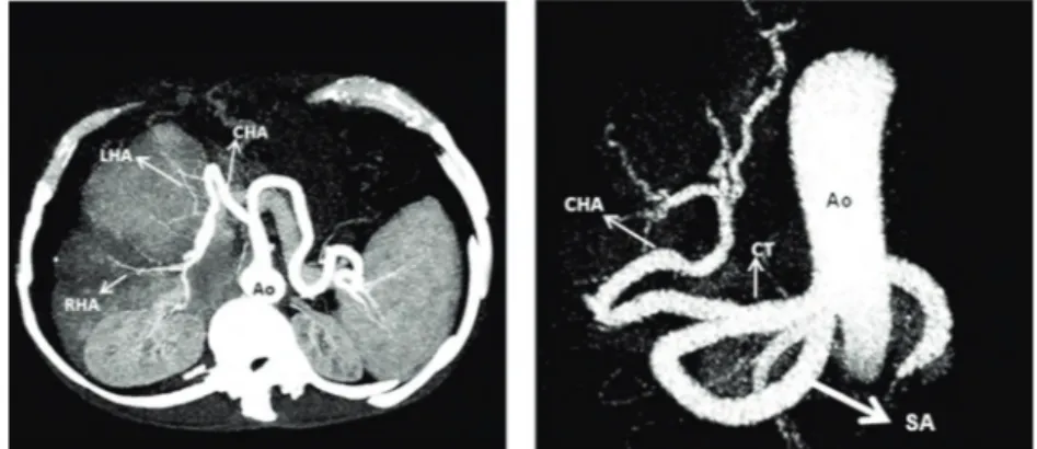
All the Gd-CTA images were found as adequate for illustration of the celiac, common hepatic, hepatic propia, and bilateral hepatic artery in all 17 patients (Figure 1a, b). No pathological findings with arterial system that could hinder liver transplantation were
detected. There was only one (5.8%) vasculary anatomic variant in our patient population: replace right hepatic artery that originated from the superior mesenteric artery (Table).
Adequate CT portography was obtained in all the patients (Figure 2a, b). Splenomegaly was detected in all the patients. Engorged gastric and coronary varices (>1 cm) were detected in 5 patients (29.4%). Severe splenorenal shunt (3 mm) was found in 2 patients (11.7%), in whom shunt closure during transplantation was advised. Thrombus of the portal vein was observed in 3 patients (17.6%). In 2 of these patients (11.7%) (2 male), thrombectomy was performed intraoperatively and transplantation was carried out (Figure 3a, b). In the other patient (female), thrombus was widespread; thus, thrombolitic therapy was applied and the operation was postponed. In the other 14 patients (82.3%), no findings that could prevent transplantation with respect to the portal vein were detected. There was a
Table. Pathologic findings and anatomic variants depicted by gadolinium-enhanced CTA.
Patient (n) %
Engorged gastric and coronary varices 5 29.4
Severe splenorenal shunt 2 11.7
Thrombus of portal vein 3 17.6
Hepatic artery anatomic variant 1 5.8
Splenomegaly 17 100
Portal trifurcation 1 5.8
Figure 1. A 46-year-old man who was a liver transplantation candidate. A, Axial MIP image and B, 3D Volume-rendered images show all arterial structures (celiac, common hepatic, hepatic propia, and bilateral hepatic artery) well. Ao: Aorta, CHA: common hepatic artery, RHA: right hepatic artery, LHA: Left hepatic artery, CT: Celiac truncus, SA: Splenic artery.
patient (5.8%) with portal trifurcation for which 2 separate anastomoses were needed. All CTAs of the IVC were adequate. No vascular abnormalities were found. In addition no local parenchymal lesion was detected. The radiological findings and operational findings were not discordant in any of the patients that underwent surgery.
Discussion
Preoperative imaging of both hepatic vessels and parenchyma in liver transplantation is beneficial to better surgical planning or even precludes the necessity for surgery. Computed tomography (CT), magnetic resonance (MR) imaging, and conventional angiography have been all useful for this purpose. Nonetheless, there are some problems with these techniques: angiography is an invasive procedure
using a large amount of iodinated contrast medium, which may be nephrotoxic and which finally increases the risks for liver transplantation recipients (9). The kidney is an important organ often involved in complications of liver transplantation (10-12). CT also uses iodinated contrast medium. Among the large number of patients who cannot receive iodinated IV contrast because of contraindications including renal failure or insufficiency as well as a history of a previous significant contrast reaction, there is a subset of patients who also cannot undergo MR angiography because of contraindications including pacemakers, surgical clips, uncontrollable motion, claustrophobia, or postoperative change that could affect the MR signal (7,13). In some of these patients, problems associated with acidosis such as tachypnea and inability to hold breath are observed. This is one of the obstacles to obtaining good images in MR. In 10
Figure 2. A 38-year-old woman who was a liver transplantation candidate. A, Axial MIP image and B, coronal MIP image shows all portal system. Pv: Portal vein.
Figure 3. A 54-year-old man who was a liver transplantation candidate. A, Coronal MIP image and B, axial MIP images shows thrombus of the portal vein and superior mesenteric veins. MPV: Main portal vein, SMV: Superior portal vein, SV: Splenic vein, Black arrows: Thrombus.
of the patients (58.8%) blood creatinine level was slightly higher than normal (level range: 2-4 mg/dL). Also in 7 of the patients (41.1%) blood creatinine level was higher than normal (level range: 5-8 mg/dL), but the serum creatinine level remained stable on the first day after the procedure. Another aspect in our research is that 11 of the patients (64.7%) had inability to hold breath, 5 (29.4%) had claustrophobia, and 1 (5.8%) had surgical clips. Therefore, we did not have sufficient image quality of MR examinations. Gd-CTA may be appropriate for these patients in need of noninvasive vascular imaging but who have contraindications to both iodinated contrast and MR (13).
Renal dysfunction frequently attends end-stage liver disease because of the association between primary organ disease and hemodynamic interactions (14). In addition, survival postliver transplant correlates with pretransplant renal function (15).
Gadolinium-based agents, routinely used for MR imaging, are safe, well-tolerated, and have shown no nephrotoxicity for doses up to 0.4 mmol/kg of body weight (3,16,17). In addition, because of its high atomic number (64 vs. 53 for iodine), X-ray attenuation can be expected, although the commercially available gadolinium chelate solutions are less concentrated (0.5 molar) than iodinated contrast agents (3).
The pharmacokinetic behavior of intravenously delivered Gd-DTPA is similar to that of iodinated contrast agents used in urography and angiography. Excretion is predominantly through glomerular filtration, with a plasma half-life of about 90 min (18). The plasma half-life increases in relation to the degree of renal insufficiency. Presently, there is no evidence of instability or dissociation of the gadolinium chelate during a prolonged period of retention in the body, which could lead to the release of toxic gadolinium ions. For doses, 0.5 mmol/kg of body weight, serum creatinine and serum urea–nitrogen show no deviations outside the physiologic range of variation (3).
The first clinical use of gadolinium as a contrast agent for CT was reported by Bloem and Wondergem (13,19). In their study, gadolinium was seen in the bladder. Quinn et al. (20) demonstrated adequate
enhancement of the circle of Willis in a single volunteer. Lumboldt et al. (21) achieved sufficient enhancement for the assessment of thoracic and abdominal aortic aneurysms and staging of carcinoma of the thyroid. Additional case reports followed (19,20,22,23), including successful depiction of pulmonary embolism (24). Rosioreanu et al. reported the use of multidetector Gd-CTA to create 3-dimensional images of the neck (13). Karcaaltincaba and Foley administered a nonionic preparation of gadolinium, to exclude an aortic dissection in a patient who had a pacemaker and an elevated creatinine level (6). Sam et al. studied the safety of gadolinium contrast angiography in patients with chronic renal insufficiency and concluded that, despite reports of negligible nephrotoxicity, rarely gadolinium based contrast agents can cause acute renal failure in patients with underlying chronic renal insufficiency. Patients at high risk should be identified, and prophylactic measures should be taken to reduce the risk for nephrotoxicity (25).
The use of newer CT scanners with decreased acquisition times should lower the mmol/kg used in each patient to levels of 0.4 to 0.5 mmol/kg or less, while maintaining very good or excellent opacification of the central arterial system. With the use of routine very thin sections, there should probably be good to excellent opacification of smaller arterial structures as well although this should be the subject of formal investigations. Although Gd-CTA has limitations compared with MRA and CTA with iodinated contrast, as parenchymal organs are not optimally evaluated at doses of 0.5 mmol/kg or lower, it has been a very useful problem-solving technique in patients who cannot undergo MRA or conventional CTA in our experience. Despite these concerns, we think that carefully selected and monitored patients do benefit from Gd-CTA, particularly if 16- or more detector CT scanners are used (13,26).
There is controversy about the use of high-dose gadolinium for conventional angiography and for its use in conjunction with CTA. In either instance, gadolinium is being used off-label, and this should probably be explained to patients while obtaining informed consent (27). The manufacturer (and FDA approved) recommended dose for MR is 0.1 mmol/kg body weight although up to 0.3 mmol/kg is used routinely for body MR angiographic studies (28).
Limitations about this study include the high cost of Gd-DTPA, which is approximately 4.5 to 6.5 times more than that of an iodinated nonionic contrast agent, and there is unknown toxicity in humans at high doses (0.8 mL/kg body weight), especially in patients with renal failure (3). Selective injections should therefore be favored, and examinations limited to patients with azotemia or with a serious contraindication to iodine (3).
Clinical trials and extensive clinical experience have proven that gadolinium-based contrast agents are safe in patients with normal kidney function. As renal function deteriorates, however, the safety of these agents diminishes. Nephrogenic systemic fibrosis (NSF) is associated with gadolinium chelates and occurs exclusively in patients with decreased renal function (29). Patients with liver disease and those who have undergone liver transplantation seem to be at increased risk of developing NSF (29). According to Kirchin et al. MR contrast materials at doses used for MR imaging lack the nephrotoxicity associated with iodinated ones (30). The differences in the effects of various gadolinium preparations are attributable to gadolinium’s capacity to dissociate from chelates. The longer dissociation constant of some chelates is probably a function of their cyclic structure; most other gadolinium chelates have a linear structure. Release of gadolinium from a cyclic chelate requires all 4 covalent bonds to be broken simultaneously. The more flexible structure of linear chelates, the more readily facilitates gadolinium release. This fact might account for the higher incidence of NSF associated with gadodiamide and gadopentetate dimeglumine. In patients like ours, even if MR were performed instead of CT, the use of Gd chelates would be required. The Gd preparation we used is in cyclic form and, to our knowledge, there is no NSF case reported to be associated with gadoterate meglumin. Thus, in our study, this agent was preferred for use.
Occluded portal vein, the most common pathologic entity that complicates liver transplantation, is manageable with newer surgical techniques such as direct venous graft, jump graft, and even hemiportocaval anastomosis (31-34). A different and more complicated surgical technique concerning vascular anastomosis is needed
(compared with cadaveric liver transplantation) in the presence of congenital vascular anomalies (35). However, these complicated procedures may cause high morbidity or even require a second transplantation (36). Portosystemic shunt is a complicated condition encountered in liver transplantation. Decreased portal inflow to the graft during the operation would result in graft failure (37). Demonstration of the size and distribution of the shunt with adjacent anatomic indications in the abdominal and retroperitoneal cavity facilitate the ligation for both superficially and deeply located shunts (36).
In our experience, all the CT examinations performed with gadolinium-based contrast agents achieved appropriate vascular enhancement and allowed radiologists to get accurate diagnostic information. Based on our results involving 17 patients evaluated with a 16-detector-row CT scanner, we conclude that Gd-CTA can be considered an alternative technique for the evaluation of the hepatic circulation in certain patients with contraindications to traditional iodinated contrast agents and MR. The increased speed of data acquisition allows marked vascular contrast and less motion artifacts. It is performed in an outpatient setting. Furthermore, gadolinium-based contrast enhanced CT examinations are useful for evaluation of hepatic parenchyma such as vascular structure.
Our preliminary results showed good diagnostic accuracy of Gd-CTA in comparison with operative findings. Nevertheless, further research with a larger sample of patients is needed to validate these encouraging results.
Conclusion
This clinical experience with gadolinium-enhanced multislice 3D CTA in our institute was acceptable, definitive, noninvasive, fast, and easy. Although gadolinium chelates have been reported to be able to cause NSF, we think that the use of 3D Gd-CTA with macrocyclic chelates in selected cases and settings for evaluation of parenchyma and vascular structure may be an alternative for MRI and iodinated CT in pretransplantation evaluations.
1. Zibari GB, Edwin D, Wall L, Diehl A, Fair J, Burdick J et al. Liver transplantation for alcoholic liver disease. Clin Transplant 1996; 10: 676-9.
2. Diem HV, Evrard V, Vinh HT, Sokal EM, Janssen M, Otte JB et al. Pediatric liver transplantation for biliary atresia: results of primary grafts in 328 recipients. Transplantation 2003; 75: 1692-7.
3. Hammer FD, Malaise J, Goffette PP, Mathurin P. Gadolinium dimeglumine: an alternative contrast agent for digital subtraction angiography in patients with renal failure. Transplant Proc 2000; 32: 432-3.
4. Yang X, Manninen H, Soimakallio S. Carbon dioxide in vascular imaging and intervention. Acta Radiol 1995; 36: 330-7. 5. Hawkins IF Jr, Wilcox CS, Kerns SR, Sabatelli FW. CO2 digital
angiography: a safer contrast agent for renal vascular imaging? Am J Kidney Dis 1994; 24: 685-94.
6. Karcaaltincaba M, Foley WD. Gadolinium-enhanced multidetector CT angiography of the thoracoabdominal aorta. J Comput Assist Tomogr 2002; 26: 875-8.
7. Gupta AK, Alberico RA, Litwin A, Kanter P, Grossman ZD. Gadopentetate dimeglumine is potentially an alternative contrast agent for three-dimensional computed tomography angiography with multidetector-row helical scanning. J Comput Assist Tomogr 2002; 26: 869-74.
8. Carrascosa P, Merletti PG, Capuñay C, Goldsmit A, Bettinotti M, Carrascosa J. New approach to noninvasive coronary angiography by multidetector computed tomography: initial experience using gadolinium. J Comput Assist Tomogr 2007; 31: 441-3.
9. Yan SX, Liang TB, Fujii M, Kawamitsu H, Sugimura K, Zheng SS. Modified magnetic resonance angiography of the liver using sensitivity encoding in comparison with digital subtraction angiography and CT arterial portography. Hepatobiliary Pancreat Dis Int 2005; 4: 185-91.
10. Wang X, Li G, Li X, Zhang F, Qian J, Kong L et al. Multimodal approach to clinical liver transplantation. Zhonghua Wai Ke Za Zhi 2002; 40: 758-61.
11. Cohen AJ, Stegall MD, Rosen CB, Wiesner RH, Leung N, Kremers WK et al. Chronic renal dysfunction late after liver transplantation. Liver Transpl 2000; 8: 916-21.
12. Bilbao I, Charco R, Balsells J, Lazaro JL, Hidalgo R, Llopart L et al. Risk factors for acute renal failure requiring dialysis after liver transplantation. Clin Transplant 1998; 12: 123-9.
13. Rosioreanu A, Alberico RA, Litwin A, Hon M, Grossman ZD, Katz DS. Gadolinium-enhanced computed tomographic angiography: current status. Curr Probl Diagn Radiol 2005; 34: 207-19.
14. Davis CL, Gonwa TA, Wilkinson AH. Pathophysiology of renal disease associated with liver disorders: Implications for liver transplantation. Part I. Liver Transpl 2002; 8: 91-109.
15. Gonwa TA, Klintmalm GB, Levy M, Levy M, Jennings LS, Goldstein RM, Husberg BS. Impact of pretransplant renal function on survival after liver transplantation. Transplantation 1995; 59: 361-5.
16. Haustein J, Niendorf HP, Krestin G, Louton T, Schuhmann-Giampieri G, Clauss W et al. Renal tolerance of gadolinium-DTPA/dimeglumine in patients with chronic renal failure. Invest Radiol 1992; 27: 153-6.
17. Niendorf HP, Haustein J, Louton T, Beck W, Laniado M. Safety and tolerance after intravenous administration of 0.3 mmol/kg Gd-DTPA. Results of a randomized controlled clinical trial. Invest Radiol 1991; 26 Suppl 1: S221-3: discussion S232-5. 18. Weinmann HJ, Brasch RC, Press WR, Wesbey GE.
Characteristics of gadolinium-DTPA complex: a potential NMR contrast agent. AJR Am J Roentgenol 1984; 142: 619-24. 19. Bloem JL, Wondergem J. Gd-DTPA as a contrast agent in CT.
Radiology 1989; 171: 578-9.
20. Quinn AD, O’hare NJ, Wallis FJ, Wilson GF. Gd-DTPA: an alternative contrast medium for CT. J Comput Assist Tomogr 1994; 18: 634-6.
21. Lumboldt W, De Santis M, Von Smekal A, Reiser M. Attenuation characteristics and application of gadolinium-DTPA in fast helical computed tomography. Invest Radiol 1997; 35: 690-5.
22. Peña CS, Kaufman JA, Geller SC, Waltman AC. Gadopentetate dimeglumine: a possible alternative contrast agent for CT angiography of the aorta. J Comput Assist Tomogr 1999; 23: 23-4.
23. Chryssidis S, Davies RP, TIE ML. Gadolinium-enhanced computed tomographic aortography. Australas Radiol 2002; 46: 97-100.
24. Coche EE, Hammer FD, Goffette PP. Demonstration of pulmonary embolism with dynamic gadolinium enhanced spiral CT. Eur Radiol 2001; 11: 2306-9.
25. Sam AD, Morasch MD, Collins J, Song G, Chen R, Pereles FS. Safety of gadolinium contrast angiography in patients with chronic renal insufficiency. J Vasc Surg 2003; 38: 313-8. 26. Wicky S, Greenfield A, Fan CM, Geller SC, Hamberg LM,
Hoffmann U et al. Aortoiliac gadoliniumenhanced CT angiography: improved results with a 16-detector row scanner compared with a four detector row scanner. J Vasc Interv Radiol 2004; 15: 947-54.
27. Spinosa DJ, Kaufmann JA, Hartwell GD. Gadolinium chelates in angiography and interventional radiology: a useful alternative to iodinated contrast media for angiography. Radiology 2002; 223: 319-25.
28. Gierada DB, Bae KT. Gadolinium as a CT contrast agent: assessment in a porcine model. Radiology 1999; 210: 829-34. 29. Penfield JG, Reilly RF JR. What nephrologists need to know
about gadolinium? Nat Clin Pract Nephrol 2007; 3: 654-68. References
30. Kirchin MA, Runge VM. Contrast agents for magnetic resonance imaging: safety update. Top Magn Reson Imaging 2003; 14: 426-35.
31. Shinkai M, Ohhama Y, Nishi T, Yamamoto H, Fujita S, Take H et al. Congenital absence of the portal vein and role of liver transplantation in children. J Pediatr Surg 2001; 36: 1026-31. 32. Tzakis A, Todo S, Stieber A, Starzl TE. Venous jump grafts for
liver transplantation in patients with portal vein thrombosis. Transplantation 1989; 48: 530-1.
33. Tzakis AG, Kirkegaard P, Pinna AD, Jovine E, Misiakos EP, Maziotti A et al. Liver transplantation with cavoportal hemitransposition in the presence of diffuse portal vein thrombosis. Transplantation 1998; 65: 619-24.
34. Castaldo P, Langnas AN, Stratta RJ, Lieberman RP, Wood RP, Shaw BW Jr. Successful liver transplantation in a patient with a thrombosed portomesenteric system after multiple failed shunts. Am J Gastroenterol 1991; 86: 506-8.
35. Lilly JR, Starzl TE. Liver transplantation in children with biliary atresia and vascular anomalies. J Pediatr Surg 1974; 9: 707. 36. Cheng YF, Chen CL, Jawan B, Huang TL, Chen TY, Chen YS et
al. Multislice computed tomography angiography in pediatric liver transplantation. Transplantation 2003; 76: 353-7. 37. De Carlis L, Del Favero E, Rondinara G, Belli LS, Sansalone CV,
Zani B et al. The role of spontaneous portosystemic shunts in the course of orthotopic liver transplantation. Transpl Int 1992; 5: 9-14.