See discussions, stats, and author profiles for this publication at: https://www.researchgate.net/publication/323174084
Journal of Clinical and Analytical Medicine |
Article · May 2017 DOI: 10.4328/JCAM.5041 CITATIONS 0 READS 40 1 author:
Some of the authors of this publication are also working on these related projects:
retinaView project
corneaView project Nurgül Örnek
Kirikkale University
74PUBLICATIONS 355CITATIONS SEE PROFILE
All content following this page was uploaded by Nurgül Örnek on 14 February 2018. The user has requested enhancement of the downloaded file.
O
h
r
c
i
r
g
a
in
e
a
s
l
R
e
Gülşah Usta1, Reyhan Oğurel2, Zafer Onaran2, Zeynep Pekcan3, Tevfik Oğurel2, Birkan Karslı3, Nurgül Örnek2, Nesrin Büyüktortop Gökçınar2 1Göz Hastalıkları Kliniği, Gölhisar Devlet Hastanesi,Burdur, 2Göz Hastalıkları Anabilim Dalı, Kırıkkale Üniversitesi Tıp Fakültesi, Kırıkkale, 3Veterinerlik Fakültesi, Kırıkkale Üniversitesi, Kırıkkale, Türkiye
Autologous serum effect to corneal endothelial damage
Autologous serum effect on corneal endothelial
damage in the phacoemulsification rabbit model
Tavsan fakoemulsıfıkasyon modelinde kornea
endotel hasarına otolog serumun etkisi
DOI: 10.4328/JCAM.5041 Received: 21.04.2017 Accepted: 03.05.2017 Printed: 01.12.2017 J Clin Anal Med 2017;8(suppl 4): 330-5 Corresponding Author: Gülşah Usta, Göz Hastalıkları Kliniği, Gölhisar Devlet Hastanesi, 15400, Gölhisar, Burdur, Türkiye.
GSM: +905305539990 F.: +90 2483254 113007 E-Mail: [email protected]
Öz
Amaç: Tavşanlarda fakoemülsifikasyon ile oluşturulan kornea endotel hasa-rına ön kamaraya verilen otolog serumun muhtemel etkilerinin speküler mik-roskopi ile değerlendirilmesi amaçlanmıştır. Gereç ve Yöntem: Çalışmada do-kuz adet 5 aylık (yaklaşık 3 kg ağırlığında) Yeni Zellanda cinsi albino erkek tavşanın etik kurul onayı ile her iki gözü kullanıldı. Sadece 0,2 ml otolog serum uygulanan 1. grup (6 göz), sadece 20 saniye ultrasonik titreşim uygulanan 2. grup (6 göz) ve 20 saniye ultrasonik titreşimi takiben 0,2 ml otolog serum uy-gulanan 3. grup (6 göz) oluşturuldu. Girişim öncesinde, işlem sonrası 1. gün ve 7. gün speküler mikroskopi, göziçi basıncı ve pakimetri ölçümleri yapıla-rak veriler istatistiksel olayapıla-rak karşılaştırıldı. Bulgular: Postop 1. hafta sadece FAKO yapılan grupta (Grup 2) kornea kalınlığının 385µ’dan 445µ’a yükseldi-ği görülürken, FAKO + otolog serum verilen grupta (Grup 3) 398µ’dan 402µ’a yükseldiği ancak bu farkın istatistiksel olarak anlamlı olmadığı görüldü. Pos-top 1. gün 2. (FAKO) ve 3. (FAKO+otolog serum) grupların speküler mikrosko-pi parametrelerinden standart sapma (p<0.05) ve varyasyon katsayısı değer-lerinin (p<0.05) 1.gruptan yüksek olduğu gözlendi. Postop 1. hafta ise bu pa-rametrelerin her üç grupta da benzer olduğu ve preop değerlere döndüğü iz-lendi. Endotel hücre sayısı, hücre yoğunluğu, ortalama hücre alanı, en büyük ve en küçük hücre alanı ve hekzagonal hücre yüzdesi değerleri arasında ise üç zaman noktasında da gruplar arasında istatistiksel olarak anlamlı düzey-de fark saptanmadı. Tartışma: Bu çalışma, fakoemülsifikasyon işleminin ar-dından ön kamaraya verilen otolog serumun speküler mikroskopi ile teyit edi-lebilecek morfolojik değişikliklere sebep olmamakla birlikte endotel pompa fonksiyonları üzerinde muhtemel olumlu etkileri ile kornea ödemini azaltabi-leceğini göstermiştir.
Anahtar Kelimeler
Fakoemülsifikasyon; Kornea Endoteli; Otolog Serum; Speküler Mikroskopi
Abstract
Aim: This study used specular microscopy to evaluate the possible effects of applying autologous serum to the anterior chamber on the corneal endo-thelial damage created by the ultrasonic vibrations of phacoemulsification (PHACO) in rabbits. Material and Method: The study, which was approved by the Ethics Committee, involved both eyes of nine 5 month-old (about 3 kg) New Zealand genus albino male rabbits. Group 1(6 eyes) received only an application of 0.2 ml autologous serum; Group 2 (6 eyes) received only the ultrasonic vibrations for 20 seconds; and Group 3 (6 eyes) received 0.2 ml autologous serum followed by ultrasonic vibrations for 20 seconds. Measure-ments were made before the intervention and at the 1st and 7th days post-operatively using specular microscopy, intraocular pressure, and pachymetry metrics, and the data were compared statistically. Results: Postoperative 1st week, the cornea thickness increased from 385 µ to 445 µ in Group 2 (PHACO only) and from 398 µ to 402 µ in Group 2 (PHACO plus autologous serum). However, this difference was not statistically significant. Postoperative 1st day standard deviation (p < 0.05) and variation coefficient (p < 0.05) values of the specular microscopy parameters for Groups 2 and 3 were higher than for Group 1. Postoperative 1st week it was observed that the parameters of the three groups were similar and had returned to the preoperational values. Between the endothelial cell numbers, the cell density, the cell space aver-age, the largest and the smallest percentage of the cell and the hexagonal cell area values, there were no statistically significant differences at the three measurement time points. Discussion: This study showed that, after the phacoemulsification process, autologous serum applied to the anterior chamber can, by positive possible effects on endothelial pump functions, re-duce corneal edema without morphological changes, which can be confirmed with specular microscopy.
Keywords
Phacoemulsification; Corneal Endothelium; Autologous Serum; Specular Mi-croscopy
I Journal of Clinical and Analytical Medicine
| Journal of Clinical and Analytical Medicine
AUTOLOGOUS SERUM EFFECT TO CORNEAL ENDOTHELIAL DAMAGE
2
Introduction
Today, the duration and the power of the ultrasonic energy used in PHACO surgery, which is the preferred treatment method for more than 90% of cataract surgeries, is associated with various corneal damages such as endothelial cell loss and burning (1). The corneal endothelial cell loss after PHACO has been reported to vary between 1.2%-16.2% [2-4]. Risk factors for this damage include advanced age, small pupil, hard or large nucleus, large infusion volume, and the total height of the ultrasound energy (5-6). To date, approaches to reducing this damage have been viscoelastics, continuous circular capsulorhexis, and bicarbon-ate in the irrigation liquid. Also, there have been experiments with different breaking techniques of the nucleus and various methods such as the cold PHACO. But still the endothelial cell damage and the visual problems that result depending on the phacoemulsification continue to be a major problem. To this end, the effectiveness of different biological agent is also being researched [7-9].
Exogenous-derived growth factors can be used on the corneal epithelium to increase the epithelialization [10]. The produc-tion of growth factors with the recombinant technology is not a cost-effective approach, but the autologous serum (AS) con-tains a large quantity of growth factor, and is a liquid which can be easily obtained. Thus, it has epitheliotrophic potential for the ocular surface [11,12]. However, the demonstrated effect upon AS on the corneal endothelium is not known.
The endothelial cells are normally exposed to the growth fac-tors found in the aqueous humor. However, in cases where the concentrations of growth factors are inadequate, while they are in the inactive form, or when aiming to induce and/or to extend the positive mitogenic signals with the damaged endothelial on the endothelial cell receptor, it is thought that they cannot connect effectively [13].
Specular microscopy permits measurement of the quality and the quantity of corneal endothelial cells, and is the standard method of in-vivo evaluation of the intensity and morphology of endothelial cells [14].
This study used specular microscopy to evaluate the effect on endothelial function of the application of AS in the anterior chamber in conjunction with PHACO surgery in rabbits.
Material and Method
The study followed the principles of the Local Ethics Commit-tee of Kirikkale University, Faculty of Veterinary medicine, and confirmed by the number: 2012/46, by Kirikkale University, Fac-ulty of Medicine Department of Ophthalmology, and with the contributions of the Faculty of Veterinary Medicine.
Nine male albino rabbits which were five months old, weighed 2500-3000 grams, and were of New Zealand genus were used. During the study period, they were kept in the appropriate cages and special nutritional conditions in the Experimental Research Center of Kirikkale University. Both eyes of all rabbits were used with the permission of the Local Ethics Committee of Kirikkale University, Faculty of Veterinary Medicine.
In accordance with the study protocol, preoperative, postop-erative first and seventh day, anterior segment examination, cornea endothelial cell analysis on the central cornea with the specular microscope (SM), central corneal thickness
measure-ment (CCT), and measuremeasure-ments of intraocular pressure with to-nopen were performed for all the rabbits. As Mencucci and col-leagues have described, the endothelium cornea damage was generated in the continuous irrigation mode and using 100% ultrasonic power [15]. The phacoemulsification application time is based on an average of the two different works respectively, as 20 sec [15, 16].
In the first phase of the study, in order to prepare the AS, after the 9 rabbits’ ear surfaces were cleaned with alcohol, we en-tered in the marginal vein with an insulin syringe of 30 gauge. 2-3 cc’s of blood were drawn and put into sterile Eppendorfs which were numbered according to the rabbit. After sitting at room temperature without the sun for two hours, and their shaped elements collapsed. Then after 10 minutes at 3000 rpm centrifuge, the serums are what remain. The serum of each rab-bit was immediately placed into the sterile insulin injectors for use in surgical process.
After putting the rabbits on the operating table, the ocular sur-face and fornix to operate are washed with 5% povidone iodine. After securing the sterile field with a disposable drape, we put the speculum into the eyelid.
All operations were performed on the same day by the same surgeon. The eyes were divided into three group such that no rabbit had both eyes in the same group.
Group 1 (autologous serum, n= 6 eyes): We entered into the an-terior chamber via upper temporal cornea-limbal junction and 0.2 cc autologous serum was given using the 30 gauge insulin injector.
Group 2 (PHACO, n= 6 eyes): Using a 3.2 mm knife we made a transparent corneal tunnel incision via upper temporal cornea-limbal junction. The continuous irrigation mode was opened. We entered into the anterior chamber by tunnel with the stan-dard sleeve phaco probe. The phaco probe was maintained at the central (without a contact with any ocular structure, includ-ing lens and cornea), the U/S modulation worked 20 seconds with 100% power and continuously on irrigation (25 ml/min.). The wound was closed by a stromal hydration and after check-ing the sealcheck-ing, we put on the cornea a 10/0 nylon suture. 3rd group (PHACO + autologous serum, n= 6 eyes): We followed the PHACO procedure described above. The autologous serum prepared before the procedure was given 0.2 ml into the ante-rior chamber taken from the insulin injector and on the same incision.
All eyes in the postoperative period were administered 1% dexamethasone (Maxidex®, Alcon®, United Kingdom) and 0.3 % ofloxacin (Siprogut®, Biofarma®, Turkey), topically applied one drop 5 times a day.
None of the cases were given anesthetic, miotic, and mydriatic agents that may produce angiogenesis toxic effects in the eye. For irrigation, balanced salt solution (BSS ®, USA) was used. Preoperative, first postoperative and seventh day, SM (NIDEK ® CEM-530 Specular Microscope, Japan) measurements were made by the same practitioner. While analyzing the specular images obtained, we selected the most net one from eight im-ages, and ensured that within the framework of analysis at least 50 cells were counted.
In our study we measured the quantity of the endothelial cells (NUM), the cell density (ECD), the average cell field (ACF), the
Journal of Clinical and Analytical Medicine I 331
AUTOLOGOUS SERUM EFFECT TO CORNEAL ENDOTHELIAL DAMAGE
standard deviation (SD), the coefficient of variation (CV), the largest cell field (MAX), the smallest cell field (MİN), the hex-agonal cell percentage (HEX), the corneal thickness (CCT), and the intraocular pressure (IOP) at three points in time. In order to evaluate each parameter as a whole, we made a curve six area calculation (CAC).
At the IOP measurements made by the tonopen (Reichert To-no-Pen AVIA, USA), we took the average of 10 measurements made automatically and consecutively.
Statistical analysis
All data were analyzed using the software Statistical Package for Social Science (SPSS) Windows version 11.5 (Chicago, IL, USA). After the descriptive statistical analyses (average ± stan-dard deviation; median [minimum-maximum]), the evaluation of the normal distribution conformity was done with Shapiro Wilks Test. For the continuous variables, for the comparison of three groups the Kruskal Wallis test was used and for the comparison of two groups the Mann Whitney U test was used. For con-tinuous variables, for the comparison of the one group values on three times point we used the Friedman test; and for the comparison of two times point analysis the Wilcoxon Signed-rank test was used. P < 0.05 value was considered statistically significant.
Results
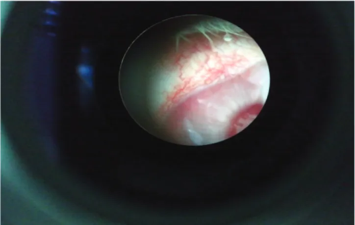
After PHACO and the injection, there was no infection, the mon-itoring deletion and the corneal edema in the anterior chamber and all rabbits had sectoral limbal hyperemia on the incision [Figure 1].
The standard deviation (SD) is the standard deviation of the values of the cell field. Among the three groups values for SD-T0 (p=0.664), SD-T2 (p=0.339), and SDCAC (p=0.203), there was no statistically significant difference, but regarding the SD-T1 val-ues of groups we found a statistically significant difference (p = 0.024). This was due to the fact the Group 1 SD-T1 values (p =. 041) were higher than those of Group 2 and Group 3 (respec-tively p=0. 041, p=0.009). There was no statistically significant difference between Group 2 and Group 3 SD-T1 values (p > 0.05) [Table 1 and Figure 2].
There was no statistically significant difference between the SS values of Group 1 (p=0.135), Group 2 (p=0.607), and Group 3 (p=0.311) at the three time points (Freidman test).
The variation (variability) coefficient (CV) is an objective crite-rion of the polymegathism that shows the variability between the cell areas (CV: SD/AVG). Among the CV-T0 (p=0.680), CV-T2 (p=0.318), and CVCAC (p=0.245) values for the three groups, there was no statistically significant difference, but we found a statistically significant difference for CV-T1 (p = 0.013). This was because the values of Group 1 were higher than those of Groups 2 and 3 (respectively p =0.026, p = 0.004). There was no statistically significant difference between the Group 2 and Group 3 CV-T1 values (p > 0.05) [Table 2 and Figure 3].
There was no statistically significant difference between the CV values of Group 1 (p=0.084), Group 2 (p=0.738) and Group 3 (p=0.311) at the three time points (Freidman test).
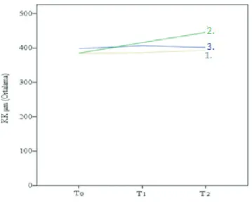
There was no statistically significance difference in the three groups regarding the corneal thickness (CCT)-T0 (p=0.605), CCT-T1 (p=0.313), and CCTCAC (p=0.291) values measured by optical pachymetry. However, there was statistically significant
Figure 1. The sectoral limbal hyperemia on the incision
Table 1. The comparison of SD measurements at three time points for the three groups Standard deviation(µm2) GROUPS P Group 1 (Average ±SD) Group 2 ( Average ±SD) Group 3 (Average ±SD) SD -T0 SD -T1 57.33±22.17 50.50±9.27 60.67±12.96 70.00±16.04 61.17±22.34 74.50±17.71 0664 0.024 SD -T2 70.00±15.82 62.83±16.17 55.17±13.95 0.339 SD CAC b2 294.92±46.57 331.00±41.55 327.17±44.49 0.203
Kruskal Wallis Test, SD=Standard deviation
Figure 2. The measuring line chart of three groups SD measurements at three time points
Table 2. The comparison of CV measurements at three time points for the three groups
Variation
coefficient (%) GROUPSGroup 1 P (Average ±SD) Group 2 (Average ±SD) Group 3 (Average ±SD) CV -T0 15.67±5.85 16.67±3.72 16.50±5.09 0.680
CV -T1 13.17±2.40 18.67±4.55 20.67±3.33 0.013
CV -T2 19.67±4.63 16.67±3.67 15.67±4.23 0.318
CV CAC b2 80.08±13.80 88.33±11.59 91.25±11.67 0.245
| Journal of Clinical and Analytical Medicine
AUTOLOGOUS SERUM EFFECT TO CORNEAL ENDOTHELIAL DAMAGE
4
difference for CCT-T2 values (p = 0.048). This difference was due to the fact that the Group 2 CCT-T2 values were statisti-cally significantly higher than the Group 1 values. There was no statistically significant difference in other binary comparisons (p > 0.05) [Table 3 and Figure 4].
There was no statistically significant difference between the CCT values of Group 1 (p=0.846), Group 2 (p=0.054), and Group 3 (p=1.000) at the three points in time (Freidman test). There was no statistically significant difference in the three
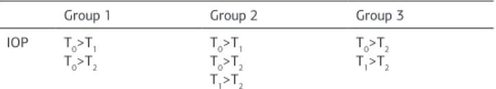
groups between the IOP-T0 (p=0.419), IOP-T1 (p=0.119), and IOPCAC (p=0.058) values, but the IOP-T2 values of groups we found a statistically significant between the level of difference (p = 0.031). This difference was due to the fact that Group 2 (p= 0.026) IOP-T2 values were statistically significantly less than the Group 1 values. There was no statistically significant differ-ence in other binary comparisons (p > 0.05) [Table 4 and Figure 5].
There was no statistically significant difference in Group 1 (p=0.006) and Group 3 (p=0.008) between the IOP values at the three points in time (Freidman test). The IOP-T0 (p=0.027) val-ues were higher than the IOP-T1 (p=0.027) and IOP-T2 (p=0.046) in Group 1. The IOP-T0 values were higher than the IOP-T1 val-ues (p=0.027), and the IOP-T0 (p=0.027) and IOP-T1 (p=0.027) values were higher than the T2 values in Group 2. The IOP-T0 (p=0.027) and IOP-T1 (p=0.042) values were higher than the IOP-T2 values in Group 3 [Table 5].
Figure 3. The measuring line chart of the three groups CV measurements at three points in time
Figure 4. The measuring line chart of three groups CCT measurements at three points in time
Figure 5. The measuring line chart of three groups IOP measurements at three points in time
Table 3. The comparison of KK measurements at three time points for the three groups Corneal thickness (µm) GROUPS P Group 1 (Average ±SD) Group 2 (Average ±SD) Group 3 (Average ±SD) CCT-T0 µm 384.67±30.89 385.33±12.56 398.83±28.72 0.605 CCT-T1 µm 386.83±19.14 416.00±44.24 406.50±34.22 0.313 CCT-T2 µm 393.67±49.19 445.50±53.11 402.17±45.85 0.048 CCTCAC b2 1946.75±151.83 2123.67±190.54 2020.00±158.51 0.291
Kruskal Wallis Test, CCT: Corneal thickness
Table 4. The comparison of IOP measurements at three time points for the three groups Intraocular pressure (mmHg) GROUPS P Group 1
(Average ±SD) (Average ±SD)Group 2 (Average ±SD)Group 3 IOP-T0 mmHg 13.00±4.69 11.33±2.07 12.83±3.06 0.419
IOP -T1 mmHg 9.17±1.47 7.33±2.25 9.67±1.75 0.119
IOP -T2 mmHg 8.17±1.33 6.50±0.84 7.33±0.52 0.031
IOP CAC b2 5.75±5.95 32.83±13.69 46.75±5.93 0.058
Kruskal Wallis Test, IOP: Intraocular pressure
Table 5. The comparison of the IOP values at three points in time in the three groups
GIB GIB-T0- GIB-T1 GIB-T0- GIB-T2 GIB-T1- GIB-T2
Group 1 0.026 0.046 ˃0.05
Group 2 0.027 0.027 0.027
Group 3 0.074 0.027 0.042
IOP: Intraocular pressure
Journal of Clinical and Analytical Medicine I 333
AUTOLOGOUS SERUM EFFECT TO CORNEAL ENDOTHELIAL DAMAGE
Discussion
We analyzed many parameters related the corneal endothelial cells and compared the values of three groups at three points in time (preoperative, post operative1st day and post operative 1st week). Within the groups and between the groups, we did not find statistically significant differences between the endo-thelial cells numbers, the cell density, the average cell area, the percentage of the largest and smallest cells, and the percent-age of the hexagonal cell values. However, across groups we determined the following:
At T1 time point (post operative 1st day):
• The standard deviation and the variation coefficient values of Group 2 (PHACO) and Group 3 (PHACO + AS) groups higher than Group 1 (AS).
At T2 time point (post operative 1st week):
• In Group 2 (PHACO) the corneal thickness increased from 385µ to 445µ, while in Group 3 (PHACO + AS) it increased from 398µ to 402µ. However, this difference was not statistically sig-nificant.
• Group 2 CT (corneal thickness) values were higher than those of Group 1 (much higher to be statistically significant from Group 3), and the IOP values were less than those of Group 1 (much less to be statistically significant from the Group 3). The increase of the variation coefficient which shows the high polymegathism (the difference between the cell areas) due to the increase of the standard deviation, is an expected finding (CV=SD/AVG×100). In Groups 2 and 3, for which we applied PHACO, it shows that the phacoemulsification operation caused the high polymegathism as expected. But because this differ-ence was not maintained through the T2 time point, it means a reduction in variability between the cell areas; on the 1st week after a surgery that not applied high PHACO, have been due to completing the normal healing process of the endothelial cells. At the T2 time point, the Group 2 CT values were higher than those of Group 1. This might have occurred because the en-dothelium pump functions were more affected without the ap-plication of AS into the eyes. Non apap-plication of phacoemulsifi-cation operation in Group 1, and then the phacoemulsifiphacoemulsifi-cation process in Group 3, to give the AS to anterior segment not occur the morphologic differences that we can confirm by SM, may not result in statistically significant increase (corneal edema) of the corneal thickness because of the possible positive effects on the endothelial pump functions.
The reason that Group 2 IOP values were lower than in Group 1 is due to the smaller incision in Group 1. (The entry is by a 3,2 knife into the anterior segment in Groups 2 and 3 and by an insulin injector 30 gauge in Group 3.) There was also a differ-ence (that did not reach statistical significance) in IOP between Group 1 and Group 3 which may be due to possible positive effects of AS on the incision or to random effect. But when EGF is added to the cell culture environment of the cornea en-dothelium cells, the cells present the fibroblast view and take a spindle shape [17]. Still the transformation of the polygonal endothelium cells to the elongated multilayered fibroblast-like cells by the basic fibroblast growth factor (FGF), and the in-crease of the endothelium cell quantity [8], we can say almost the positive effects of AS.
In our analysis there was no statistically significant difference
between groups regarding the endothelial cell count, the cell density, the average cell area, the percentage of largest and smallest cells, the hexagonal cell area, and the corneal thick-ness values, but in Group 3 the IOP measurement of the 1st day was higher than on the 7th day. In Group 2 the IOP measure-ment of the 0 day was higher than the 1st and 7th days, and the measurement of 1st day was higher than the 7th day. In Group 2 the IOP measurement of the 0 day was higher than the 1st and 7th days (Table 6). In this case 1st next week, we see that the leak increases, and we think that the IOP can be low due to the saturation could not be stressful enough or because of the topical drops or finally the mechanical trauma that the rabbit’s own implements.
The results of this study have shown that generally among the
three groups the parameters measured with SM did not change along the trace. There are several reasons why this may be. The first reason is that the rabbit corneal endothelial damage done by phacoemulsification may not be sufficient to demon-strate an effect. In a study made by Rothschild and his col-leagues the average duration of phacoemulsification was de-termined as 13.9 ± 5.0 mm (up to a maximum of 6-7 min of ultrasonic energy is the amount of time that is running) [18]. In our study, the processing time was limited to 20 seconds, whereas in the literature it ranges from 10-30 seconds [15, 19]. Also, we didn’t do the lens emulsification and lens implantation in rabbits. During the emulsification it’s possible the insufficient endothelial damage in the challenge applied because of the tracked nucleus parts shock, and the endothelium wasn’t ex-posed to stress factors such as the liquid turbulence and surge. The second possible reason is that the properties obtained in our measurements are the specifications of paracentral and pericentral cornea regions depending on the difficulty of evalu-ating the central cornea on the rabbits during SM assessment (because of the fixation impossibility). Hence each measure-ment was taken from a different point in the image is likely to be, also if in the short term in the rabbits made the GAA measuring cornea must remain open, and therefore the quality of the measurement can be reduced.
The third reason is that, given the small sample size (due to guidelines of the Ethics Committee to use the minimum experi-mental animal quantity), the results may not reach statistical significance.
The fourth reason is that the growth factors may have different effects in different experimental animals. For example, there is some evidences that the transformable growth factor (TGF)-β1 increases the DNA synthesis in human [20] and bovine corneal endothelial cell cultures [21], but it is ineffective in rabbit cor-neal endothelial cells [22]. Also, there are some studies indicat-ing that the effect of inhibitory [23] onto the cells.
Table 6. The comparison of the IOP values intergroup at the different time points
Group 1 Group 2 Group 3
IOP T0>T1 T0>T2 T0>T1 T0>T2 T1>T2 T0>T2 T1>T2
IOP: Intraocular pressure, T0: Preoperative IOP, T1: Postoperative 1st day IOP,
| Journal of Clinical and Analytical Medicine
AUTOLOGOUS SERUM EFFECT TO CORNEAL ENDOTHELIAL DAMAGE
6
Freitas Valbon et al. evaluated the central corneal thickness for PHACO patients in the preoperative period, and the postopera-tive first and seventh day. They found that the central corneal thickness shows a decline in the postoperative period compared to the preoperative period [24]. Another study found that the central corneal thickness for PHACO patients increases in the postoperative first hour and stays high on the first day measure-ments, but by the end of the postoperative week has rebounded to the preoperative levels [25]. In our study, we found that the Group 2 corneal thickness values were higher than the Group 1 values only at the T2 time point. In the intergroup evalua-tion, we did not find a significant difference in corneal thickness between the three time points between the groups receiving PHACO (Groups 2 and 3) and the group that did not (Group 1). Conclusion
The observed loss of endothelial cells after cataract surgery is an inevitable result of the procedure. The important steps during cataract surgery and the perioperative care attention can reduce the endothelial cell loss, but further reduction of the endothelial cell loss will positively affect the results of this commonly-performed procedure.
We can expect that body fluids which contain growth factors will have positive effects in healing the corneal endothelium. This study was done in order to evaluate the OS’s effects on the corneal endothelium which applied on the OK after the phaco-emulsification operation. It showed that although the damage created on the rabbit’s corneal endothelium by the phacoemul-sification did not reach a statistically significant level, the cor-neal edema can be reduced with the possible positive effects on the endothelial pump functions.
In this case, our work can be attached within the methodologi-cal problems in the interventional and evolutional framework, but also can be connected to the type II fault (in reality, there is a situation that cannot be in working order) which can occurred depending on the sample size.
In future studies to evaluate the OS effects on corneal endo-thelial damage resulting from phacoemulsification, increasing the sample size and doing the preliminary tests in terms of the methodological issues will provide clearer results.
Competing interests
The authors declare that they have no competing interests. References
1. Sippel KC, Pineda R Jr. Phacoemulsification and thermal wound injury. Semin Ophthalmol 2002;17(34):102-9.
2. Gogate P, Ambardekar P, Kulkarni S, Deshpande R, Joshi S, Deshpande M. Com-parison of endothelial cell loss after cataract surgery: phacoemulsification versus manual small-incision cataract surgery: six-week results of a randomized control trial. J Cataract Refract Surg 2010;36(2):247-53.
3. Thakur SK, Dan A, Singh M, Banerjee A, Ghosh A, Bhaduri G. Endothelial cell loss after small incision cataract surgery. Nepal J Ophthalmol 2011;3(2):177-80. 4. Moschos MM, Chatziralli IP, Sergentanis TN. Viscoat versus Visthesia during phacoemulsification cataract surgery: corneal and foveal changes. BMC Ophthal-mol 2011;11:9.
5. Hayashi K, Hayashi H, Nakao F, Hayashi F. Risk factors for corneal endothelial injury during phacoemulsification. J Cataract Refract Surg 1996;22(8):1079-84. 6. Wong T, Hingorani M, Lee V. Phacoemulsification time and power requirements in phaco chop and divide and conquer nucleofractis techniques. J Cataract Refract Surg 2000;26(9):1374-8.
7. Rieck P, Oliver L, Engelmann K, Fuhrmann G, Hartmann C, Courtois Y. The role of exogenous/endogenous basic fibroblast growth factor (FGF2) and transforming growth factor beta (TGF beta-1) on human corneal endothelial cells proliferation
in vitro. Exp Cell Res 1995;220(1):36-46.
8. Kay EP, Gu X, Ninomiya Y, Smith RE. Corneal endothelial modulation: a factor released by leukocytes induces basic fibroblast growth factor that modulates cell shape and collagen. Invest Ophthalmol Vis Sci 1993;34(3):663-72.
9. Imanishi J, Kamiyama K, Iguchi I, Kita M, Sotozono C, Kinoshita S. Growth fac-tors: importance in wound healing and maintenance of transparency of the cor-nea. Prog Retin Eye Res 2000;19(1):113-29.
10. Foreman DM, Pancholi S, Jarvis-Evans J, McLeod D and Boulton ME, A simple organ culture model for assessing the effects of growth factors on corneal re-epithelialization. Exp Eye Res 1996;62(5):555-64.
11. Esquenazi S, He J, Bazan HE, Bazan NG. Use of autologous serum in corneal epithelial defects post-lamellar surgery. Cornea 2005;24(8):992-7.
12. Shahriari HA, Tokhmehchi F, Reza M, Hashemi NF. Comparison of the effect of amniotic membrane suspension and autologous serum on alkaline corneal epithe-lial wound healing in the rabbit model. Cornea 2008;27(10):1148-50.
13. Schultz G, Cipolla L, Whitehouse A, Eiferman R, Woost P, Jumblatt M. Growth factors and corneal endothelial cells: III. Stimulation of adult human corneal endo-thelial cell mitosis in vitro by defined mitogenic agents. Cornea 1992;11(1):20-7. 14. Phillips C, Mannis, E.J. Holand, Editors Elsevier Philadelphia 2005;261-81. 15. Mencucci R, Ambrosini S, Ponchietti C, Marini M, Vannelli GB, Menchini U. Ul-trasound thermal damage to rabbit corneas after simulated phacoemulsification. J Cataract Refract Surg, 2005;31(11):2180-6.
16. Murano N, Ishizaki M, Sato S, Fukuda Y, Takahashi H. Corneal endothelial cell damage by free radicals associated with ultrasound oscillation. Arch Ophthalmol 2008;126(6):816-21.
17. Neufeld AH, Jumblatt MM, Matkin ED, Raymond GM. Maintenance of corneal endothelial cell shape by prostaglandin E2: effects of EGF and indomethacin. In-vest Ophthalmol Vis Sci 1986;27(10): 1437-42.
18. Rothschild PR, Grabar S, Le Du B, Temstet C, Rostaqui O and Brezin AP, Pa-tients’ subjective assessment of the duration of cataract surgery: a case series. BMJ Open 2013;3(5).
19. Li D, Zhang C, Song F, Lubenec I, Tian Y, Song QH. VEGF regulates FGF-2 and TGF-beta1 expression in injury endothelial cells and mediates smooth muscle cells proliferation and migration. Microvasc Res. 2009;77(2):134-42.
20. Plouet J, Gospodarowicz D. Transforming growth factor beta-1 positively mod-ulates the bioactivity of fibroblast growth factor on corneal endothelial cells. J Cell Physiol 1989;141(2):392-9.
21. Rieck P, Oliver L, Engelmann K, Fuhrmann G, Hartmann C, Courtois Y. The role of exogenous/endogenous basic fibroblast growth factor (FGF2) and transforming growth factor beta (TGF beta-1) on human corneal endothelial cells proliferation in vitro. Exp Cell Res 1995;220(1):36-46.
22. Hongo M, Itoi M, Yamaguchi N, Imanishi J. Distribution of epidermal growth factor (EGF) receptors in rabbit corneal epithelial cells, keratocytes and endothe-lial cells, and the changes induced by transforming growth factor-beta 1. Exp Eye Res 1992;54(1):9-16.
23. Lee YS and Kay EP, TGF-/3 stimulates synthesis of FGF-2 in keratocytes and is inhibited by mannose-6 phosphate. Invest Ophthalmol Vis Sci 1997;38(4):503. 24. de Freitas Valbon B, Ventura MP, da Silva RS, Canedo AL, Velarde GC and Ambrosio R, Jr.,Central corneal thickness and biomechanical changes after clear corneal phacoemulsification. J Refract Surg 2012 ;28(3):215-9.
25. Salvi SM, Soong TK, Kumar BV and Hawksworth NR, Central corneal thick-ness changes after phacoemulsification cataract surgery. J Cataract Refract Surg 2007;33(8):1426-8.
How to cite this article:
Usta G, Oğurel R, Onaran Z, Pekcan Z, Oğurel T, Karslı B, Örnek N, Gökınar NB. Au-tologous Serum Effect on Corneal Endothelial Damage in the Phacoemulsıfıcatıon Rabbıt Model. J Clin Anal Med 2017;8(suppl 4): 330-5.
Journal of Clinical and Analytical Medicine I 335
Autologous serum effect to corneal endothelial damage
View publication stats View publication stats