..
..
..
..
..
..
..
..
..
..
..
..
..
..
..
..
..
Imaging for screening cardiovascular
involvement in patients with systemic
rheumatologic diseases: more questions
than answers
Leyla Elif Sade
1* and Ali Akdogan
21
Department of Cardiology, Baskent University, 10. Sokak No: 45 Bahcelievler, 06490 Ankara, Turkey; and2
Division of Rheumatology, Department of Internal Medicine, Hacettepe University, Ankara, Turkey
Received 29 April 2019; editorial decision 31 May 2019; accepted 4 June 2019; online publish-ahead-of-print 23 June 2019
Cardiovascular involvement due to systemic rheumatologic diseases (SRDs) remains largely underdiagnosed despite causing excess mortality and limiting the favourable effect of therapeutic developments on survival. Traditional risk scoring systems are poorly cali-brated for SRD patients. There is an unmet need to develop a cardiovascular (CV) risk stratification tool and screening algorithm for CV involvement dedicated to asymptomatic patients with SRDs. Even though accelerated atherosclerosis is the most prominent cause of major CV events, a more comprehensive approach is crucial to detect different pathological processes associated with SRDs that are leading to CV complications. In that regard, incorporation of imaging parameters obtained from echocardiography and carotid ultrasound (CUS) might help to improve risk models, to detect and monitor subclinical CV involvement. These two imaging modalities should be an integral part of screening SRD patients with suspicion of CV involvement on top of electrocardio-gram (ECG). Cardiac magnetic resonance and multi-slice computerized tomography angiography and nuclear imaging modalities seem very important to complement echocardiography and CUS for further evaluation. However, to answer the question ‘Should asymptomatic patients with SRDs undergo screening with echocardiography and CUS on top of ECG?’ necessitates large studies performing cardiac screening with a standard approach by using these imaging methods to obtain longitudinal data with hard CV outcomes.
... Keywords rheumatologic diseases
•
risk assessment•
cardiovascular complications•
multimodality imagingIntroduction
Cardiovascular (CV) system can be involved by many aspects during
the course of systemic rheumatologic diseases (SRDs) (Table1). CV
complications are associated with excess mortality and limit the fa-vourable effect of therapeutic developments on survival in SRDs. Yet,
CV involvement remains underestimated or silent most of the time.1
Monitorization of subclinical CV diseases and early diagnosis might protect from the dismal course related to CV complications by
modi-fying the therapeutic approach.2This review focuses on the rational
and potential yields of imaging as part of screening CV involvement in SRDs and highlights gaps in evidence.
Rationale for screening
atherosclerotic CV disease
Accelerated atherosclerosis has long been known as the leading cause of excess mortality in SRDs, particularly in those with
inflam-matory arthritis.3 Inflammation acts synergistically with traditional
risk factors to promote atherosclerosis and the control of inflamma-tory activity has been shown to reduce the residual risk of recurrent
CV events on top of optimal control of traditional risk factors.2,4
There is higher rate of incident CV events,5case fatality,6,7and
recur-rences after an index event, even when patients are managed similarly
* Corresponding author. Tel:þ90 532 474 4998; Fax: þ90 312 223 8697. E-mail: [email protected]
Published on behalf of the European Society of Cardiology. All rights reserved.VCThe Author(s) 2019. For permissions, please email: [email protected].
..
..
..
..
..
..
..
..
..
..
..
..
..
..
..
..
..
..
..
..
..
..
..
..
..
..
..
..
..
..
..
..
..
..
..
..
..
..
and despite having similar classical risk factors in rheumatoid arthritis
(RA) patients as compared to their non-RA counterparts.8Likewise,
patients with systemic lupus erythematosus (SLE) are at increased
risk for myocardial infarction and stroke.9Importantly, female gender
is preponderant and patients with SRDs are younger than the general
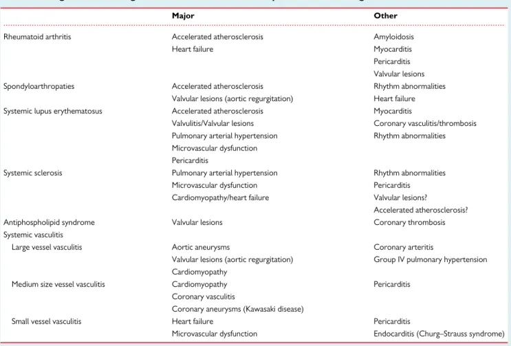
population having comparable severity of atherosclerosis10(Figure1,
Supplementary dataonline, Video S1). Of note, the excess risk of mor-tality from CVevents in patients with RA and SLE was shown to be
comparable to the risk associated with type 2 diabetes mellitus.6,11
This malignant course is partly explained by vulnerable plaques prone to rupture with excess inflammatory activity as shown in
pathologic specimens, despite less severe stenotic lesions.12
Consequently, the presentation is atypical with more frequent silent myocardial infarction, sudden cardiac death despite less frequent
angina,1explaining the poor outcome after CV events and limited
success of secondary prevention.4Vigilant and aggressive assessment
of risk and subclinical CV involvement in patients with SRDs seems therefore indispensable. However classical risk scoring systems are poorly calibrated for SRD patients, reaching almost 50%
underesti-mation by Framingham score in patients with RA and SLE.13,14
Patients who are mostly women with persistently elevated inflamma-tory markers are placed at low risk categories by age and gender giv-ing a false sense of security. Although not validated, the proposed solution by EULAR (European League Against Rheumatism) to im-prove risk stratification is to introduce a multiplication factor of 1.5
when using the Framingham or European SCORE systems for
patients with RA.15This approach is extended to all SRDs as Class IIb
indication in European Society of Cardiology (ESC) guidelines for CV
prevention16despite the lack of use of any multiplier for SRDs in
American College of Cardiology/American Heart Association (ACC/
AHA) prevention guidelines.17
Imaging for screening
atherosclerotic CV disease
There is low level of agreement for the use of carotid ultrasound (CUS) for risk stratification and screening asymptomatic carotid pla-ques in patients with RA in EULAR recommendations and no imaging
is recommended for screening CV disease for SRD in general.15Yet,
in two large studies, CUS findings re-classified RA patients into more
appropriate CV risk groups.18,19In addition, evidence shows that; (i)
carotid intima media thickness is a robust marker of atherosclerosis
in large populations,18(ii) carotid plaques are prevalent in patients
with SRDs, have rapid progression, particularly in association with
high levels of systemic inflammation,20and (iii) carotid atherosclerosis
predicts incident acute coronary syndromes in RA.21On the other
hand, higher coronary calcium scores have been found in patients
with established RA and SLE than those with early or no disease.22,23
However, coronary calcium score is not sensitive for vulnerable
...
Table 1 Targets for screening cardiovascular involvement in systemic rheumatologic diseases
Major Other
Rheumatoid arthritis Accelerated atherosclerosis Amyloidosis Heart failure Myocarditis
Pericarditis Valvular lesions Spondyloarthropaties Accelerated atherosclerosis Rhythm abnormalities
Valvular lesions (aortic regurgitation) Heart failure Systemic lupus erythematosus Accelerated atherosclerosis Myocarditis
Valvulitis/Valvular lesions Coronary vasculitis/thrombosis Pulmonary arterial hypertension Rhythm abnormalities Microvascular dysfunction
Pericarditis
Systemic sclerosis Pulmonary arterial hypertension Rhythm abnormalities Microvascular dysfunction Pericarditis
Cardiomyopathy/heart failure Valvular lesions?
Accelerated atherosclerosis? Antiphospholipid syndrome Valvular lesions Coronary thrombosis Systemic vasculitis
Large vessel vasculitis Aortic aneurysms Coronary arteritis
Valvular lesions (aortic regurgitation) Group IV pulmonary hypertension Cardiomyopathy
Medium size vessel vasculitis Cardiomyopathy Pericarditis Coronary vasculitis
Coronary aneurysms (Kawasaki disease)
Small vessel vasculitis Heart failure Pericarditis
Microvascular dysfunction Endocarditis (Churg–Strauss syndrome)
Figure 1A 48-year-old women, suffering from rheumatoid arthritis for 10 years, apparently asymptomatic with severe functional disability due to joint pain and no traditional risk factors for atherosclerosis (A). Mildly reduced global longitudinal strain with subtle segmental heterogeneity, no visual wall motion abnormality. (B) Multi-slice computerized tomography angiography and (C) conventional angiography confirms severe three-vessel disease. (D) Carotid plaques of the patient (arrow).
..
..
..
..
..
..
..
..
..
..
..
..
..
..
..
..
..
..
..
..
..
..
..
..
..
..
..
..
..
..
..
..
..
..
..
..
..
..
..
..
..
..
..
..
..
..
..
..
..
..
..
..
..
..
..
..
..
..
..
..
..
..
..
..
..
..
..
..
..
..
..
..
..
..
..
..
..
..
..
..
..
..
..
..
..
..
.
mixed or non-calcified plaques that are frequent in patients with
SRDs;23hence, clinical judgment about risk should prevail. Primary
prevention guidelines of ESC and ACC/AHA recommend to con-sider carotid plaques and coronary calcium score, respectively, as additional risk prediction tools to reclassify risk estimate among
individuals at borderline and intermediate risk.17Whether CUS or
calcium score for screening and monitoring subclinical atheroscler-osis in SRD patients modifies the outcome awaits further investigation.
Arterial stiffness by pulsed-wave velocity and endothelial function by flow-mediated dilatation have also been used to assess athero-sclerosis in RA, SLE, and systemic athero-sclerosis (SSc) in several small ser-ies that consistently showed functional abnormalitser-ies. However, the predictive value of these vascular tests to detect CV events in SRDs
is not established.24
Multi-slice computerized tomography angiography (MCTA) is the best non-invasive modality to assess coronary lesions anatomically and plaque composition and reliably shows increased incidence of si-lent coronary atherosclerosis in patients with RA, SLE, and psoriatic
arthritis.23Radiation exposure limits its use for serial follow-up in
relatively young, asymptomatic patients. Furthermore, relative impli-cations of anatomical vs. functional assessment are unknown. Several fold increased risk of myocardial ischaemia in exercise echocardiog-raphy and increased mortality in patients with ischaemia were shown
in RA.25However, wall motion score at rest is insensitive while stress
echocardiography and stress-rest single-photon emission tomog-raphy are moderately sensitive to detect ischaemia. Importantly, not only flow limiting coronary artery disease but also inflammation,
microvascular dysfunction, or fibrosis can cause perfusion
abnormalities.26
Rational for screening
non-atherosclerotic vascular
impairment
Non-atherosclerotic mechanisms including microvascular dysfunc-tion, obliterative vasculopathy, thrombosis, and small vessel vasculitis are added to CV risk in all SRDs, particularly in SSc. Vasculopathic process in small coronary vessels triggers endothelial dysfunction, in-timal proliferation, immune system activation, and thrombogenicity, all leading to obliterative vasculopathy. Intermittent spasms of intra-myocardial coronary arteries with ischaemia–reperfusion episodes (intramyocardial Raynaud) are responsible from patchy fibrosis in
early stages.27 Later, obliterative vasculopathy of intramyocardial
arteries is irreversible and leads to extensive fibrosis, irrespective of coronary territories. These changes are associated with rapid
deteri-oration of ventricular function, life threatening arrhythmias (Figure2,
Supplementary dataonline, Videos S2.1-4), and increased risk of death in patients with SSc.28
Primary systemic vasculitis (i.e. Churg–Strauss Syndrome, granulo-matosis with polyangiitis, and polyarteritis nodosa) can also cause se-vere myocardial damage due to intramyocardial coronary vasculitis
besides epicardial coronary arteritis.29
Importantly, early perfusion defects caused by non-atherosclerotic
vascular impairments may be reversible with treatment.26
Imaging for screening
non-atherosclerotic vascular
impairment
The assessment of coronary flow reserve (CFR) by transthoracic Doppler echocardiography is readily available and reflects micro-vascular dysfunction in the absence of epicardial coronary lesions. CFR is the ratio of hyperaemic to baseline peak diastolic velocity of the coronary flow during adenosine or dipyridamole infusion. Reduced CFR has been shown in patients with SRDs independently
of traditional atherosclerotic risk factors.30Significant impairment of
CFR can occur due to inflammation, intramyocardial fibrosis, and
vas-cular damage.26Although, CFR by Doppler echocardiography seems
safe and feasible for screening microcirculatory disturbances, more data are needed to understand its prognostic and therapeutic implica-tions in asymptomatic, low–moderate risk SRD patients. Perfusion defects can be assessed more accurately by contrast-enhanced CMR and impaired myocardial blood flow reserve by positron emission tomography (PET) with rest-stress quantification of myocardial blood flow in the absence of flow limiting atherosclerotic lesions in patients
with SRDs.27,31,32 However, rigorous assessment of ischaemia by
advanced imaging tools should be driven by a judicious use of resting echocardiography with strain imaging and CUS or calcium score in
asymptomatic patients with low to intermediate CV risk.26
Rational for screening myocardial
involvement
Heart failure (HF) due to ischaemic and non-ischaemic causes, is a common complication and an important cause of CV mortality in
SRDs.33Inflammation can damage the myocardium directly by
trig-gering fibroblast activity, leading to myocardial remodelling by react-ive fibrosis. Chronic inflammation, drug toxicity, antiphospholipid antibodies, immune complex deposition, vasculitis, microvascular dysfunction, and renal dysfunction can all underlie primary myocardial damage. Myocarditis is a serious but an uncommon manifestation of SRDs. Although recovery is possible with early diagnosis and treat-ment, irreversible HF, ventricular arrhythmias, and sudden cardiac
death may occur.34Despite being subclinical most of the time, the
presentation and progression are variable and unpredictable (Figure
3,Supplementary dataonline, Video S3A, B). Myocarditis is associated with poor prognosis and can be the initial manifestation; particularly in SLE.34,35In postmortem series, histologic evidence of myocarditis has been reported up to 57% of SLE patients which is far more
fre-quent than recognized clinically.35
Symptoms and electrocardiogram (ECG) are of limited value to detect myocardial involvement, therefore adequate size studies with imaging modalities are needed.
Imaging for screening myocardial
involvement
Echocardiography is the first line imaging tool to detect myocardial morphological and functional changes. Speckle-tracking imaging (STI)
..
..
..
..
..
..
..
..
..
is a method that increases the sensitivity of echocardiography for detecting subclinical myocardial dysfunction when ejection fraction is still preserved, irrespective of underlying mechanism. Preliminary data suggest that echocardiography with STI could be a promising tool for screening early myocardial involvement and for monitoring
the effects of anti-inflammatory treatment in SRDs.36,37
CMR, PET, and PET/CMR enable tissue characterization and help to differentiate inflammation, fibrosis, and oedema from ischaemic
lesions (Figure 4, Supplementary data online, Video S4).34,38–40
However, there is not enough evidence to counterbalance the cost and limited availability of CMR or PET as screening and serial follow-up tools to assess myocardial involvement in asymptomatic SRD
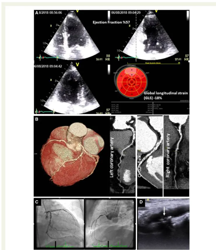
Figure 2(A) A 36-year-old women with palpitation, suffering from systemic sclerosis for 15 years, had multiple episodes of non-sustained
ventricu-lar tachycardia on Holter. (B) Severe regional wall motion abnormalities in left (LV) and right ventricle (RV) (arrows). (C, D) Global longitudinal strain (GLS) shows severe regional LV and RV systolic dysfunction. (E) Cardiac magnetic resonance shows enlarged RV and transmural late gadolinium hyperenhancement on both LV and RV walls (arrows) despite no significant epicardial coronary lesion or pulmonary hypertension.
..
..
..
..
..
..
..
..
..
..
..
..
..
..
..
..
.
patients. Of note, these tools can be more accurate than echocardi-ography in SRD patients with predominant right ventricular (RV) in-flammation, fibrosis, or dysfunction due to pulmonary hypertension
(PH).36
Rational for screening valvular
involvement
Valvular involvement is common in SRD patients reaching a
fre-quency of 80% in SLE by transoesophageal echocardiography.41
Valvular regurgitation is the most frequent consequence.
Progression to surgery is around 4–10%.42 Antiphospholipid
anti-body positivity significantly increases the risk of valvulitis and
endo-carditis.43 Libman–Sacks endocarditis (non-bacterial vegetations)
that can be encountered in up to 11% of SLE patients,44is an
inde-pendent cause of stroke and associated with mortality in SLE and
antiphospholipid syndrome (APLS) (Figure 5, Supplementary data
online, Video S5).41,45Aortic regurgitation is an important complica-tion of ankylosing spondylitis and large vessel vasculitis (LVV) main-ly in association with root dilatation and/or extension of fibrosis
into the valve.46
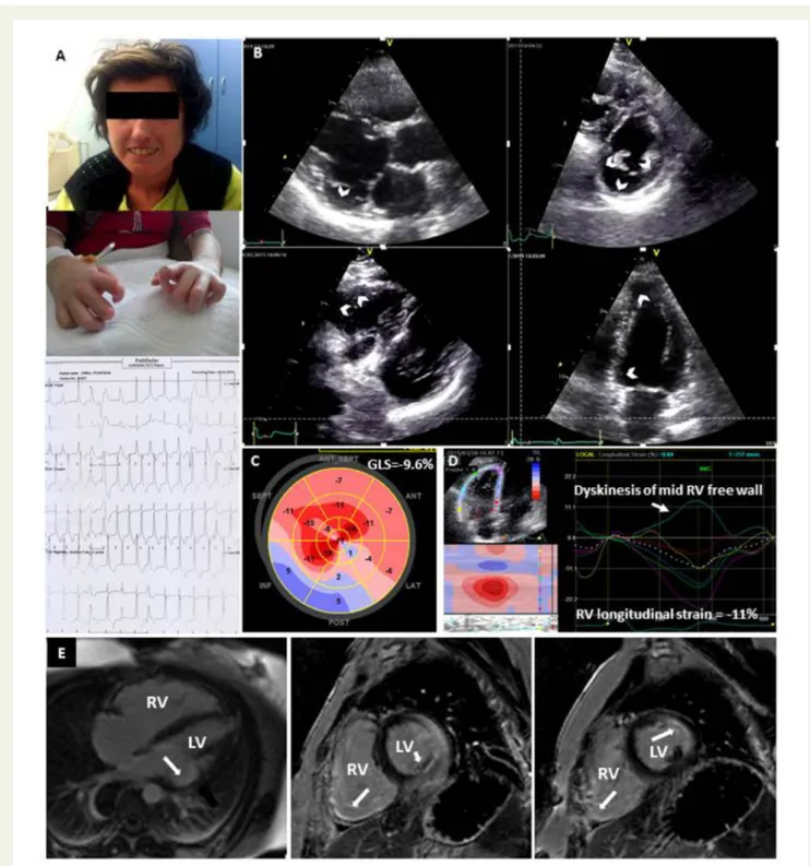
Figure 3A 50-year-old women with systemic sclerosis, clinically stable over the last 10 years. (A) Asymptomatic during next to last visit, mild wall
motion abnormality at the apex. (B) Chest pain 4 months later, severe regional wall motion abnormalities and restrictive diastolic function despite normal coronary angiography. GLS, global longitudinal strain.
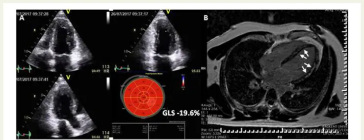
Figure 4Lupus myocarditis in a 32-year-old woman with chest pain, arrhythmias, elevated C-reactive protein, troponins, and normal coronaries.
(A) 2D echocardiography shows normal systolic function, but global longitudinal strain (GLS) is at the lower limit of normal. (B) Cardiac magnetic res-onance shows extensive mid-wall late gadolinium hyperenhancement (arrows).
..
..
..
..
..
..
..
..
..
..
..
..
..
..
..
..
..
..
..
..
..
..
..
..
..
..
..
..
..
..
..
..
..
..
..
..
..
..
..
..
..
..
..
..
..
..
..
..
..
..
..
..
..
..
..
..
..
..
..
..
..
..
..
..
..
..
..
..
..
..
..
..
..
..
..
..
..
..
..
..
..
..
..
..
..
Most of the valvular abnormalities are clinically silent and over-looked without echocardiography although acute aortic or mitral
valvulitis can be the initial presentation particularly in SLE and AS.46,47
Infective endocarditis should always be considered in these immune compromised patients particularly in those with underlying valvulop-athy in case of clinical deterioration.
Imaging for screening valvular
involvement
Echocardiography is key to detect and characterize valvular damage and has been recommended as a screening tool for valvular
patholo-gies in asymptomatic patients with APLS and SLE.43,44Typical valvular
lesions include fibrotic thickening, retraction of the leaflets, nodules, or sterile vegetations most frequently effecting mitral and aortic
valves.27,47 Tricuspid valve can also be involved (Figure 6,
Supplementary dataonline, Video S6A, B). However,no evidence sup-ports routine assessment of valvular involvement in other asymptom-atic SRD patients without relevant findings.
Rationale for screening PH
PH is a poor prognosticator irrespective of its cause. All groups of PH can be encountered as a complication of SRD. Pulmonary arterial hypertension (PAH) is an important cause of mortality particularly in
SSc affecting 6–12% of the patients.48Other than PAH, LV
dysfunc-tion and pulmonary involvement can cause PH in SSc. The presence of antiphospholipid antibodies increases the risk for chronic thrombo-embolic PH. Moreover, vasculitis of large pulmonary
arteries can cause PH in Takayasu’s arteritis (TA).49Early diagnosis by
screening PH in SSc has been shown to impact survival and patient
management.50
Imaging for screening PH
Yearly echocardiographic screening is only recommended for detect-ing PH related to SSc spectrum disorders by the European
guidelines.51,52In other SRDs, echocardiographic screening for PH is
indicated only if patients are symptomatic. Echocardiography also provides information about the LV dysfunction which is the most common cause of PH. RV adaptation to elevated pulmonary artery pressure is not uniform among patients and is the main determinant of survival in PAH including SRD patients. Other imaging modalities, particularly CMR, are complementary to echocardiography to define
RV function and to help the differential diagnosis of PH.53
Pericardial involvement
Pericardial effusion and pericarditis are frequent in SRDs, whereas haemodynamically significant effusions or chronic constrictive
peri-carditis are unlikely.47Imaging for pericardial involvement is useful for
the differential diagnosis of chest pain. Documentation of pericardial involvement could help to make the diagnosis of SRDs, particularly SLE. Pericardial effusion is a poor prognostic factor in patients with
PAH.51 However, no evidence supports screening asymptomatic
SRD patients for pericardial involvement. Computerized tomography and CMR are complementary to echocardiography to assess the size and location of effusion, pericardial thickness, or calcification when-ever needed in symptomatic patients.
Rational for screening large
vessels
Giant cell arteritis (GCA) and TA are the most common LVVs, char-acterized by involvement of aorta and its main branches. Aortic aneurysms are common in GCA than TA, both can cause vascular
stenosis and occlusions.53Stroke or acute coronary syndrome can
be the initial manifestation. Pulmonary arterial lesions can lead to
group IV PH in TA.49Cardiac complications including
cardiomyop-athy and severe hypertension are among major causes of mortality in
TA.54Behc¸et’s syndrome is another vasculitis that can cause
pulmon-ary artery and aortic aneurysms and stenoses.55Isolated aortitis or
LVV secondary to SRDs as a rare manifestation, can also be seen. As patients can be asymptomatic, early documentation of pathologies are critical for intensifying treatment such as vigorous control of hypertension or optimizing immune suppression.
Imaging for screening large
vessels
Ultrasound of temporal and axillary arteries is useful for the diagnosis of cranial GCA. Magnetic resonance is recommended for documen-tation of vasculitic involvement in aorta and its branches. PET, MCTA, and/or ultrasound may be used as alternative modalities. All of the above imaging modalities have the capability to document
in-flammation and complications of LVV.56 Addition of
echocardio-graphic screening in the initial workup might help to detect cardiac
complications of LVV (Figure7,Supplementary dataonline, Video S7A,
B) whereas no evidence supports the use of transoesophageal
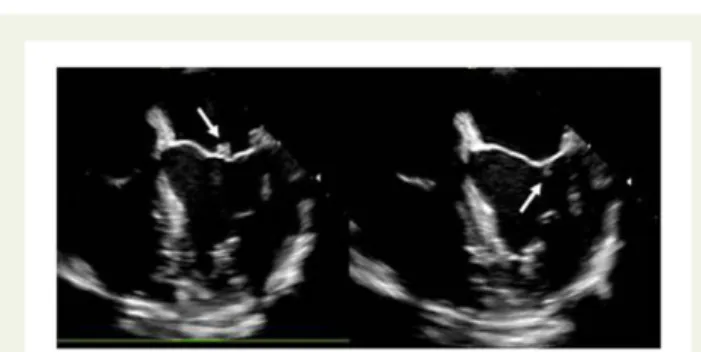
Figure 5Libman–Sacks vegetations on both sides of mitral leaflet
tips (arrows) in a 48-year-old woman with antiphospholipid syn-drome and acute ischaemic stroke.
..
..
..
..
..
..
..
..
..
..
..
..
..
..
..
..
..
..
..
..
..
..
..
..
..
..
..
..
..
..
..
..
..
..
..
..
..
..
..
..
echocardiography for screening structural abnormalities of the aorta in asymptomatic patients.
General comments
CV involvement due to SRDs remains largely underdiagnosed despite high rates of CV complications leading to poor survival. Traditional risk scoring systems underestimate CV risk in SRD patients. Primary prevention guidelines recommend clinical judgement to be applied on a case-by-case basis regarding the impact of imaging for screening
patients with SRDs, except for PH in SSc.16,17,51Rigorous CV risk
as-sessment and early referral to a cardiologist are key to optimize the outcome. Although accelerated atherosclerosis is the most promin-ent cause of major CV evpromin-ents, the contribution of other causes of
cardiac pathologies should also be considered (Figure8).
Echocardiography and CUS or calcium score, in addition to ECG, should be an integral part of screening in SRD; especially in RA, SLE, and SSc patients with suspicion of CV involvement. Echocardiography has great potential to detect subclinical myocardial dysfunction, valvular, macro- and microvascular damage by means of its versatile use at a low cost, free of radiation, and nephrotoxicity. The addition of CUS to traditional risk scoring systems, improves risk estimation for atherosclerotic complications in these patients. CMR, MCTA and nuclear imaging complement echocardiography and CUS to obtain more data about CV complications. They should also be considered
whenever the level of suspicion remains high despite inconclusive
echocardiographic and/or CUS findings (Figure9).
Future research and gaps in
evidence
Development of a CV risk stratification tool and screening algorithm dedicated to asymptomatic patients with SRDs is an unmet need. In that regard, imaging parameters obtained from echocardiography and CUS or calcium scoring might help to improve risk models, to detect, and monitor subclinical CV involvement. Certainly, some CV abnormalities are more progressive and life threatening than others and necessitate closer follow-up. But those have been incompletely defined so far. Existing imaging studies in SRDs have heterogeneous outcome measurements, inclusion criteria, follow-up durations, and different patient populations and do not allow to pool the data to de-fine the prevalence, incidence, and progression of CV diseases related to SRDs and to conduct insightful meta-analyses. So far, the answer to ‘Should asymptomatic patients with SRDs undergo screening with echocardiography and CUS on top of ECG?’ remains an extrapola-tion of the pieces of evidence from heterogeneous studies. Obviously, large studies performing cardiac screening with a standard approach by using echocardiography and CUS to obtain longitudinal data with hard endpoints are awaited in order to implement imaging
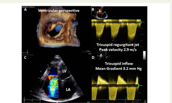
Figure 6Lupus valvulitis. (A) Thickened, fibrotic, retracted tricuspid leaflets with (B, C) severe regurgitation and (D) mild stenosis. Mitral leaflets
were less severely affected in this patient. LA, left atrium; LV, left ventricle.
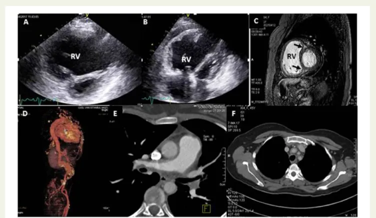
Figure 7 A 44-year-old woman with Takayasu’s arteritis. Cardiac involvement is secondary to pulmonary hypertension which is caused by pul-monary vasculitis. (A, B) Right ventricle (RV) is severely dilated with poor function and pericardial effusion. (C) Cardiac magnetic resonance confirms RV dilatation and shows late gadolinium hyperenhancement at interventricular junctions (arrows), (D) Multi-slice computerized tomography angiog-raphy shows aneurysms and narrowings along the aorta, (E) enlarged main pulmonary artery and narrowing of left and right pulmonary arteries by se-vere wall thickening, and (F) narrowing of aortic arch branches by sese-vere wall thickening.
Figure 8Causes of cardiac pathologies in systemic rheumatologic diseases.
..
..
..
..
..
..
..
..
..
..
.
as part of screening, patient monitorization, and formulate a follow-up timetable. Consequently, future research should aim to:
•
test whether incorporating echocardiography and CUS intoscreening protocols favourably impacts the outcome,
•
define high risk subgroups who need routine screening andmoni-torization with imaging,
•
answer when imaging should be repeated if no CV involvement isdetected initially, and when it should be repeated if any CV abnor-mality is detected,
•
explore whether asymptomatic cardiac involvement impactstreat-ment strategy and is useful to monitor response to treattreat-ment,
•
evaluate and validate cost-effective imaging protocols for screeningand monitoring subclinical CV involvement.
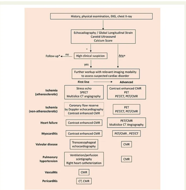
Figure 9Proposal of screening algorithm for cardiovascular involvement with imaging in systemic rheumatologic diseases. CFR, coronary flow
reserve; CMR, cardiac magnetic resonance; CT, computerized tomography; PET, positron emission tomography; SPECT, single-photon emission computed tomography. (*Follow-up should be individualized except for PH related to SSc spectrum disorders.)
..
..
..
..
..
..
..
..
..
..
..
..
..
..
..
..
..
..
..
..
..
..
..
..
..
..
..
..
..
..
..
..
..
..
..
..
..
..
..
..
..
..
..
..
..
..
..
..
..
..
..
..
..
..
..
..
..
..
..
..
..
..
..
..
..
..
..
..
..
..
..
..
..
..
..
..
..
..
..
..
..
..
..
..
.
In conclusion, more questions than answers remain to be explored to formulate CV prevention guidelines dedicated to SRD patients and to optimize the outcome where CV imaging seems to be pivotal.
Supplementary data
Supplementary dataare available at European Heart Journal - Cardiovascular Imaging online.
Conflict of interest: none declared.
References
1. Maradit-Kremers H, Crowson CS, Nicola PJ, Ballman KV, Roger VL, Jacobsen SJ. Increased unrecognized coronary heart disease and sudden deaths in rheumatoid arthritis: a population-based cohort study. Arthritis Rheum 2005;52:402–11. 2. Myasoedova E, Gabriel SE, Matteson EL, Davis JM 3rd, Therneau TM, Crowson
CS. Decreased cardiovascular mortality in patients with incident rheumatoid arthritis in recent years: dawn of a new era in cardiovascular disease in RA? J Rheumatol 2017;44:732–9.
3. Han C, Robinson DW, Hackett MV, Paramore LC, Fraeman KH, Bala MV. Cardiovascular disease and risk factors in patients with rheumatoid arthritis, psoriatic arthritis, and ankylosing spondylitis. J Rheumatol 2006;33:2167–72. 4. Ridker PM, Everett BM, Thuren T, MacFadyen JG, Chang WH, Ballantyne C et al.
Antiinflammatory therapy with canakinumab for atherosclerotic disease. N Engl J Med 2017;377:1119–3.
5. Avina-Zubieta JA, Thomas J, Sadatsafavi M, Lehman AJ, Lacaille D. Risk of incident cardiovascular events in patients with rheumatoid arthritis: a meta-analysis of ob-servational studies. Ann Rheum Dis 2012;71:1524–9.
6. Peters MJ, van Halm VP, Voskuyl AE, Smulders YM, Boers M, Lems WF et al. Does rheumatoid arthritis equal diabetes mellitus as an independent risk factor for cardiovascular disease? A prospective study. Arthritis Rheum 2009;61:1571–9. 7. Mantel A, Holmqvist M, Jernberg T, Wa˚llberg-Jonsson S, Askling J. Rheumatoid
arthritis is associated with a more severe presentation of acute coronary syn-drome and worse short-term outcome. Eur Heart J 2015;36:3413–22. 8. Douglas KM, Pace AV, Treharne GJ, Saratzis A, Nightingale P, Erb N et al. Excess
recurrent cardiac events in rheumatoid arthritis patients with acute coronary syndrome. Ann Rheum Dis 2006;65:348–53.
9. Bartels CM, Buhr KA, Goldberg JW, Bell CL, Visekruna M, Nekkanti S et al. Mortality and cardiovascular burden of systemic lupus erythematosus in a US population-based cohort. J Rheumatol 2014;41:680–7.
10. Roman MJ, Shanker BA, Davis A, Lockshin MD, Sammaritano L, Simantov R et al. Prevalence and correlates of accelerated atherosclerosis in systemic lupus eryth-ematosus. N Engl J Med 2003;349:2399–406.
11. Wajed J, Ahmad Y, Durrington PN, Bruce IN. Prevention of cardiovascular dis-ease in systemic lupus erythematosus–proposed guidelines for risk factor man-agement. Rheumatology (Oxford) 2004;43:7–12.
12. Aubry MC, Maradit-Kremers H, Reinalda MS, Crowson CS, Edwards WD, Gabriel SE. Differences in atherosclerotic coronary heart disease between sub-jects with and without rheumatoid arthritis. J Rheumatol 2007;34:937–42. 13. Crowson CS, Matteson EL, Roger VL, Therneau TM, Gabriel SE. Usefulness of
risk scores to estimate the risk of cardiovascular disease in patients with rheuma-toid arthritis. Am J Cardiol 2012;110:420–4.
14. Petri MA, Kiani AN, Post W, Christopher-Stine L, Magder LS. Lupus ATHEROSCLEROSIS prevention study. Ann Rheum Dis 2011;70:760–5. 15. Agca R, Heslinga SC, Rollefstad S, Heslinga M, McInnes IB, Peters MJ et al.
EULAR recommendations for cardiovascular disease risk management in patients with rheumatoid arthritis and other forms of inflammatory joint disorders: 2015/ 2016 update. Ann Rheum Dis 2017;76:17–28.
16. Piepoli MF, Hoes AW, Agewall S, Albus C, Brotons C, Catapano AL et al. 2016 European guidelines on cardiovascular disease prevention in clinical practice. Eur Heart J 2016;37:2315–81.
17. Arnett DK, Blumenthal RS, Albert MA, Buroker AB, Goldberger ZD, Hahn EJ et al. ACC/AHA guideline on the primary prevention of cardiovascular disease. Circulation 2019;doi:10.1161/CIR.0000000000000678.
18. Corrales A, Gonza´lez-Juanatey C, Peiro´ ME, Blanco R, Llorca J, Gonza´lez-Gay MA. Carotid ultrasound is useful for the cardiovascular risk stratification of patients with rheumatoid arthritis: results of a population-based study. Ann Rheum Dis 2014;73:722–7.
19. Daı¨en CI, Tubery A, Cailar GD, Mura T, Roubille F, Morel J et al. Application of the 2015/2016 EULAR recommendations for cardiovascular risk in daily practice: data from an observational study. Ann Rheum Dis 2018;77:625–6.
20. Giles JT, Post WS, Blumenthal RS, Polak J, Petri M, Gelber AC et al. Longitudinal predictors of progression of carotid atherosclerosis in rheumatoid arthritis. Arthritis Rheum 2011;63:3216–25.
21. Evans MR, Escalante A, Battafarano DF, Freeman GL, O’Leary DH, del Rinco´n I. Carotid atherosclerosis predicts incident acute coronary syndromes in rheuma-toid arthritis. Arthritis Rheum 2011;63:1211–20.
22. Chung CP, Oeser A, Raggi P, Gebretsadik T, Shintani AK, Sokka T et al. Increased coronary-artery atherosclerosis in rheumatoid arthritis: relationship to disease duration and cardiovascular risk factors. Arthritis Rheum 2005;52: 3045–53.
23. Shen J, Wong KT, Cheng IT, Shang Q, Li EK, Wong P et al. Increased prevalence of coronary plaque in patients with psoriatic arthritis without prior diagnosis of coronary artery disease. Ann Rheum Dis 2017;76:1237–44.
24. Szucs G, Tı´ma´r O, Szekanecz Z, De´r H, Kerekes G, Szamosi S et al. Endothelial dysfunction precedes atherosclerosis in systemic sclerosis—relevance for pre-vention of vascular complications. Rheumatology 2007;46:759–62.
25. Saghir MK, Attenhofer Jost C, Warrington KJ, Cha SS, Pellikka PA. Exercise echo-cardiography in rheumatoid arthritis: a case-control study. J Am Soc Echocardiogr 2009;22:1228–31.
26. Faccini A, Kaski JC, Camici PG. Coronary microvascular dysfunction in chronic inflammatory rheumatoid diseases. Eur Heart J 2016;37:1799–806.
27. Allanore Y, Meune C, Kahan A. Systemic sclerosis and cardiac dysfunction: evolv-ing concepts and diagnostic methodologies. Curr Opin Rheumatol 2008;20: 697–702.
28. Vacca A, Montisci R, Garau P, Siotto P, Piga M, Cauli A et al. Prognostic impact of coronary microcirculation abnormalities in systemic sclerosis: a prospective study to evaluate the role of non-invasive tests. Arthritis Res Ther 2013;15:R8. 29. Mukhtyar C, Brogan P, Luqmani R. Cardiovascular involvement in primary
sys-temic vasculitis. Best Pract Res Clin Rheumatol 2009;23:419–28.
30. Kakuta K, Dohi K, Sato Y, Yamanaka T, Kawamura M, Ogura T et al. Chronic Inflammatory disease is an independent risk factor for coronary flow velocity re-serve impairment unrelated to the processes of coronary artery calcium depos-ition. J Am Soc Echocardiogr 2016;29:173–80.
31. Rodrı´guez-Reyna TS, Morelos-Guzman M, Herna´ndez-Reyes P, Montero-Duarte K, Martı´nez-Reyes C, Reyes-Utrera C et al. Assessment of myocardial fibrosis and microvascular damage in systemic sclerosis by magnetic resonance imaging and coronary angiotomography. Rheumatology 2015;54:647–54.
32. Recio-Mayoral A, Mason JC, Kaski JC, Rubens MB, Harari OA, Camici PG. Chronic inflammation and coronary microvascular dysfunction in patients with-out risk factors for coronary artery disease. Eur Heart J 2009;30:1837–43. 33. Mantel A¨ , Holmqvist M, Andersson DC, Lund LH, Askling J. Association between
rheumatoid arthritis and risk of ischemic and nonischemic heart failure. J Am Coll Cardiol 2017;69:1275–85.
34. Hinojar R, Foote L, Sangle S, Marber M, Mayr M, Carr-White G et al. Native T1 and T2 mapping by CMR in lupus myocarditis: disease recognition and response to treatment. Int J Cardiol 2016;222:717–26.
35. Comarmond C, Cacoub P. Myocarditis in auto-immune or auto-inflammatory diseases. Autoimmun Rev 2017;16:811–6.
36. Fine NM, Crowson CS, Lin G, Oh JK, Villarraga HR, Gabriel SE. Evaluation of myocardial function in patients with rheumatoid arthritis using strain imaging by speckle-tracking echocardiography. Ann Rheum Dis 2014;73:1833–9.
37. Ikonomidis I, Tzortzis S, Lekakis J, Paraskevaidis I, Andreadou I, Nikolaou M et al. Lowering interleukin-1 activity with anakinra improves myocardial deformation in rheumatoid arthritis. Heart 2009;95:1502–7.
38. Hachulla AL, Launay D, Gaxotte V, de Groote P, Lamblin N, Devos P et al. Cardiac magnetic resonance imaging in systemic sclerosis: a cross-sectional ob-servational study of 52 patients. Ann Rheum Dis 2009;68:1878–84.
39. Mavrogeni S, Vassilopoulos D. Is there a place for cardiovascular magnetic reson-ance imaging in the evaluation of cardiovascular involvement in rheumatic dis-eases? Semin Arthritis Rheum 2011;41:488–96.
40. Ntusi NAB, Piechnik SK, Francis JM, Ferreira VM, Matthews PM, Robson MD et al. Diffuse myocardial fibrosis and inflammation in rheumatoid arthritis: insights from CMR T1 mapping. JACC Cardiovasc Imaging 2015;8:526–36.
41. Turiel M, Muzzupappa S, Gottardi B, Crema C, Sarzi-Puttini P, Rossi E. Evaluation of cardiac abnormalities and embolic sources in primary antiphospholipid syn-drome by transesophageal echocardiography. Lupus 2000;9:406–12.
42. Foroughi M, Hekmat M, Ghorbani M, Ghaderi H, Majidi M, Beheshti M. Mitral valve surgery in patients with systemic lupus erythematosus. ScientificWorldJournal 2014;2014:1.
43. Zuily S, Regnault V, Selton-Suty C, Eschwe`ge V, Bruntz JF, Bode-Dotto E et al. Increased risk for heart valve disease associated with antiphospholipid antibodies
..
..
..
..
..
..
..
..
..
..
..
..
..
..
..
..
..
..
..
..
..
.
in patients with systemic lupus erythematosus: meta-analysis of echocardiograph-ic studies. Circulation 2011;124:215–24.44. Moyssakis I, Tektonidou MG, Vasilliou VA, Samarkos M, Votteas V, Moutsopoulos HM. Libman-Sacks endocarditis in systemic lupus erythematosus: prevalence, associations and evolution. Am J Med 2007;120:636–42.
45. Roldan CA, Sibbitt WL, Qualls CR, Jung RE, Greene ER, Gasparovic CM et al. Libman-Sacks endocarditis and embolic cerebrovascular disease. JACC Cardiovasc Imaging 2013;6:973–83.
46. Klingberg E, Svea¨lv BG, Ta¨ng MS, Bech-Hanssen O, Forsblad-d’Elia H, Bergfeldt L. Aortic regurgitation is common in ankylosing spondylitis: time for routine echocardiography evaluation? Am J Med 2015;128: 1244–50.
47. Corrao S, Messina S, Pistone G, Calvo L, Scaglione R, Licata G. Heart involve-ment in rheumatoid arthritis: systematic review and meta-analysis. Int J Cardiol 2013;167:2031–8.
48. Launay D, Sobanski V, Hachulla E, Humbert M. Pulmonary hypertension in sys-temic sclerosis: different phenotypes. Eur Respir Rev 2017;26:170056.
49. Sari A, Sener YZ, Firat E, Armagan B, Erden A, Oksul M et al. Pulmonary hyper-tension in Takayasu’s arteritis. Int J Rheum Dis 2018;21:1634–9.
50. Humbert M, Yaici A, de Groote P, Montani D, Sitbon O, Launay D et al. Screening for pulmonary arterial hypertension in patients with systemic sclerosis: clinical characteristics at diagnosis and long-term survival. Arthritis Rheum 2011; 63:3522–30.
51. Galie` N, Humbert M, Vachiery JL, Gibbs S, Lang I, Torbicki A et al. 2015 ESC/ ERS Guidelines for the diagnosis and treatment of pulmonary hypertension. Eur Heart J 2016;37:67–119.
52. Frost A, Badesch D, Gibbs JSR, Gopalan D, Khanna D, Manes A et al. Diagnosis of pulmonary hypertension. Eur Respir J 2019;53:1801904.
53. Kermani TA. Takayasu arteritis and giant cell arteritis: are they a spectrum of the same disease? Int J Rheum Dis 2019;22(Suppl. 1):41–8.
54. Kim ESH, Beckman J. Takayasu arteritis: challenges in diagnosis and management. Heart 2018;104:558–65.
55. Tuzun H, Seyahi E, Guzelant G, Oz B, Batur S, Demirhan O et al. Surgical treat-ment of pulmonary complications in Behc¸et’s Syndrome. Semin Thorac Cardiovasc Surg 2018;30:369. 78
56. Dejaco C, Ramiro S, Duftner C, Besson FL, Bley TA, Blockmans D et al. EULAR recommendations for the use of imaging in large vessel vasculitis in clinical prac-tice. Ann Rheum Dis 2018;77:636–43.