Evaluation of the Patients with Congenital Rickets
Enver Atay
Department of Pediatrics, Medipol University Hospital, Istanbul, Turkey
Introduction: The present study aims to evaluate the clinical findings and biochemical properties of the patients who were followed up in the neonatal intensive care unit due to congenital rickets.
Methods: The data of patients who were followed up in our neonatal intensive care unit between March 2015 and March 2020 due to congenital rickets were analyzed retrospectively from the patient’s files and registration forms. The findings of physical examination and biochemical tests of the patients were recorded at the time of admission.
Results: Six patients were included in this study. Four of the patients were girls. The mean age of application was 19±6 days. All patients were admitted with convulsion. The average calcium at the first application was 5.5±0.7 mg/dL; ionized calcium 0.7±0.02 mmol/L; phosphorus 6.9±1.2 mg/dL; alkaline phosphatase 747±148 U/L; parathyroid hormone 265±40 pg/mL and mean 25-OH vitamin D 3.4±0.4 ng/mL. All cases were hospitalized and treated and diagnosed with congenital rickets. Discussion and Conclusion: Despite vitamin D prophylaxis, rickets can still be seen in our country. With this study, we aimed to draw attention to the significance of vitamin D prophylaxis.
Keywords: Congenital rickets; hypocalcemia; Vitamin D.
T
o prevent the development of rickets due to vitamin D deficiency in Turkey, daily doses of 400 UI vitamin D are administered to all newborn babies from the 15th day of birth by the Ministry of Health since 2005[1]. In addition, to prevent maternal/perinatal vitamin D deficiency, vitamin D supplementation (1200 U/day) has been recommended to every pregnant (except in cases where vitamin D will not be applied) since 2011[2]. However, despite all these ap-proaches, severe maternal vitamin D deficiency continues to be a problem frequently seen in Turkey. This is an impor-tant risk factor for congenital rickets[3–5]. The present study aims to determine the clinical and biochemical properties of patients who received inpatient treatment in the neona-tal intensive care unit with the diagnosis of congenineona-tal rick-ets and to draw attention to the importance of vitamin D prophylaxis once again.Materials and Methods
In this study, the data of the patients who were followed up in our neonatal intensive care unit between March 2015 and March 2020 due to congenital rickets were analyzed retrospectively from patient files and patient registration forms. Physical examination findings and biochemical tests of the patients were recorded at the time of the admission. Ethics committee approval was received for this study (No: 2019-201).
Physical examinations of the patients were performed on admission. Clinical findings were recorded. Blood calcium (Ca), phosphorus (P), magnesium (Mg), alkaline phos-phatase (ALP), 25-OH vitamin D, parathyroid hormone (PTH) levels of the patients and their mothers were
mea-DOI: 10.14744/hnhj.2020.79847
Haydarpasa Numune Med J 2020;60(3):284–287
hnhtipdergisi.com
HAYDARPAŞA NUMUNE MEDICAL JOURNAL
ORIGINAL ARTICLE
Abstract
Correspondence (İletişim): Enver Atay, M.D. Medipol Universitesi Hastanesi, Cocuk Sagligi ve Hastaliklari Anabilim Dali, Istanbul, Turkey Phone (Telefon): +90 505 394 52 93 E-mail (E-posta): [email protected]
Submitted Date (Başvuru Tarihi): 21.04.2020 Accepted Date (Kabul Tarihi): 09.05.2020
Copyright 2020 Haydarpaşa Numune Medical Journal
285
Atay, Evaluation of the Patients with Congenital Rickets / doi: 10.14744/hnhj.2020.79847
sured. A 25-OH vitamin D level of <12 ng/mL was consid-ered to be a deficiency, 12-20 ng/mL to be insufficiency, and 20-100 ng/mL to be normal.
Statistical Evaluation
The data obtained were analyzed using the statistics pro-gram (SPSS) (Version 17, Chicago, IL, USA) prepared for social sciences. Descriptive statistics (mean, standard de-viation, median value, minimum, maximum, number and percentile) were given for the variables in this study.
Results
Between March 2015 and March 2020, six patients were admitted to our neonatal intensive care unit with the di-agnosis of congenital rickets. Four of these patients were female, and two were male. The mean age of the patients was 19±6 days. All patients applied with the complaints of convulsions. All patients were term except one case. There was a wide fontanelle in two patients and an ankle enlarge-ment in one patient. No other features were detected in the physical examination of the patients. The clinical features of the cases are presented in Table 1.
When the biochemical findings of the patients were examined, mean calcium (5.5±0.7 mg/dL); albumin (4.2±0.3 g/dL), ionized calcium (0.7±0.02 mmol/L);
phos-phorus (6.9±1.2 mg/dL); alkaline phosphatase (747±148 U/L); parathyroid hormone (265±40 pg/mL) and 25-OH vitamin D (3.4±0.4 ng/mL) values were measured as in-dicated. No radiological findings other than increased metaphyseal width were detected on the radiographs of the patients. The biochemical findings of the cases are presented in Table 2.
When the biochemical findings of the mothers of the patients were examined, the mean calcium (7.9±1 mg/ dL), phosphorus (3.7±0.7 mg/dL); alkaline phosphatase (189±84 U/L), parathyroid hormone (214±103 pg/mL) and 25-OH vitamin D (4.8±2.3 ng/mL) values were determined as indicated. None of the mothers used vitamin D during pregnancy. The biochemical findings of the mothers of the cases are presented in Table 3.
Discussion
Congenital rickets is the term given to the fetus born with the clinical features of rickets, but newborns that do not have prominent clinical features but show the biochemi-cal findings of rickets can also be considered as congenital rickets[6]. Placental calcium transfer is thought to protect the fetus from rickets[6]. Therefore, clinical and radiolog-ical findings may be uncertain in cases of rickets seen in the neonatal period, and the disease may occur only with hypocalcemia or hypocalcemic seizure as in our cohort[6, 7]. Table 1. Clinical findings of the patients
No Gender Gestational age Birth weight (g) Age at admission (days) Admission complaint Nutrition Vitamin D prophylaxis
1 Female Term 3410 12 Convulsion BM No
2 Male Term 3240 25 Convulsion AS Irregular
3 Male Preterm 2200 28 Convulsion AS+ formula Irregular
4 Female Term 3010 17 Convulsion AS+formula No
5 Female Term 2950 21 Convulsion BM Irregular
6 Female Term 2870 14 Convulsion AS No
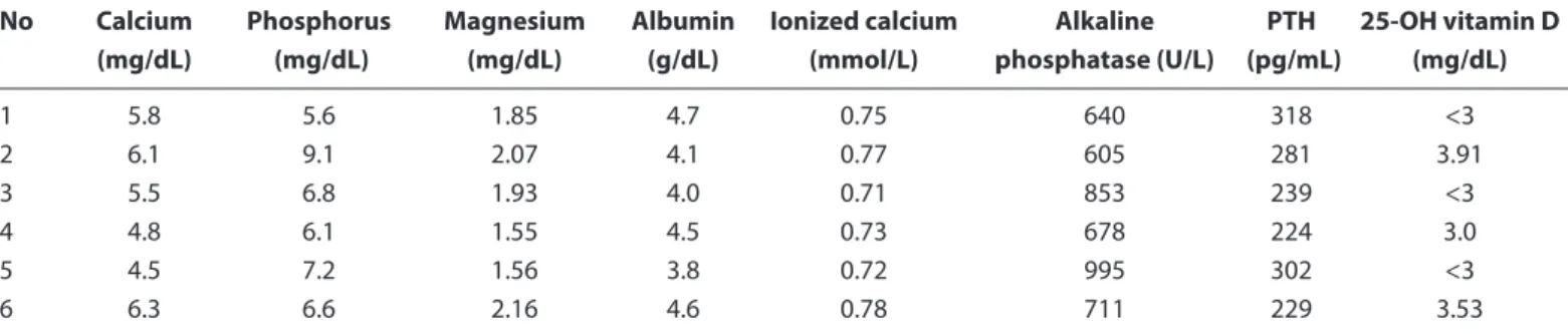
Table 2. Laboratory findings of the patients
No Calcium Phosphorus Magnesium Albumin Ionized calcium Alkaline PTH 25-OH vitamin D (mg/dL) (mg/dL) (mg/dL) (g/dL) (mmol/L) phosphatase (U/L) (pg/mL) (mg/dL)
1 5.8 5.6 1.85 4.7 0.75 640 318 <3 2 6.1 9.1 2.07 4.1 0.77 605 281 3.91 3 5.5 6.8 1.93 4.0 0.71 853 239 <3 4 4.8 6.1 1.55 4.5 0.73 678 224 3.0 5 4.5 7.2 1.56 3.8 0.72 995 302 <3 6 6.3 6.6 2.16 4.6 0.78 711 229 3.53
Normal values: Calcium; 8.0-10.8 mg/dL phosphorus 4.8-8.2 mg/dL, magnesium;1.7-2.7 mg/dL, Alkaline phosphatase; 150-420 U/L, PTH;15-87 pg/mL, 25-OH vitamin D; 20-100ng/mL: BM, breast milk.
286 Atay, Evaluation of the Patients with Congenital Rickets / doi: 10.14744/hnhj.2020.79847
In congenital rickets, hypotonia, severe tremor, serum al-kaline phosphatase elevation, 25-OH vitamin D deficiency, secondary hyperparathyroidism, hypocalcemia and resis-tant convulsions due to hypocalcemia may be seen[8]. In addition, unlike classical rickets, laboratory findings may accompany hypophosphatemia, as well as hypophos-phatemia in cases of congenital rickets[7]. All these condi-tions may cause difficulty in the differential diagnosis. In this single-center retrospective study, the clinical and laboratory findings of the six patients who were treated in the neonatal intensive care unit for congenital rickets were examined. It is noteworthy that the mothers of all patients had low 25-OH vitamin D levels and did not receive the appropriate doses of vitamin D prophylaxis during preg-nancy. Laboratory and radiological findings of all our pa-tients were compatible with congenital rickets. In addition to hypocalcemia, hyperphosphatemia, all patients had in-creased ALP, and PATHOLOGY, but dein-creased 25 (OH) vita-min D levels. In addition, mothers of the patients had lower 25 (OH) vitamin D levels.
The only source of vitamin D for the fetus is vitamin D, which passes through the placenta from the mother. A low 25-OH vitamin D level was found in the cord blood of in-fants of mothers with maternal vitamin D deficiency[9, 10]. In addition, it has been seen in many studies conducted to date that maternal vitamin D deficiency is the most im-portant risk factor for vitamin D deficiency in the neonatal period and early infancy[11]. This increases the risk of con-genital rickets in babies born to mothers with low 25-OH vitamin D levels.
As in our study, congenital rickets may present with life-threatening hypocalcemic seizures. Congenital rickets are a very important preventable public health problem con-cerning causing permanent complications. The require-ment and dosage of vitamin D supplerequire-mentation for
preg-nant women may vary according to countries and regions. Despite all the developments in the field of public health in our country, it is seen that clinical problems related to vitamin D deficiency remain an important issue today. Given that clinical and radiological findings of congenital rickets may not appear over time, it is also important to evaluate children who are admitted to the hospital con-cerning rickets findings, especially in regions with low so-cioeconomic status[12].
In conclusion, our experience with these six patients has shown that maternal vitamin D deficiency is still seen in our country and remains important as a severe health problem. Thus, to prevent maternal/perinatal vitamin D deficiency, appropriate doses of vitamin D supplementation are very important for every pregnant woman.
Ethics Committee Approval: Istanbul Medipol University Ethics
Committee, No: 2019-201.
Peer-review: Externally peer-reviewed. Conflict of Interest: None declared.
Financial Disclosure: The authors declared that this study
re-ceived no financial support.
References
1. TC Sağlık Bakanlığı. Bebeklerde D Vitamini Yetersizliğinin Ön-lenmesi ve Kemik Sağlığının Geliştirilmesi Programı: http:// www.saglik.gov.tr/TR/belge/1-11576/bebeklerde-dvitamini-yetersizliginin-onlenmesi-ve-kemik-.html;1 Ocak 2005 2. T.C. Sağlık Bakanlığı, Ana Çocuk Sağlığı ve Aile Planlaması
Genel Müdürlüğü. Gebelere D Vitamini Destek Programı. Available at: http://www.saglik.gov.tr/TR/belge/1-12656/ gebelere-d-vitamini-destek-programi.html; Accessed January 1, 2011.
3. Bereket A. Rickets in developing countries. Endocr Dev 2003;6:220–32.
4. Pehlivan I, Hatun S, Aydoğan M, Babaoğlu K, Gökalp AS.
Ma-Table 3. Laboratory findings of the mothers of the patients
No Calcium Phosphorus Alkaline Phosphatase PTH 25-OH vitamin D Use of vitamin D
(mg/dL) (mg/dL) (U/L) (pg/mL) (mg/dL) during pregnancy
1 6.7 5 344 397 <3 No 2 8.4 3.9 130 190 3.3 No 3 8.5 3.1 208 212 9.1 No 4 8.7 3.3 103 106 4.0 No 5 8.9 3.0 166 133 3.9 No 6 6.8 4.1 184 248 3.9 No
Normal values: Calcium; 8.0-10.8 mg/dL phosphorus 4.8-8.2 mg/dL, magnesium;1.7-2.7 mg/dL, Alkaline phosphatase; 150-420 U/L, PTH;15-87 pg/mL, 25-OH vitamin D; 20-100ng/mL.
287
Atay, Evaluation of the Patients with Congenital Rickets / doi: 10.14744/hnhj.2020.79847
ternal vitamin D deficiency and vitamin D supplementation in healthy infants. Turk J Pediatr 2003;45:315–20.
5. Alagöl F, Shihadeh Y, Boztepe H, Tanakol R, Yarman S, Azizlerli H, Sandalci O. Sunlight exposure and vitamin D deficiency in Turkish women. J Endocrinol Invest 2000;23:173–7.
6. Elidrissy AT. The Return of Congenital Rickets, Are We Missing Occult Cases? Calcif Tissue Int 2016;99:227–36.
7. Ramavat LG. Vitamin D deficiency rickets at birth in Kuwait. Indian J Pediatr 1999;66:37–43.
8. Mutlu GY, Özsu E, Oruç M, Çizmecioğlu F, Hatun Ş. Maternal D vitamini eksikliğine bağlı hipokalsemik nöbet: nasıl ön-lenebilir? Çocuk Sağlığı ve Hastalıkları Dergisi 2011;54:79–82.
9. Brooke OG, Brown IR, Cleeve HJ, Sood A. Observations on the vitamin D state of pregnant Asian women in London. Br J Ob-stet Gynaecol 1981;88:18–26.
10. Hollis BW, Wagner CL. Assessment of dietary vitamin D re-quirements during pregnancy and lactation. Am J Clin Nutr. 2004;79:717–26.
11. Andiran N, Yordam N, Ozön A. Risk factors for vitamin D de-ficiency in breast-fed newborns and their mothers. Nutrition 2002;18:47–50.
12. Hatun S. Günümüzde D vitamini eksikliği ve nutrisyonel rikets. Çocuk Sağlığı ve Hastalıkları Dergisi 2003;46:224–41.