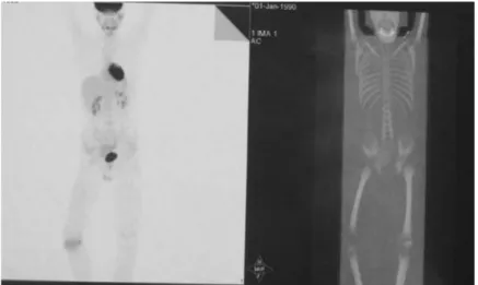
Primary epiphyseal ewing sarcoma: A case report
Tam metin
Şekil

Benzer Belgeler
答:青光眼睫狀體炎危象(Glaucomatocyclitic crisis)又稱為 Posner-Schlossman
神農本草經 陽湖孫星衍撰 原文 著本草者,代有明哲矣,而求道者必推本於神農,以
Surface cleaning by ion b ombardment and surface modification by chemical polymerization of plasma treatments are be lieved to remove contamination on titanium surfaces and
The most commonly reported dermatoscopical findings for PG were reddish structureless (homogeneous) areas, collarette sign, white intersecting lines, ulceration,
An extremely rare lung tumor of a young adult: Primary synovial sarcoma Genç bir yetişkinin çok nadir bir akciğer tümörü: Primer sinoviyal sarkom.. Ali Özdil 1 , Huriye
Low-grade fibromyxoid sarcoma is usually seen in young adults in the form of large masses characterized by fibrous, myxoid areas and may be located in the
Yukarıdaki veriler bize Türkiye’deki emek piyasalarının OECD’nin söylemiş olduğu gibi katı değil, ter- sine çalışma saatlerinin çok yüksek olduğu, enformel sektörün
UNDP, “Making Global Trade Work for People”, United Nations Development Programme, Earthscan Publications, London, 2003, s.127-129.. deki özelleĢtirme sürecinde önemli bir
