CASE REPORT
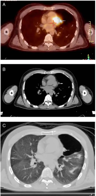
Rare presentation of a localised malignant pleural
mesothelioma with cranial metastasis
Gulhan Ertan,
1Abdulkadir Eren,
1Sila Ulus
21Department of Radiology,
Medipol University School of Medicine, Istanbul, Turkey
2Acıbadem University School
of Medicine, Istanbul, Turkey Correspondence to Dr Sila Ulus, silaulus@hotmail.com Accepted 19 September 2016
To cite: Ertan G, Eren A, Ulus S. BMJ Case Rep Published online: [please include Day Month Year] doi:10.1136/bcr-2016-217348
SUMMARY
Mesothelioma is an uncommon malignant neoplasm and a localised form of the pleura is especially very rare. Diagnosis of localised malignant pleural mesothelioma (LMPM) is very challenging. Histopathological verification is the gold standard, and studies such as CT, positron emission tomography (PET) and thoracoscopy are very valuable tools in assisting diagnosis. We report a case of histopathologically proven LMPM, which was discovered as a well circumscribed solitary subpleural nodule on PET-CT after presentation with cranial metastasis. This case shows that LMPM can present with uncommon radiological and clinical appearances, and imaging tools such as PET-CT have a very important role in diagnosis.
BACKGROUND
Malignant mesothelioma (MM) is a rare neo-plasm, yet it is the most common primary malig-nancy of the pleura. It typically presents with pleural effusion or pleural thickening and masses.1
Most of the mesotheliomas present as a diffuse form, but localised forms are also described. Diffuse form is usually associated with asbestos exposure. Localised malignant pleural mesotheli-oma (LMPM) is a rare tumour,2–4 which usually appears as solitary circumscribed nodule or mass attached, either in a sessile manner or peduncu-lated manner to the surface of the pleura.5LMPM
should be distinguished from diffuse malignant pleural mesotheliomas, because a good prognosis may be obtained by surgical resection.2 6 Diagnostic imaging involves: standard X-rays, CT, MRI and positron emission tomography (PET). Combination of chest CT and PET shows a high specificity. The best methods for diagnosis are videothoracoscopy and histopathological veri fica-tion. We present a case of a male patient with a well circumscribed solitary subpleural nodule dis-covered on PET-CT scan after presentation with cranial metastasis, who underwent video-assisted thoracic surgery (VATS), which was histopatho-logically proven to be a LMPM.
CASE PRESENTATION
Three months ago, a 44-year-old male patient was examined because of diplopia and a small parie-tooccipital cerebral mass detected on MRI at another medical centre. There, he was operated and histopathological examination result was con-cordant with metastasis. The patient was referred to our clinic for the search of primary malignancy.
INVESTIGATIONS
Initially PET-CT scan, gastroscopy and colonoscopy were performed to rule out lung and gastrointes-tinal system neoplasms. Gastroscopy and colonos-copy findings were normal. However, PET-CT examination revealed a hypermetabolic subpleural nodule in the left upper lobe lingular segment (figure 1A). Thoracic CT examination showed that the size of the nodule was 15×8 mm and it was oval shaped with smooth contours (figure 1B,C). In addition, emphysematous changes characterised with bullae in the thorax parenchyma more prom-inent on the upper lobes were noted. The nodule was protruding into a large bulla. There was no pleural thickening or calcification on imaging. The patient had 30 pack-year smoking history. The patient was an accountant and there was no history of asbestos exposure.
DIFFERENTIAL DIAGNOSIS
The lesion appeared highly suspicious and was pre-sumed to be the primary malignancy focus. Differential diagnosis included adenocarcinoma, mesothelioma and benign sequel pleural focal thickening.
TREATMENT
VATS left upper lobectomy was performed because there was very thin parenchyma remaining due to the bulla. Frozen section procedure was carried out during the surgery and tumour within the bulla was detected as a papillary structure. Lymphadenectomy was also performed. No parietal pleural nodule or thickening was seen during VATS.
OUTCOME AND FOLLOW-UP
On histopathological examination, the lesion was diagnosed as LMPM and it had the same immuno-phenotype with the cranial resection material. No subtype was reported. GATA 3, CK 7 and Kalretinin were positive in the immunohistochem-ical study confirming the diagnosis. The immuno-histochemical expression of thyroid transcription factor-1 was also evaluated and it was negative. Lymphadenectomy dissection results were consist-ent with reactive lymph nodes with anthracosis. The patient underwent chemotherapy and radio-therapy after the surgery. Three months after the operation diffuse metastases were detected in the brain, liver and bones. Biopsy of the lesions from the liver was consistent with MM metastates. Bilateral pleural effusion and ascites occurred. Radiotherapy was performed for the metastatic brain lesions. The patient died a few months later. Ertan G, et al. BMJ Case Rep 2016. doi:10.1136/bcr-2016-217348 1
DISCUSSION
MM is a very aggressive and treatment-resistant tumour making early diagnosis very important.7 MM usually presents with pleural thickening, calcification and effusion and it is usually associated with long-term asbestos exposure. MM has two forms; diffused and localised. LMPMs are very rare and could present with diffuse parenchymal infiltration without clinically or radiologically detectable pleural lesions.8Localised forms are difficult to diagnose.9The patients can be asymptomatic or can present with metastasis. In our case, the patient had a solitary subpleural nodule and did not have any pleural thickening or calcification on PET-CT scan. He presented with cranial
metastasis, which is identified in about 3% of cases in recent postmortem studies.10He also had no history of asbestos
expos-ure. CT scan is thefirst-line diagnostic tool,11but PET-CT scan is the gold standard modality for the evaluation of metastatic disease. In our case PET-CT scan was used as the first step to search for the primary tumour. Since early diagnosis is essential, our case shows the importance of recognising that LMPM can present with uncommon radiological and clinical appearance such as a solitary subpleural nodule or with its metastasis. Imaging tools, especially PET-CT have a very significant role in the establishment of this rare diagnosis.
Learning points
▸ It should be kept in mind that localised malignant pleural mesothelioma can present with uncommon radiological and clinical appearance such as a solitary subpleural nodule or with its metastasis.
▸ Imaging tools, especially positron emission tomography CT have a very significant role in the diagnosis of localised malignant pleural mesothelioma.
▸ Early diagnosis is essential for the treatment of localised malignant pleural mesothelioma.
Contributors GE, AE and SU made substantial contributions to the conception or design of the work, or the acquisition, analysis or interpretation of data. SU and GE contributed by drafting the work or revising it critically for important intellectual content. GE, AE and SU contributed by approving thefinal version of the manuscript published.
Competing interests None declared. Patient consent Obtained.
Provenance and peer review Not commissioned; externally peer reviewed.
REFERENCES
1 Tamer Dogan O, Salk I, Tas F, et al. Thoracic computed tomographyfindings in malignant mesothelioma.Iran J Radiol2012;9:209–11.
2 Nakano T, Hamanaka R, Oiwa K, et al. Localized malignant pleural mesothelioma.
Gen Thorac Cardiovasc Surg2012;60:468–74.
3 Crotty TB, Myers JL, Katzenstein AL, et al. Localized malignant mesothelioma. A clinicopathologic andflow cytometric study.Am J Surg Pathol1994;18:357–63. 4 Galateau-Salle F, Churg A, Roggli V, et al. World Health Organization Committee
for tumors of the Pleura. The 2015 World Health Organization classification of tumors of the Pleura: advances since the 2004 Classification.J Thorac Oncol
2016;11:142–54.
5 Haithcock BE, Zagar TM, Zhang L, et al. Chapter 73: diseases of the pleura and mediastinum. In: Niederhuber JE, Armitage JO, Dorshow JH, et al., eds. Abeloff’ s clinical oncology. 5th edn. Philadelphia, PA: Elsevier, 2014.
6 Papadopoulos A, Porfyridis I, Christodoulides G, et al. A rare clinical case—solitary fibrous tumor of the pleura.Respir Med Case Rep2015;16:117–19.
7 Hassan I, Goolam-Mahomed A. An unusual cause of an anterior mediastinal mass.
Respir Med Case Rep2014;12:52–4.
8 Hasegawa M, Sakai F, Sato A, et al. FISH analysis of intrapulmonary malignant mesothelioma without a clinically detectable primary pleural lesion: an autopsy case.Jpn J Clin Oncol2014;44:1239–42.
9 Klawiter A, Damaszke T. Pleural mesothelioma—case report. Pol J Radiol 2010;75:61–3.
10 Finn RS, Brims FJ, Gandhi A, et al. Postmortemfindings of malignant pleural mesothelioma: a two-center study of 318 patients.Chest2012;142:1267–73. 11 Yao W, Yang H, Huang G, et al. Massive localized malignant pleural mesothelioma
(LMPM): manifestations on computed tomography in 6 cases. Int J Clin Exp Med 2015;8:18367–74.
Figure 1 Axial PET-CT image (A) revealing a hypermetabolic subpleural nodule in the left upper lobe lingular segment. The nodule is seen in the mediastinal (B) and parenchymal (C) windows of the thorax CT examination. Note the large bulla adjacent to the nodule in the parenchymal window. PET-CT, positron emission tomography CT.
2 Ertan G, et al. BMJ Case Rep 2016. doi:10.1136/bcr-2016-217348
Copyright 2016 BMJ Publishing Group. All rights reserved. For permission to reuse any of this content visit http://group.bmj.com/group/rights-licensing/permissions.
BMJ Case Report Fellows may re-use this article for personal use and teaching without any further permission. Become a Fellow of BMJ Case Reports today and you can:
▸ Submit as many cases as you like
▸ Enjoy fast sympathetic peer review and rapid publication of accepted articles ▸ Access all the published articles
▸ Re-use any of the published material for personal use and teaching without further permission For information on Institutional Fellowships contact consortiasales@bmjgroup.com
Visit casereports.bmj.com for more articles like this and to become a Fellow
Ertan G, et al. BMJ Case Rep 2016. doi:10.1136/bcr-2016-217348 3