E269 © 2019 Forum Multimedia Publishing, LLC
ABSTRACT
Advanced diagnostic systems and healthcare screening programs enabled increased diagnosis of congenital cardio-vascular anomalies, including variations in coronary arteries. Single coronary artery is a rare congenital cardiovascular mal-formation in which all three main coronary arteries originate from a single coronary trunk.
In this report, we present a patient with single coronary ostium giving rise to the left and right coronary artery systems, which was diagnosed incidentally with coronary computerized tomography and confirmed with conventional angiography.
INTRODUCTION
Cardiovascular problems—especially coronary artery dis-ease—constitute the leading cause of death in the developed world [Yaymaci 2010]. The number of patients screened with non-invasive diagnostic methods for the diagnosis of coronary artery pathologies increase rapidly in the current era. As a result, congenital coronary variations are more fre-quently encountered.
In this report, we present single coronary trunk rising from the left and right main coronary artery systems.
Case Report: The patient was a 63-year-old male admit-ted to the institution, complaining about a burning sensa-tion in his chest following exercise. He had been a smoker for more than 25 years. He was obese (BMI: 28.9 kg/m2) and
hypertensive (145/90mmHg).
Electrocardiography and echocardiography were unre-markable. He underwent stress during his electrocardiogra-phy, which ceased after six minutes because he was tired and hypertensive. We decided to perform coronary computerized tomography angiography to evaluate the patient’s coronary arteries. This revealed single coronary ostium (right) giving rise to the left main coronary artery and right coronary artery (Figure 1).
The Heart Surgery Forum #2018-2319 22 (3), 2019 [Epub June 2019] doi: 10.1532/hsf.2319
Coronary Arteries Arising from Single Coronary Ostium: A Case Report
Cenk Conkbayir,
1Ugur Coskun,
2Didem Melis Oztas,
3Metin Onur Beyaz,
4Mert Meric,
5Murat Ugurlucan
51Department of Cardiology, Near East University School of Medicine, Nicosia, Cyprus; 2Department of Cardiology, Kyrenia University School of Medicine, Karakum, Kyrenia, Cyprus; 3Department of Cardiovascular Surgery, Bagcilar Education and Research Hospital, Istanbul, Turkey; 4Department of Cardiovascular Surgery, Sultanbeyli State Hospital, Istanbul, Turkey; 5Department of Cardiovascular Surgery, Istanbul Medipol University Medical Faculty, Istanbul, Turkey
Received November 25, 2019; received in revised form March 29, 2018; accepted April 27, 2019
Correspondence: Cenk Conkbayir, MD, Near East University, Department of Cardiology, Nicosia/Cyprus; +90 533 877 50 42 (e-mail: cenkconk@ hotmail.com).
Online address: http://journal.hsforum.com
Figure 1. Coronary computerized tomography angiography showing single coronary artery originating from the right coronary sinus and giving rise to the coronary arteries.
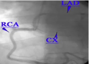
Figure 2. Conventional coronary angiography showing the solitary coro-nary artery giving rise to all branches.
The Heart Surgery Forum #2018-2319
E270
There were calcifications leading to significant and minor stenosis at the course of the circumflex and left anterior descending coronary arteries, respectively. Fur-ther diagnostic work up with angiography confirmed right solitary coronary ostium giving rise to the dominant right coronary artery, circumflex coronary artery, and left anterior descending artery without high grade stenosis (Figure 2). We decided to perform regular follow-up and started anti-hypertensive therapy with beta-blockers, Aspirin 100mg/ day, and life style modifications.
DISCUSSION
Single coronary artery anomaly has an incidence of 0.026– 0.4% in the population [Yaymaci 2010; Desmet 1992; Lipton 1979] and accounts for < 3% of congenital coronary anomalies [Yaymaci 2010] with no gender preference. They may accom-pany congenital cardiac pathologies. They are coincidentally detected during coronary investigations in adult populations. Coronary anomalies are challenging, during percutaneous coronary interventions and surgery.
There is no strong association between the solitary coronary artery and myocardial events. It is usually clinically silent and does not affect a patient’s life expectancy or quality of life [Saglam 2017]. Anomalous origin of one coronary artery from the pul-monary artery may mimic solitary coronary artery, but it leads to severe suppression of myocardial functions in early years of life.
Cardiovascular anomalies are more commonly encoun-tered with the frequent use of radiographic diagnostic tools for the diagnosis and screening of patients. Especially the noninvasive nature of the computerized tomography and enhanced image quality enabled computerized tomography is a more frequently used tool in patients highly suspected for coronary artery disease. However, conventional coronary angiography is the gold standard method to precisely detect coronary artery disease [Bayram 2008].
There is no definite information whether single coronary artery anomaly leads to cardiac problems in the literature. Garg et al indicate solitary coronary artery is not associated with increased risk for coronary atherosclerosis [Garg 2000]. On the other hand, Porto et al state that risk of atherosclerosis is increased in the case of single coronary artery resulting from acute angles [Porto 2004].
Coronary spasm [Yamamoto 1981], angina pectoris, and myocar-dial infarction are also reported in patients with single coronary artery in the absence of coronary stenosis [Joswig 1978]. As a result, percutaneous angioplasty and stenting in the presence of single coronary artery has also been reported [Porto 2004].
In conclusion, solitary coronary artery is a rare congenital anomaly. In our case, all three coronary arteries arose from the right coronary sinus and followed their usual courses on the epicardium.
REFERENCES
Bayram E, Kocatürk H, Kantarci M, Fil F, Colak MC. 2008. Anomalous origin of the right coronary artery arising from the left anterior descend-ing artery in a case with sdescend-ingle coronary artery anomaly: multi-detector computer tomography imaging. Anadolu Kardiyol Derg. 8(5):385-6. Desmet W, Vanhaecke J, Vrolix M, Van de Werf F, Piessens J, Willems J, de Geest H. 1992. Isolated single coronary artery: a review of 50,000 consecutive coronary angiographies. Eur Heart J. 13:1637-40.
Garg N, Tewari S, Kapoor A, Gupta DK, Sinha N. 2000. Primary con-genital anomalies of the coronary arteries: A coronary arteriographic study. Int J Cardiol 74:39–46.
Joswig BF, Warren SE, Vieweg WV, Hagan AD. 1978. Transmural myo-cardial infarction in the absence of coronary arterial luminal narrowing in a young man with single coronary arterial anomaly. Cathet Cardiovasc Diagn 4:297–301.
Lipton MJ, Barry WH, Obrez I, Silverman JF, Wexler L. 1979. Isolated single coronary artery: Diagnosis, angiographic classification and clinical significance. Radiology 130:39–47.
Porto I, Banning AP. 2004. Unstable angina in a patient with single coro-nary artery. Heart 90:858.
Saglam M, Dogan D, Sahin S, Turkkan C, Kula O. 2017. Single right coronary artery with absence of the left main coronary artery, left anterior descending artery, and circumflex artery. Echocardiography. 34(9):1401-1403.
Yamamoto K, Koiwaya Y, Tajimi T, Inou T, Mitsutake A, Orita Y, Takeshita A, Nakamura M. 1981. Coronary arterial spasm in single coro-nary artery. Circulation 64:1287–1290.
Yaymaci B, Ugurlucan M, Basaran M, Selimoglu O, Kocailik A, Akyildiz S, Coskun O, Us M. 2010. Solitary coronary artery nourishing the entire heart. Ann Saudi Med. 30(1):81-3.