Address for correspondence: Dr. İlker Kemal Yücel, Dr. Siyami Ersek Göğüs Kalp ve Damar Cerrahisi Eğitim ve Araştırma Hastanesi, Selimiye Mahallesi, Tıbbiye Cad. No:13, 34668 Üsküdar/İstanbul-Türkiye
Phone: +90 216 542 44 44 Fax: +90 216 418 33 17 E-mail: [email protected] Accepted Date: 11.02.2018 Available Online Date: 21.03.2018
©Copyright 2018 by Turkish Society of Cardiology - Available online at www.anatoljcardiol.com DOI:10.14744/AnatolJCardiol.2018.61257
Emine Hekim Yılmaz, Mustafa Orhan Bulut, Mehmet Küçük, İlker Kemal Yücel, Abdullah Erdem
1, Ahmet Çelebi
Department of Pediatric Cardiology, Dr. Siyami Ersek Thoracic and Cardiovascular Surgery Training and Research Hospital; İstanbul-Turkey
1Department of Pediatric Cardiology, Medipol University Hospital; İstanbul-Turkey
Use of covered stents in simultaneous management of coarctation of
the aorta and patent ductus arteriosus
Introduction
The diagnosis and management of aortic coarctation is usu-ally established during infancy or childhood. Occasionusu-ally, ado-lescent or adult asymptomatic patients may be diagnosed dur-ing investigations for hypertension. Dependdur-ing on the patient’s age and the coarctation anatomy, treatment options for aortic coarctation include balloon angioplasty, stent implantation, and surgery.
Patent ductus arteriosus (PDA) is accepted as a congenital heart disease when its persistency continues beyond the third month of life in term infants. Clinical manifestations of PDA vary from asymptomatic heart murmur to congestive heart failure, and even Eisenmenger syndrome. The clinical setting of PDA can be managed medically by percutaneous interventions or surgery. Aortic coarctation is often associated with PDA. The accept-ed treatment modality for a combination of these two
patholo-gies is surgery during infancy. However, use of interventional procedures in either sequential or simultaneous sessions has been reported in previous studies (1-3). Use of covered stents as a primary treatment modality for relieving coarctation accompa-nied by PDA has been reported in older children, adolescents, and adults (4, 5). In this study, we report 12 patients who received a covered stent for the treatment of aortic coarctation and con-current PDA. To the best of our knowledge, this report includes the largest series of patients with concurrent aortic coarctation and PDA treated via a transcatheter route.
Methods
Study cohortA single center database was retrospectively evaluated to obtain data of patients with combined aortic coarctation and
Objective: To report clinical and procedural characteristics of twelve patients who received a covered stent for the treatment of aortic coarcta-tion and concurrent patent ductus arteriosus (PDA).
Methods: A single center database was retrospectively evaluated to obtain data of patients with combined aortic coarctation and PDA. We selected patients in whom a covered stent was used for the treatment of both pathologies. The stent length was chosen so as to cover the entire length of the lesion from healthy to healthy tissue and also cover the ampulla of PDA.
Results: The median age of the patients was 15 (range, 6.5-35) years. The diameter of the coarctated segment increased from a median of 8.4 (range, 2.6-10.8) mm to 16 (range, 9-24) mm (p<0.005), whereas the pressure gradient decreased from a median of 43 (range, 10-71) mm Hg to 0 (range, 0-8) mm Hg (p<0.005). Fourteen covered stents were used for 12 patients. Following deployment, seven stents were flared with larger and low-pressure balloons because of the gap between the distal end of the stent and the poststenotic dilated segment of the aorta, which caused residual PDA shunts and/or instability of the stent. After the procedure, no residual PDA shunt was present in any patient.
Conclusion: To the best of our knowledge, this study includes the largest series of patients reported in literature in whom covered CP stents were used for simultaneous percutaneous treatment of coarctation and PDA. The procedure was successful and stable results were obtained during follow-up in all cases. (Anatol J Cardiol 2018; 19: 00-00)
Keywords: aortic coarctation, patent ductus arteriosus, covered stent
A
BSTRACTPDA since 2007. We selected only those patients in whom a cov-ered stent was used for the treatment of both pathologies for the study. All patients had a native coarctation, except one who had previously undergone balloon angioplasty for aortic coarcta-tion. Because a covered stent implantation procedure has been deemed suitable for patients weighing ≥30 kg, the study group comprised older children, adolescents, and adults.
Catheterization technique
Informed consent was received from the patients or their legal guardians. All patients underwent catheterization under general anesthesia. Heparin (100 IU/kg, max 5000 IU) was intra-venously administered immediately after the right femoral artery and vein were accessed. The coarctated segment was retro-gradely crossed with a Judkins right coronary catheter (JR4) and a 0.035-inch floppy guide wire (Terumo®). After placement of a
standard 0.035-inch exchange guidewire in the ascending aorta through the JR4 catheter, it was exchanged with a pigtail cath-eter. Hemodynamic assessment and serial angiograms were obtained using the pigtail catheter. The diameter and length of the stenotic area, diameter of the transverse arch, diameter of the aorta at the level of the subclavian artery, descending aorta diameter at the level of the diaphragm, the minimum diameter and the largest diameter (usually at the aortic ampulla) of PDA, and the length of PDA were measured. The anatomy of PDA was determined according to the classification defined by Krichenko et al. (6).
The profile of the balloon used for implantation was chosen after considering the diameter of the isthmus at the level of the left subclavian artery take-off and so that it would not exceed the diameter of the aorta at the level of the diaphragm. The stent length was chosen so that it would cover the entire length of the lesion from healthy to healthy tissue and also cover the ampulla of PDA. The chosen balloon length was longer than the stent length. In all patients, except two in whom a Z Med balloon (NuMED Inc., Hopkinton, NY, USA) was utilized, Balloon-in-Balloon (BIB) catheters (NuMED Inc., Cornwall, Ontario, Canada) were used. After placing an extra stiff guidewire into the ascending aorta or the right subclavian artery, a long sheath of appropriate size for the profile of the desired balloon catheter was advanced. A covered Cheatham-platinum (CCP) (NuMED Inc., Hopkinton, NY, USA) stent was then manually crimped onto the delivery balloon and advanced through the long sheath. A 90% platinum/10% iridium 0.013-inch wire formed the frame of the CCP stent. It was covered by a polytetrafluoroethylene (PTFE) covering, glued at 0° and 180° at each end of the stent. The expanded PTFE cover-ing could be dilated up to 26 mm before tearcover-ing. These stents are available in lengths of 16, 22, 28, 34, 39, and 45 mm. After ensuring that the stent layout was appropriate, the balloon was manually inflated, according to the manufacturer’s recommendations, and the stent was deployed. Angiograms were performed during the stent placement through the side arm of the sheath to evaluate the results and the presence of any dissection or rupture.
Hemo-dynamic studies and serial angiograms were repeated after the procedure. A gap remained between the dilated segment of the descending aorta and the distal end of the covered stent, lead-ing to a residual shunt from PDA. Flarlead-ing and seallead-ing the distal end of the stent to the aortic wall using larger low-pressure bal-loons is required. Heparin’s effect was monitored by activated clotting time and neutralized when it was >200 seconds at the end of the procedure. Manual compression to the groin provided adequate hemostasis for all patients. During follow-up, physi-cal examination, blood pressure measurement, electrocardiog-raphy, and echocardiography were performed at every clinical visit at 1, 3, 6, and 12 months. All patients underwent a computed tomographic scan after the first year of intervention.
Statistical analysis
SPSS 18.0 was used (SPSS Inc, Chicago, IL, USA) for statisti-cal analysis. Values are expressed as median (minimum-maxi-mum). Wilcoxon signed-rank test was used for comparison of measurements obtained before and after the procedures. A p-value of <0.05 was considered statistically significant.
Results
The median age of the patients was 15 (range, 6.5-35) years. Six of the patients were males. All patients had upper extremity hypertension before the procedure (the median blood pressure measured from the right arm was 165±15/105±12 mm Hg). Five of the patients were still hypertensive after the procedure and
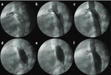
cur-a d b e c f
Figure 1. Angiogram of a patient with a mild to moderate coarctation and large PDA (a); a 34-mm covered stent mounted on a 16-mm balloon was advanced across the coarctation segment (b) and then implanted (c); however, there was significant shunt through the duct after the im-plantation due to the gap between the distal end of the stent and the poststenotic dilated segment of the aorta (c); postdilation of the distal part of the stent with 20-mm and 23-mm low-pressure balloons (d, e); closed the gap between the aortic wall and covered stent. Finally, there was no residual shunt through PDA (f)
rently use dual antihypertensive medication. The type of aortic coarctation was discrete in all patients, except in one who had a long segment coarctation. All patients had an associated PDA. Six of the PDAs were small, two were medium-sized, and four were large. According to Krichenko’s classification system, six of the PDAs were Type E, four were type C, one was type A, and one was type B (Table 1). The diameter of coarctated segment increased significantly from a median of 8.4 (range, 2.6-10.8) mm to 16 (range, 9-24) mm (p<0.005), whereas the pressure gradient across this segment decreased significantly from a median of 43 (range, 10-71) mm Hg to 0 (range, 0-8) mm Hg (p<0.005). The me-dian fluoroscopy time was 19.5 (range, 12-37) min. Prior to inter-vention, four patients had associated pulmonary hypertension, which was found to be severe in two patients. In one patient, pulmonary hypertension remained severe after the procedure, and this patient currently uses appropriate medication. Four-teen covered stents were used for 12 patients. The stent lengths ranged from 22 to 45 mm. BIB and Z med balloon catheters were used for stent deployment in ten and two patients, respectively. Following deployment, seven stents were flared with larger and low-pressure balloons because of the gap between the distal end of the stent and the poststenotic dilated segment of the aorta, which caused residual PDA shunts and/or instability of the stent (Fig. 1). After completion of the process, no residual PDA shunt was present in any patient. In one patient, while flaring the stent, a non-progressive extravasation occurred. It persisted on the control CT angiograms after the procedure; hence, the patient underwent another catheterization and received two additional covered stents 20 days after the initial procedure. All patients were stable, without the need for additional intervention during the follow-up period.
Discussion
Patient age and coarctation anatomy together with the size and shape of PDA are very important variables when consider-ing treatment options for patients with aortic coarctation and concurrent PDA. Generally, the preferred treatment modality for a combination of the two pathologies is surgery during infancy, because the incidence of recoarctation after balloon dilation is significantly higher in infants (7).
Geggel et al. (2) suggested that treatment of infants with aortic coarctation and a small PDA was possible through per-cutaneous interventions. However, whether these procedures should be performed in the same session or in sequential ses-sions has not been discussed. The disadvantages of a sequen-tial catheterization for the child and their family include cost and the need for a second hospitalization and a second intervention. However, because balloon angioplasty could disrupt the intimal and medial layers of the aorta, manipulations for PDA occlusion after balloon dilation could increase the risk of aortic dissection, mediastinal hematoma, and aneurysm formation at the newly dilated aortic segment, when performed in single sitting. More-over, when a balloon angioplasty is performed after coil occlu-sion of PDA, risk of coil embolization increases significantly be-cause the size of the PDA can change after balloon angioplasty of the aorta (5).
Singhi and Sivakumar (8) reported three different transcath-eter strategies for three of their cases with coarctation and PDA. One of the patients was a 13-year-old girl with a severe aortic coarctation and a large PDA; she was treated simultaneously via percutaneous implantation of a covered stent. The second Table 1. Demographic characteristics and procedural data
Age Weight PDA Additional PHT Preprocedural Postprocedural Balloon size Balloon Stent type Stent length
(years) (kg) type findings Systemic HT Systemic HT (mm) type (mm)
Case 1 26 28 C VSD, BAV + + + 16x4 BIB Covered CP 45
Case 2 6.5 19 B - - + - 10x3 Z Med Covered CP 34
Case 3 16 55 E BAV - + + 12x4 BIB Covered CP 34
Case 4 12 38 C VSD+ASD + + - 16x4 BIB Covered CP 34
Case 5 35 73 E BAV - + - 18x4 BIB Covered CP 45
Case 6 22 84 E - - + - 20x3.5 BIB Covered CP 28
Case 7 12 19 E VSD (small) - + - 13x3 Z Med Covered CP 28
Case 8 25 60 A - - + + 20x3.5 BIB Covered CP 34
Case 9 10 26 C BAV + + - 14x3 BIB Covered CP 22
Case 10 15 53 C BAV+SAR + + + 20x4 BIB Covered CP 39
Case 11 13 42 E - - + + 16x4 BIB Covered CP 34
Case 12 14 57 E - - + - 18x4 BIB Covered CP 39
BAV - bicuspid aortic valve; PHT - pulmonary hypertension; HT – hypertension; PDA - patent ductus arteriosus; BIB - balloon-in-balloon; CP - cheatham-platinum; SAR - subaortic ridge; VSD - ventricular septal defect
patient was a nine-year-old girl with PDA and a mild isthmus hypo-plasia. The PDA was occluded with an Amplatzer® duct occluder. When the pressure gradient across the coarctation rose during the seven-year follow-up, the coarctation was stented. The third patient was a 10-year-old who had PDA and a discrete aortic co-arctation. After occlusion of the PDA with an Amplatzer® duct
oc-cluder, coarctation was relieved by balloon angioplasty (8). Simultaneous closure of PDA with dilation of the coarctation can be achieved using covered CP stents. Using a single device in a single sitting would reduce the fluoroscopy and procedure times. Sadiq et al. (4) defined the use of a covered CP stent to treat coexistent aortic coarctation and PDA. Kulkarni et al. (5) reported a 20-year-old girl with severe coarctation, a large PDA, and pulmonary hypertension. By implanting a covered stent dur-ing a sdur-ingle session, both pathologies were treated; pulmonary artery pressure decreased, and the patient was discharged on the following day after the procedure (5).
While some physicians prefer using covered stents only for curative purposes, such as following a complication like aneu-rysm formation or a tear, others prefer the prophylactic use of covered stents to avoid late aneurysm formation. Chang et al. (9) reported 25 cases in whom covered stents were used as the primary modality for the treatment of native coarctation. Five of their cases had associated PDAs that were completely occluded when the coarctation was stented. The risk of occlusion of the side branches of the aorta and spinal artery during implantation of covered stents has been a major concern (10). Because the spinal artery arises below the level of the ninth thoracic ver-tebra, placing a covered stent at the usual site of coarctation seems safe. Among the major side branches of the aorta, sac-rificing the left subclavian artery causes no significant morbid-ity. Another concern has been the long-term fate of the covered stents because they may need redilation before and after full growth of the patient.
Use of BIB catheters allows proper stent positioning and decreases the risk of migration. The inner and outer balloons of BIB catheters can be inflated separately, and the position of the stents can be checked at that time; hence, a more controlled deployment can be achieved. In our study, for the deployment of stents in the desired position, BIB catheters were used in ten patients. A high-pressure Z Med balloon was used because the correctly sized BIB catheters were not available during the procedures for two patients. We chose the size of the delivery balloon by taking into consideration the diameter of the distal transverse arcus and taking care not to exceed the size.
After deployment of the stents, high-pressure balloons were used for redilation of the residual stenosis. Low-pressure bal-loons, less than the diameter of the aorta at the level of the dia-phragm, were utilized for flaring the stents against the dilated vessel wall. Because the space between the implanted stent and the dilated aortic wall could result in a distal endoleak and a persistent shunt through the ductus, especially in patients with marked poststenotic dilatation, adequate sealing of the stent to
the aorta by flaring the stent against the vessel wall with larger in size and low-pressure balloons is essential. Additionally, provid-ing an adequate seal and avoidprovid-ing residual shunts through PDA, flaring of the stents is advised to decrease resistance within tube grafts and precludes proximal infolding of the stent (11).
All covered stents, except two, were implanted, with excel-lent acute outcomes. In one patient, aged six and a half years, the stent protruded into the transverse arcus causing a pressure gradient of 10 mm Hg, which did not require reintervention. In an-other patient, aged 34 years, we noticed a residual flow through the PDA caused by a gap between the descending aortic wall and the distal end of the covered stent. During the flaring of the stent, an extravasation that did not compromise the patient he-modynamically had occurred. After 20 days, two additional cov-ered stents were implanted telescopically to cover the ruptured segment in this patient.
The use of covered stents, instead of bare stents, can reduce the potential complications mentioned above, but not the overall risk. Any tear, dissection, or rupture would be covered because of the PTFE covering of the CCP stents. However, particular at-tention must be paid to make sure that both ends of the stents are placed over healthy aortic tissue. It should be noted that the covering of the CCP stent does not reach the edges of the stent, leaving uncovered parts that may tear or traumatize the vessel wall when the stent is fully expanded.
Diagnostic cardiac catheterization revealed that two of our patients had severe pulmonary hypertension (patients 1 and 4). Vasoreactivity testing was found to be positive and the Qp/Qs ratio was >1.5 in the patient who had a large ventricular septal defect (VSD) (patient 1). As published previously, PDA closure is advised when the shunt is from left to right and pulmonary vas-cular reactivity is present in patients with severe pulmonary hy-pertension (12). After the procedure, an immediate decrease in pulmonary artery pressure was observed. In the follow-up, pul-monary artery pressure, estimated by echocardiographic evalu-ation via VSD jet, showed that the pulmonary hypertension had been completely resolved. In the setting of a PDA with Eisen-menger syndrome, closure of the duct is not advised because it may cause right ventricular failure (13). The other patient with severe pulmonary hypertension was 12 years old and had aortic coarctation, a large PDA, large VSD, and a medium-sized ASD that had resulted in Eisenmenger syndrome (patient 4). Because the accompanying large VSD, which would permit a bi-direc-tional shunt for the relief of pulmonary artery hypertension, was present, the coarctated segment was dilated and the PDA was simultaneously closed with a covered stent. In this way, both the left and right ventricular pressures and thus the systemic pres-sure were reduced. In the follow-up, however, medications for pulmonary arterial hypertension were continued.
Aneurysm formation and recoarctation are possible compli-cations after surgical and endovascular repair of aortic coarcta-tion. In our study group, after discharge from hospital, regular evaluation using echocardiography did not reveal any need for
reintervention for any of the patients. CT angiography, which was performed after the first year of intervention in all patients, showed mild dilation at the poststenotic segment of the de-scending aorta in four patients. No other pathology, such as re-coarctation or aneurysm formation, was detected in any patient. During a median follow-up period of 85 (range, 12-110) months, no adverse event such as aneurysm formation, stent migration, stent fracture, or dissection was found in any patient.
All patients were hypertensive prior to the interventions. Seven patients became normotensive after the procedure, whereas five patients still needed antihypertensive medications. It is known that hypertension can be an issue during the follow-up period of patients with aortic coarctation even after a suc-cessful repair and in the absence of restenosis. Canniffe et al. (14) reviewed 26 articles related to residual hypertension after aortic coarctation repair. They reported the median prevalence of hypertension after coarctation repair to be 32.5% (range, 25%-68%) in this review of 26 studies. Our results with residual hypertension were consistent with these findings: Five patients (41.6%) were still hypertensive after the procedures. Although age at the time of repair, present age, method used to measure blood pressure, and description of the hypertension all influence the prevalence, hypertension continues to be a common and po-tentially serious problem even after successful repair (14).
Study limitations
The retrospective design was the main limitation of the pres-ent study. Additionally, the sample size was relatively small due to restriction of the population to older children, adolescents, and adults as a result of the requirement of larger sheaths for covered stent implantation.
Conclusion
To the best of our knowledge, this study includes the larg-est series of patients reported in literature in whom covered CP stents were used for simultaneous percutaneous treatment of coarctation and PDA. The procedure was successful and stable results were obtained during follow-up in all cases. We believe that if stent implantation is possible, use of a covered stent en-ables treatment of both conditions in one setting, is safe and simple, and avoids use of an additional device to close the duc-tus. Besides, the fluoroscopy and procedure times as well as the number of future interventions reduced. We find the use of BIB catheters convenient in this subgroup of patients because these allow proper stent positioning and decrease the risk of migra-tion. We should keep in mind that high-pressure balloons may be needed for further dilation after the deployment to close residual shunts through PDA.
Conflict of interest: None declared. Peer-review: Externally peer-reviewed.
Authorship contributions: Concept – E.H.Y., M.O.B., M.K., İ.K.Y., A.E., A.Ç.; Design – E.H.Y., M.O.B., M.K., İ.K.Y., A.E., A.Ç; Supervision – E.H.Y., M.O.B., M.K., İ.K.Y., A.E., A.Ç; Fundings – E.H.Y., M.O.B., M.K., İ.K.Y., A.E., A.Ç; Materials – E.H.Y., M.O.B., M.K., İ.K.Y., A.E., A.Ç; Data collection &/ or processing – E.H.Y., M.O.B., M.K., İ.K.Y., A.E., A.Ç; Analysis &/or in-terpretation – E.H.Y., M.O.B., M.K., İ.K.Y., A.E., A.Ç; Literature search – E.H.Y., M.O.B., M.K., İ.K.Y., A.E., A.Ç; Writing – E.H.Y., M.O.B., M.K., İ.K.Y., A.E., A.Ç; Critical review – E.H.Y., M.O.B., M.K., İ.K.Y., A.E., A.Ç.
References
1. Celebi A, Yalcin Y, Erdem A, Zeybek C, Akdeniz C, Polat TB. Suc-cessful transcatheter balloon dilatation of coarctation of aorta and coil occlusion of patent ductus arteriosus in a single catheteriza-tion procedure. Turk J Pediatr 2007; 49: 94-7.
2. Geggel RL, Hijazi ZM, Rhodes J. Interventional cardiac catheter-ization therapy for combined coarctation of the aorta and patent ductus arteriosus: successful outcome in two infants. Cathet Car-diovasc Diagn 1996; 38: 67-70.
3. Hakim F, Hawelleh AA, Goussous Y, Hijazi ZM. Simultaneous stent implantation for coarctation of the aorta and closure of patent duc-tus arteriosus using the Amplatzer duct occluder. Cathet Cardio-vasc Interv 1999; 47: 36-8.
4. Sadiq M, Malick NH, Qureshi SA. Simultaneous treatment of native coarctation of the aorta combined with patent ductus arteriosus using a covered stent. Catheter Cardiovasc Interv 2003; 59: 387-90. 5. Kulkarni S, Vimala J, Parmar R. Single therapeutic catheterization
for treatment of native coarctation of aorta and large patent ductus arteriosus using a covered stent. Indian Heart J 2005; 57: 713-6. 6. Krichenko A, Benson LN, Burrows P, Moes CAF, McLaughlin P,
Free-dom RM. Angiographic classification of the isolated, persistently patent ductus arteriosus and implications for percutaneous cath-eter occlusion. Am J Cardiol 1989; 67: 877-80.
7. Rao PS, Koscik R. Validation of risk factors in predicting recoarcta-tion after initially successful balloon angioplasty for native aortic coarctation. Am Heart J 1995; 130: 116-21.
8. Singhi AK, Sivakumar K. Different transcatheter strategies for aor-tic coarctation associated with patent ductus arteriosus. Indian Heart J 2012; 64: 423-6.
9. Chang ZP, Jiang SL, Xu ZY, Zhang GJ, Huang LJ, Zhao SH, et al. Use of covered Cheatham-Platinum stent as the primary modality in the treatment for native coarctation of the aorta. Chin Med J (Engl) 2012; 125: 1005-9.
10. Khan MS, Moore JW. Treatment of abdominal aortic pseudoaneu-rysm with covered stents in a pediatric patient. Catheter Cardio-vasc Interv 2000; 50: 445-8.
11. Vanagt WY, Cools B, Boshoff DE, Frerich S, Heying R, Troost E, et al. Use of covered Cheatham-Platinum stents in congenital heart disease. Int J Cardiol 2014; 175: 102-7.
12. Baruteau AE, Hascoet S, Baruteau J, Boudjemline Y, Lambert V, Angel CY, et al. Transcatheter closure of patent ductus arteriosus: past, present and future. Arch Cardiovasc Dis 2014; 107: 122-32. 13. Bhalgat PS, Pinto R, Dalvi BV. Transcatheter closure of large
pat-ent ductus arteriosus with severe pulmonary arterial hypertension: Short and intermediate term results. Ann Pediatr Cardiol 2012; 5: 135-40.
14. Canniffe C, Ou P, Walsh K, Bonnet D, Celermajer D. Hypertension after repair of aortic coarctation--a systematic review. Int J Cardiol 2013; 167: 2456-61.