© TÜBİTAK
E-mail: [email protected] doi:10.3906/sag-1002-588
Cardiac complications of secondary hyperparathyroidism in
chronic hemodialysis patients
Hülya ÇİÇEKÇİOĞLU1, İhsan ERGÜN2, Özgül UÇAR1, Cüneyt YÜKSEL3, Alper AZAK4, Ekrem ABAYLI4, Mehmet Deniz AYLI5
Aim: To evaluate the eff ects of intact parathormone (iPTH) on left ventricular function using transthoracic echocardiography on chronic hemodialysis (HD) patients with secondary hyperparathyroidism. In HD patients, mortality is high and is frequently due to cardiac complications. Secondary hyperparathyroidism, high levels of phosphate (PO4), and high calcium phosphate product (Ca × PO4) are related to cardiac complications.
Materials and methods: We examined 20 patients with normal iPTH levels (Group 1) and 20 patients with high iPTH levels (Group 2). Intact parathormone levels were measured in serum with a Coat-A-Count kit (Diagnostic Products Corporation, Los Angeles, CA, USA) using an immunoradiometric assay. Th e normal level of iPTH was 0.8-5.2 pmol/L. In patients with end-stage renal disease, iPTH levels should be 1.5 to 3 times higher than the normal range in order to maintain the bone mass; thus, patients with iPTH levels 4 or more times higher than the normal range (PTH ≥ 20.8 pmol/L) were defi ned as Group 2 while patients who had normal iPTH levels were defi ned as Group 1.
Results: In both groups, Doppler parameters indicated diastolic dysfunction. However, mitral annular E velocity was lower in Group 2 than in Group 1 (6.1 ± 1.1 cm/s and 7.5 ± 1.6 cm/s, respectively; P = 0.034). It is well known that left ventricular hypertrophy (LVH) increases mortality rates. Left ventricle mass index and relative wall thickness are parameters refl ecting LVH, and both were higher in Group 2 (294.4 ± 103.0 g/m² and 53.5 ± 11.7%) when compared with Group 1 (179.2 ± 104.2 g/m² and 41.8 ± 8.9%). Th ese diff erences were found to be statistically signifi cant (P < 0.001).
Conclusion: Th is study demonstrates that high levels of iPTH contribute to diastolic dysfunction and LVH in hemodialysis patients.
Key words: Hemodialysis, parathormone, chronic renal failure, echocardiography, tissue Doppler echocardiography
Kronik hemodiyaliz hastalarında ikincil hiperparatiroidinin
kardiyak komplikasyonları
Amaç: Kronik hemodiyaliz hastalarında mortalite yüksektir ve sıklıkla kardiyak komplikasyonlara bağlıdır. Bu komplikasyonlara ikincil hiperparatroidizm, yüksek fosfat (PO4) ve yüksek kalsiyum fosfat çarpımı (Ca × PO4) eşlik eder. Biz ikincil hiperparatroidizmli hemodiyaliz hastalarında intakt parathormonun (iPTH) sol ventrikül fonksiyonları üzerindeki etkilerini araştırmak amacıyla transtorasik ekokardiyografi uyguladık.
Original Article
Received: 07.02.2010 – Accepted: 08.11.2010
1 Department of Cardiology, Ankara Numune Education and Research Hospital, Ankara - TURKEY 2 Department of Nephrology, Faculty of Medicine, Ufuk University, Ankara - TURKEY
3 Department of Nephrology, Ankara Oncology Education and Research Hospital, Ankara - TURKEY 4 Department of Internal Medicine, Ankara Numune Education and Research Hospital, Ankara - TURKEY 5 Department of Nephrology, Ankara Dışkapı Education and Research Hospital, Ankara - TURKEY
Correspondence: Özgül UÇAR, Harbiye Mahallesi, Dikmen Caddesi, No: 176/69, 06460 Çankaya, Ankara - TURKEY
Introduction
Th e prevalence of cardiovascular disease (CVD) in patients with end-stage renal disease (ESRD) is high and is associated with a higher risk of mortality. In addition to common traditional risk factors (smoking, dyslipidemia, hypertension, diabetes mellitus, and positive family history), uremic factors including hyperparathyroidism, elevated serum PO4, and Ca × PO4 product levels are thought to increase the risk of CVD (1-3). Th e eff ect of intact parathormone (iPTH) on the development and progression of cardiovascular disease is not clearly understood and this subject still needs to be evaluated by further research. In this study, we aimed to evaluate the eff ect of high iPTH levels on cardiac functions and left ventricular hypertrophy (LVH) indices by using transthoracic echocardiography on chronic hemodialysis patients.
Materials and methods
Patients with ESRD on hemodialysis therapy for at least 6 months were included in the study. Patients who had parathyroidectomy, coronary artery disease, aortic stenosis (transaortic fl ow velocity > 2.5 m/s), pericardial eff usion, anemia (Hb < 10 g/dL), uncontrolled hypertension, atrial fi brillation, diabetes mellitus, and/or another systemic disease were excluded from the study. Th e local ethics committee of Ankara Numune Education and Research Hospital approved the study protocol and informed consent
was obtained from all patients participating in the study.
Intact parathormone levels were measured in serum with a Coat-A-Count kit (Diagnostic Products Corporation, Los Angeles, CA, USA) using an immunoradiometric assay. Th e normal level of iPTH ranged between 0.8 and 5.2 pmol/L (conversion factor: pg/mL × 0.1053 = pmol/L). At least 2 measurements were made per patient. Th e intraassay coeffi cient of variation was 6% and the interassay coeffi cient of variation was 6.6%. Patients were divided into 2 groups according to iPTH levels. In patients with ESRD, iPTH levels should be 1.5 to 3 times higher than the normal range in order to maintain the bone mass (4); thus, patients with iPTH levels 4 or more times higher than the normal range (PTH ≥ 20.8 pmol/L) were defi ned as Group 2 while patients who had normal iPTH levels were defi ned as Group 1.
Between May 2008 and July 2009, a total of 107 patients from the hemodialysis unit of our institution were evaluated. Due to the requirements of the nature of our investigation, the study excluded 2 patients who had undergone parathyroidectomy, 13 patients with documented coronary artery disease, 4 patients with aortic stenosis (transaortic fl ow velocity > 2.5 m/s), 4 patients with pericardial eff usion, 10 patients with anemia (Hb < 10 g/dL), 11 patients with uncontrolled hypertension, 5 patients with atrial fi brillation, 14 patients with diabetes mellitus, 2 Yöntem ve gereç: Yirmi normal iPTH düzeyine sahip hasta (grup 1) ve 20 yüksek iPTH düzeyine sahip hasta (grup 2) çalışmaya dahil edildi. İntakt parathormon düzeyleri serumda Coat-A-Count kiti (Diagnostic Products Corporation, Los Angeles, CA, USA) kullanılarak immunoradyometrik yöntem ile ölçüldü. Normal iPTH düzeyleri 0,8-5,2 pmol/ L idi. Son dönem böbrek hastalarında kemik kütlesini korumak için iPTH düzeyleri normalden 1,5-3 kat yüksek olmalıdır, dolayısıyla iPTH düzeyleri normalden 4 kat yüksek olanlar (PTH ≥ 20,8 pmol/ L) grup 2, normal iPTH düzeyleri olanlar grup 1 olarak tanımlandı.
Bulgular: Her iki grupta ekokardiyografi k parametreler diyastolik disfonksiyon varlığını gösterdi. Doku Doppler ile bakılan mitral anular E velosite grup 2’de grup 1’e kıyasla daha düşüktü (7,5 ± 1,6 cm/sn’e karşı 6,1 ± 1,1 cm/sn, P = 0,034). Sol ventrikül hipertrofi sinin mortalite oranlarını arttırdığı iyi bilinmektedir. Sol ventrikül kitle indeksi ve rölatif duvar kalınlığı sol ventrikül hiperrtofi sini işaret eden parametrelerdir ve her ikisi de Grup 1 ile kıyaslandığında Grup 2’de daha yüksek bulundu ve bu farklılık istatistiksel olarak anlamlıydı (179,2 ± 104,2 g/m²’e karşı 294,4 ± 103,0 g/m²; % 41,8 ± 8,9’ e karşı % 53,5 ± 11,7; herbirisi için P < 0,001).
Sonuç: Bu çalışma hemodiyaliz hastalarında yüksek iPTH düzeylerinin diyastolik disfonksiyon ve sol ventrikül hipertrofi sine katkıda bulunduğunu göstermiştir.
Anahtar sözcükler: Hemodiyaliz, parathormon, kronik böbrek yetmezliği, ekokardiyografi , doku Doppler ekokardiyografi
patients with rheumatoid arthritis, and 2 patients that were unwilling to participate. Groups were created and 20 patients (10 male, 10 female; mean age: 49.7 ± 13.8 years) with normal iPTH levels were placed in Group 1 while 20 patients (10 male, 10 female; mean age: 51.2 ± 15 years) with higher iPTH levels were put into Group 2. Patient characteristics are given in Table 1.
All of the patients in the study population were undergoing a hemodialysis program consisting of 4-h sessions 3 days a week using blood fl ow rates of 250-350 mL/min and dialysate fl ows of 500 mL/min. All patients were dialyzed with a standard bicarbonate-containing dialysate bath (Na: 138 mmol/L; K: 2 mmol/L; HCO3: 33 mmol/L; Ca: 1.25 mmol/L; Mg: 0.5 mmol/L). Standard heparinization was performed with a minimum of 1.6 m2 polysulfone membranes
and Fresenius 4008-B dialysis machines were used.
Th e duration of the hemodialysis program was 49.5 ± 30.2 months in Group 1 and 69.9 ± 37.7 months in Group 2, a duration that was not statistically signifi cant (P = 0.067). Most patients (90% of those in Group 1 and 95% of those in Group 2) used phosphate binders, mainly calcium carbonate and calcium acetate.
Blood samples were withdrawn before hemodialysis therapy and levels of total calcium, inorganic phosphorus, Ca × PO4 product, hemoglobin, and iPTH were measured by standardized clinical laboratory methods.
Patients’ blood pressure was measured aft er resting in the supine position for 5 min and immediately before the echocardiographic examination was administered by the same observer using a standard sphygmomanometer. Only patients who had controlled hypertension were enrolled in the study.
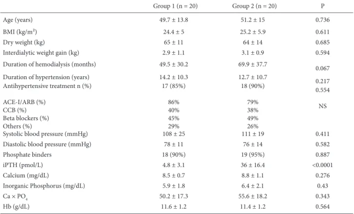
Table 1. Baseline patient characteristics.
Group 1 (n = 20) Group 2 (n = 20) P
Age (years) 49.7 ± 13.8 51.2 ± 15 0.736
BMI (kg/m²) 24.4 ± 5 25.2 ± 5.9 0.611
Dry weight (kg) 65 ± 11 64 ± 14 0.685
Interdialytic weight gain (kg) 2.9 ± 1.1 3.1 ± 0.9 0.594
Duration of hemodialysis (months) 49.5 ± 30.2 69.9 ± 37.7
0.067 Duration of hypertension (years) 14.2 ± 10.3 12.7 ± 10.7
0.217 Antihypertensive treatment n (%) ACE-I/ARB (%) CCB (%) Beta blockers (%) Others (%) 17 (85%) 86% 40% 45% 29% 18 (90%) 79% 38% 49% 26% 0.554 NS
Systolic blood pressure (mmHg) 108 ± 25 111 ± 19 0.411
Diastolic blood pressure (mmHg) 78 ± 11 76 ± 14 0.582
Phosphate binders 18 (90%) 19 (95%) 0.887 iPTH (pmol/L) 4.8 ± 3.1 36 ± 16.4 <0.0001 Calcium (mg/dL) 8.5 ± 0.7 8.8 ± 1.1 0.276 Inorganic Phosphorus (mg/dL) 5.9 ± 1.8 6.4 ± 2.1 0.43 Ca × PO4 50.2 ± 17.3 55.6 ± 18.2 0.343 Hb (g/dL) 11.6 ± 1.2 11.4 ± 1.2 0.564
BMI: Body mass index, ACE-I: angiotensin-converting enzyme inhibitor, ARB: angiotensin II receptor blocker, CCB: calcium channel blocker, iPTH: intact parathormone, Hb: hemoglobin.
All except 3 patients in Group 1 and 2 patients in Group 2 were receiving antihypertensive treatment. Th e duration of hypertension and antihypertensive regimens were similar between the groups (Table 1).
In both groups, left ventricular systolic and diastolic functions, the left ventricular mass index (LVMI), and relative wall thickness (RWT) were determined by M-mode echocardiography. Echocardiographic examinations were performed on the day aft er hemodialysis when patients were at dry weight. Echocardiographic measurements were performed with VingMed System Five (GE VingMed System Five, Horton, Norway) equipment using 2.5-mHz transducer probes. Echocardiographic measurements were taken through the standard echocardiographic windows while the patient was lying in a left lateral decubitus position as per the criteria of the American Society of Echocardiography (5). Left ventricular ejection fraction (EF) and the left ventricular end-systolic volumes and end-diastolic volumes were calculated by using the modifi ed Simpson method. Left ventricular diastolic functions were examined by the fl ux of blood through the edges of the mitral leafl ets and in the middle of the left ventricular cavity, using pulsed wave Doppler. Maximum E v elocity (E), maximum A velocity (A), and mitral E deceleration time (DT) were calculated by mitral
fl ux samples. Th e period from the end of the aortic fl ux to the starting of the mitral fl ux was measured as isovolumic relaxation time (IVRT). Tissue Doppler mitral annular velocities were derived from the lateral mitral annulus by pulsed wave Doppler. In the apical 4-chamber view, a 2-mm pulse wave Doppler sample gate was placed at the lateral mitral annulus to obtain the peak early diastolic (Ea), atrial (Aa), and systolic tissue velocities (Sa). Th e average was taken from 3 consecutive beats. Using the parasternal long axis images obtained by 2-dimensional and M-mode echocardiography, interventricular septum thickness during diastole (IVSd), posterior wall thickness during diastole (PWd), and left ventricular end-diastolic diameter (LVEDD) were calculated. Th e left ventricular mass was calculated by the Devereux formula (6) and this left ventricular mass was divided by the body surface area in order to calculate the left ventricle mass index. Relative wall thickness was calculated as (2 × PWd / LVEDD) × 100.
Statistics of all parameters were analyzed with the Kolmogorov-Smirnov test and all parameters showed normal distribution. Student’s t-test was used for independent groups. Th e results were given as mean ± standard deviation for continuous variables. Findings were considered signifi cant at P < 0.05.
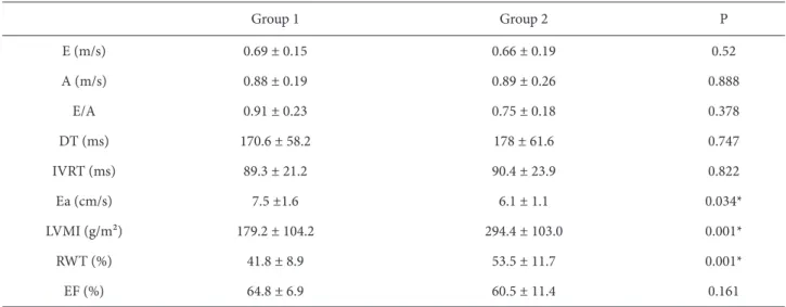
Table 2. Comparison of echocardiographic and clinical parameters between the 2 groups.
Group 1 Group 2 P E (m/s) 0.69 ± 0.15 0.66 ± 0.19 0.52 A (m/s) 0.88 ± 0.19 0.89 ± 0.26 0.888 E/A 0.91 ± 0.23 0.75 ± 0.18 0.378 DT (ms) 170.6 ± 58.2 178 ± 61.6 0.747 IVRT (ms) 89.3 ± 21.2 90.4 ± 23.9 0.822 Ea (cm/s) 7.5 ±1.6 6.1 ± 1.1 0.034* LVMI (g/m²) 179.2 ± 104.2 294.4 ± 103.0 0.001* RWT (%) 41.8 ± 8.9 53.5 ± 11.7 0.001* EF (%) 64.8 ± 6.9 60.5 ± 11.4 0.161
E: Maximum E velocity, A: maximum A velocity, DT: mitral E deceleration time, IVRT: isovolumic relaxation time, Ea: mitral annular E velocity, LVMI: left ventricular mass index, RWT: relative wall thickness, EF: ejection fraction.
Results
Th e echocardiographic and clinical comparisons are given in Table 2. Left ventricular hypertrophy indicators, left ventricular mass index (Group 1: 179.2 ± 104.2 g/m²; Group 2: 294.4 ± 103.0 g/m²; P < 0.001), and relative wall thickness (Group 1: 41.8 ± 8.9%; Group 2: 53.5 ± 11.7%; P < 0.001) were signifi cantly higher in Group 2. Th ere was no signifi cant diff erence between groups in terms of mean age, body mass index, dry weight, interdialytic weight gain, duration of hemodialysis, systolic and diastolic blood pressures, calcium, inorganic PO4, Ca × PO4 product, or hemoglobin levels (Table 2).
Th e mean EF of Group 2 was 60.5 ± 11.4% and it was 64.8 ± 6.9% in Group 1, a diff erence that was not found to be statistically signifi cant (P > 0.05).
When parameters indicating diastolic dysfunction (maximum E velocity, maximum A velocity, E/A ratio, DT, and IVRT) were compared, no statistical signifi cance was identifi ed between the groups.
Mitral annular E velocities measured by tissue Doppler imaging was signifi cantly lower in Group 2 compared to Group 1 (6.1 ± 1.1 cm/s compared to 7.5 ± 1.6 cm/s, P = 0.034).
Discussion
We investigated the infl uence of elevated iPTH levels on left ventricular functions and left ventricular hypertrophy indicators in chronic hemodialysis patients. Patients with uncontrolled hypertension, atrial fi brillation, coronary artery disease, aortic stenosis, pericardial eff usion, anemia, or diabetes mellitus were excluded. We demonstrated that patients with higher iPTH levels had lower mitral annular E velocities and higher indices of left ventricular hypertrophy such as left ventricular mass index or relative wall thickness.
Elevated iPTH levels were found to be an independent risk factor for left ventricular hypertrophy (7). PTH acts on cardiomyocytes by binding to the PTH/PTHrP receptor, which induces a rise in the intracellular levels of calcium. Increased calcium levels activate protein kinase C and mediate hypertrophic as well as metabolic eff ects (8,9). Many published experiments report that iPTH contributes
to cardiac fi broblast activation and the fi brosis of intermyocardiocytes, which is a prerequisite of diastolic dysfunction (10). In addition, higher levels of calcium due to hyperparathyroidism have been shown to induce arrhythmia (11).
Previous studies have shown that patients with hyperparathyroidism have diastolic dysfunction while systolic functions remain normal (12-14). Our study is in accordance with previous research indicating that systolic functions were preserved in both groups without statistical diff erence.
Echocardiographic parameters of diastolic dysfunction were analyzed and mitral annular E velocities measured by tissue Doppler were found to be lower in patients with high iPTH levels than in patients with normal iPTH levels. It is known that tissue Doppler mitral Ea is a more sensitive parameter for abnormal relaxation than conventional mitral infl ow measurements, and, by using this method, diastolic dysfunction can be more easily detected in patients (15,16). In patients with hyperparathyroidism, the energy-consuming diastolic relaxation may be aff ected. However, left ventricular mass index or relative wall thickness parameters were also higher in patients with high iPTH levels, and lower diastolic mitral annular E velocities may be a consequence of this fi nding.
Park et al. demonstrated that in hemodialysis patients, partial treatment of secondary hyperparathyroidism by intravenous calcitrol reduced myocardial hypertrophy without infl uencing the heart rate or the peripheral resistance (17). In addition, improvements were observed in terms of cardiac function, reduction in blood pressure, and regression of left ventricular hypertrophy following parathyroidectomy (18,19).
In conclusion, our study shows that high levels of iPTH are associated with diastolic dysfunction and left ventricular hypertrophy in hemodialysis patients. Th e reduction of iPTH to normal ranges may reduce left ventricular hypertrophy and improve cardiac function. Th us, we believe that one of the early measures for preventing cardiac hypertrophy in patients with ESRD is to reduce the high level of iPTH. In order to confi rm this result, large multicenter studies are required.
References
1. Foley RN, Levin A. Cardiovascular disease in chronic renal insuffi ciency. Am J Kidney Dis 2000; 36: S24-S30. 2. Levey AS, Beto JA, Coronado BE, Eknoyan G, Foley RN,
Kasiske BL et al. Controlling the epidemic of cardiovascular disease in chronic renal disease: What do we know? What do we need to learn? Where do we go from here? Am J Kidney Dis 1998; 32: 853-906.
3. Linder A, Charra B, Sherrard DJ, Scribner BH. Accelerated atherosclerosis in prolonged maintenance hemodialysis. N Engl J Med 1974; 290: 697-701.
4. Brenner BM, Rector FC. Brenners and Rector’s Th e Kidney. 6th ed. Philadelphia: W.B. Saunders Company; 2000. p.2121-52.
5. Park SH, Shub C, Nobrega TP, Bailey KR, Seward JB. Two-dimensional echocardiographic calculation of left ventricular mass as recommended by the American Society of Echocardiography: correlation with autopsy and M-mode echocardiography. J Am Soc Echocardiogr 1996; 9: 119-28.
6. Devereux RB, Alonso DR, Lutas EM, Gottlieb GJ, Campo E, Sachs I et al. Echocardiographic assessment of left ventricular hypertrophy: comparison to necropsy fi ndings. Am J Cardiol 1986; 57: 450-8.
7. Randon RB, Rohde LE, Comerlato L, Ribeiro JP, Manfro RC. Th e role of secondary hyperparathyroidism in left ventricular hypertrophy of patients under chronic hemodialysis. Braz J Med Biol Res 2005; 38: 1409-16. 8. Bro S, Olgaard K. Eff ects of excess PTH on nonclassical
target organs. Am J Kidney Dis 1997; 30: 606-20.
9. Smogorzewski M, Zayed M, Zhang YB, Roe J, Massry SG. Parathyroid hormone increases cytosolic calcium concentration in adult rat cardiac myocytes. Am J of Physiology 1993; 264: H1998-2006.
10. Amann K, Ritz E, Wiest G, Klaus G, Mall G. A role of parathyroid hormone for the activation of cardiac fi broblasts in uremia. J Am Soc Nephrol 1994; 4: 1814-19. 11. McCarty MF, Barnoso-Aranda J, Contreras F. Can moderate
elevations of parathyroid hormone acutely increase risk for ischemic cardiac arrhythmias? Med Hypotheses 2009; 72: 581-3.
12. Baykan M, Erem C, Erdogan T, Ersöz HO, Gedikli O, Korkmaz L et al. Assessment of left ventricular diastolic function and the Tei index by tissue Doppler imaging in patients with primary hyperparathyroidism. Clin Endocrinol (Oxf) 2007; 66: 483-8.
13. Barletta G, De Feo ML, Del Bene R, Lazzeri C, Vecchiarino S, La Villa G et al. Cardiovascular eff ects of parathyroid hormone: a study in healthy subjects and normotensive patients with mild primary hyperparathyroidism. J Clin Endocrinol Metab 2000; 85: 1815-21.
14. Ohara N, Hiramatsu K, Shigematsu S, Hayashi Y, Ishihara F, Aizawa T et al. Eff ect of parathyroid hormone on left ventricular diastolic function in patients with primary hyperparathyroidism. Miner Electrolyte Metab 1995; 21: 63-6.
15. Nagueh SF, Appleton CP, Gillebert TC, Marino PN, Oh JK, Smiseth OA et al. Recommendations for the evaluation of left ventricular diastolic function by echocardiography. Eur J Echocardiogr 2009; 10: 165-93.
16. Hayashi SY, Rohani M, Lindholm B, Brodin LA, Lind B, Barany P et al. Left ventricular function in patients with chronic kidney disease evaluated by colour tissue Doppler velocity imaging. Nephrol Dial Transplant 2006; 21: 125-32.
17. Park C, Oh Y, Shin Y, Kim C, Kim Y, Kim S et al. Intravenous calcitriol regresses myocardial hypertrophy in hemodialysis patients with secondary hyperparathyroidism. Am J Kidney Dis 1999; 33: 73-81.
18. Hara S, Ubara Y, Arizono K, Ikeguchi H, Katori H, Yamada A et al. Relation between parathyroid hormone and cardiac function in long-term hemodialysis patients. Miner Electrolyte Metab 1995; 21: 72-6.
19. Nanasato M, Goto N, Isobe S, Unno K, Hirayama H, Sato T et al. Restored cardiac conditions and left ventricular function aft er parathyroidectomy in a hemodialysis patient. Parathyroidectomy improves cardiac fatty acid metabolism assessed by 123I-BMIPP. Circ J 2009; 73: 1956-60.