I
ntroductIonD
ental caries is one of the major public health problems, possibly greater than many other well‑known diseases such as hypertension, heart disease, and cancer.[1,2] Researchers and product developers continue to search for ways to reduce the risk of caries in patients. Recently, many remineralization techniques have been tried out, among which the well‑known is the use of fluoride.[3]Quantitative Evaluation of the Enamel Caries Which Were Treated with
Casein Phosphopeptide-amorphous Calcium Fluoride Phosphate
O Yazicioğlu, BC Yaman1, A Güler2, F Koray3
Address for correspondence: Dr. O Yazicioğlu, Department of Conservative Dentistry, Faculty of Dentistry, Istanbul University, 34093 Çapa, Istanbul, Turkey. E‑mail: [email protected] This is an open access article distributed under the terms of the Creative Commons Attribution-NonCommercial-ShareAlike 3.0 License, which allows others to remix, tweak, and build upon the work non-commercially, as long as the author is credited and the new creations are licensed under the identical terms.
For reprints contact: [email protected]
How to cite this article: Yazicioglu O, Yaman BC, Güler A, Koray F. Quantitative evaluation of the enamel caries which were treated with casein phosphopeptide-amorphous calcium fluoride phosphate. Niger J Clin Pract 2017;20:686-92.
Access this article online Quick Response Code:
Website: www.njcponline.com
DOI: 10.4103/1119-3077.180073 PMID: XXX
Objectives: The aim of this in vivo study was to quantitatively evaluate the remineralization of the enamel caries on smooth and occlusal surfaces using DIAGNOdent, after daily application of casein phosphopeptide‑amorphous calcium fluoride phosphate (CPP‑ACFP). Materials and Methods: Thirty volunteers, aged 18–30 years, with white spot lesions on the smooth and occlusal surfaces of the teeth were included in the study. These white spot lesions were visually examined and the degree of demineralization was quantitatively evaluated using DIAGNOdent. Volunteers with lesions scored as enamel caries on smooth surfaces (n = 109) and on occlusal surfaces (n = 176) were randomly divided into control and study groups. Both groups were instructed regarding oral hygiene and were asked to brush their teeth with the same tooth paste and tooth brush. In the study group, CPP‑ACFP was applied daily for 4 min on the existing enamel caries lesions. After 4 weeks, the mineralization changes in enamel caries on the smooth and occlusal surfaces were assessed by DIAGNOdent. Recorded data were statistically analyzed by Wilcoxon signed‑rank test and Mann–Whitney U‑test. Results: Comparison of DIAGNOdent values evaluated before and after the application of CPP‑ACFP showed that the remineralization of enamel caries lesions on smooth and occlusal surfaces occurred in the study group (both, P < 0.001). The control group showed no quantitative changes at the end of 4 weeks (P > 0.05). At the end of the study period, the DIAGNOdent values differed significantly between the control and study groups (P < 0.001). Conclusions: Daily local application of CPP‑ACFP for 4 min for 4 weeks results in significant remineralization of the initial caries lesions. Clinical Relevance: CPP‑ACFP can be used in the treatment of white spot lesions.
Keywords: Casein phosphopeptide-amorphous calcium phosphate, fluoride, noncavitated caries lesions, remineralization
Department of Conservative Dentistry, Faculty of Dentistry, Istanbul
University, 2Department of
Periodontology, Faculty of Dentistry, Istanbul Medipol
University, 3Department of Conservative Dentistry, Faculty of Dentistry, Yeniyüzyıl University, Istanbul, 1Department of Conservative Dentistry, Faculty of Dentistry, Osmangazi University, Eskişehir, Turkey Date of Acceptance: 18-Jan-2016
Gels, varnishes, and fluoride‑releasing materials are the professional fluoride delivery methods, which can contribute greatly to the inhibition of demineralization and can remineralize high‑risk tooth areas with oral hygiene instructions and dietary control.[4‑6] Topical
A
bstr
A
fluoride agents have been shown to decrease enamel demineralization in vitro and in clinical studies.[7,8] Fluoride ions can be incorporated into the hydroxylapatite structure of tooth enamel by the replacement of hydroxyl groups or by redeposition of dissolved hydroxyl‑apatite as less soluble fluoridated forms, such as fluorapatite or fluorhydroxyapatite.[9]
Fluoride treatment for remineralization is limited by the lack of available calcium and phosphate ions.[10] Bioactive agents based on milk protein casein‑phosphopeptide (CPP) with amorphous calcium phosphate (ACP) have been also developed to release elements that enhance remineralization of the enamel and dentin. CPP‑ACP agents may have potential in preventing caries in high‑risk patients.[11‑13] CPP‑ACP has a multifactorial anticariogenic mechanism. It has been claimed that it promotes remineralization of the carious lesions by maintaining a supersaturated state of enamel mineral by high‑concentration of calcium and phosphate ions.[14] Moreover, CPP‑ACP has been already shown to prevent enamel demineralization and to promote remineralization of subsurface enamel lesions in animal and human in in situ caries models.[12,14]
The anticaries role of CPP‑ACP is attributed to the presence of calcium and phosphate, and the negative relationship between caries and the presence of calcium and phosphate has been generally accepted.[15‑18] Further, the fluoride ion has been shown to reduce the speed of demineralization and enhance the reproduction of enamel crystals.[19‑22] When CPP‑ACP is combined with fluoride toothpaste, the fluoride ions react with CPP‑ACP to form CPP‑amorphous calcium fluoride phosphate (ACFP). Reynolds established that the nanocomplex, CPP‑ACFP, provided calcium, phosphate, and fluoride ions to the surface of the teeth and therefore had a tremendous effect on enamel remineralization.[12] For diagnosing caries in a clinic, visual inspection, detection with an explorer, and radiographic examination have been used.[23] On the other hand, diagnosis of early enamel caries using the traditional methods have been found to be insensitive and inaccurate. Furthermore, radiography has the added risk of the patient being exposed to ionizing radiation.[24] In modern dentistry, imaging techniques such as conventional and digital bitewing radiography, and electrical resistance (such as ECM), auto‑fluorescence (such as QLF) of teeth are available methods for caries detection. In caries diagnosis, the other supplemental methods such as DIAGNOdent, transillumination, and DIFOTI devices could be also used.[24] Tooth enamel is almost transparent to red light and near‑infrared light. Stained carious lesions have been shown to fluoresce under red light with the fluorescence
in the near‑infrared region.[25] DIAGNOdent is based on fluorescence measurements and produced promising results in in vitro studies.[23] Diagnosis using this device is based on the fluorescence emitted from carious surfaces when they are irradiated with the laser beam by a wavelength of 655 nm. Compared to traditional methods, the DIAGNOdent device clearly improves diagnostic accuracy of carious lesions,[26] thus facilitating the introduction of appropriate preventive measures sufficiently early. High sensitivity was observed for teeth with hidden caries, and the reproducibility of the results was such that monitoring of the course of the carious process may be possible for occlusal lesions.[25,27]
The removal of the affected tissues and replacement with a restorative material is the conventional treatment concept for all caries‑affected teeth. Optimal invasive strategies are preferred for the treatment of decayed lesions, according to the principles of minimal intervention dentistry.[28] Noncavitated lesions as well as caries extending up to the dentinoenamel junction can be arrested if the cariogenic challenges of the specific micro environment are sufficiently controlled or/and if therapeutic agents are applied for tissue healing.[29] Recently, CPP‑ACP products have been used in in vivo studies, in which volunteers were asked to apply CPP‑ACP paste twice a day after brushing their teeth with a fluoride toothpaste.[30,31] Furthermore, the aim of this study was to evaluate the effect of CPP‑ACFP paste, which was applied for 4 min/day by the dentist for 4 weeks, on the remineralization process in enamel smooth surfaces and occlusal surfaces by using laser fluorescence device.
M
AterIAls AndM
ethodsStudy population
Approval for the clinical trial was obtained from the Istanbul University Faculty of Medicine Ethics Committee (2009/3011‑124). The study was conducted in full accordance with the World Medical Association Declaration of Helsinki.
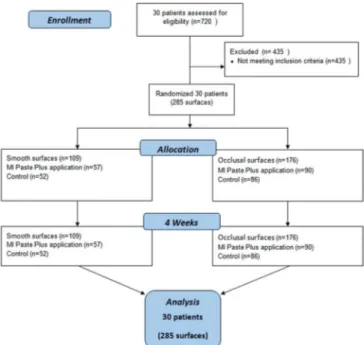
Thirty healthy volunteers (14 female and 16 male) aged 18–30 years with white spot lesions on tooth smooth surfaces and fissures were included in the study. Patients who are allergic to milk protein were excluded [Figure 1].
Visual examination and DIAGNOdent measurements
At the beginning of the investigation, teeth were categorized according to the International Caries Detection and Assessment System II (ICDAS II) criteria (www.icdas.org) by consensus between two trained and calibrated examiners (a and b) and at a later
date, the two examiners were trained and calibrated to the use of the laser fluorescence device (c and d). Kappa statistics were obtained for assessment of interobserver agreement by using 20 extracted teeth. Both examiners were encouraged to carefully re‑measure the points with maximal readings. A high level of agreement between the observers was obtained (Kappa score = 0.90). In the cases involving a disagreement between the results, the evaluation was repeated at different time intervals and the observers reached a consensus by consultation. The maximum reading for an ICDASII index score for each surface was recorded. DIAGNOdent measurements were repeated until 3 readings were within ±3 units of each other, and then recorded for that surface.
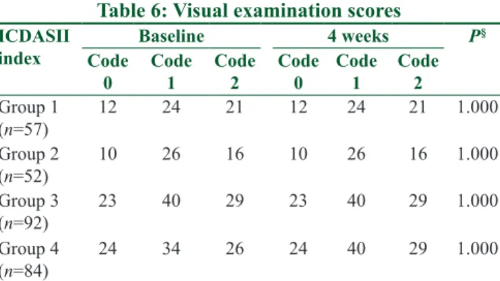
Visual examination was performed with the ICDASII index. Tooth surfaces were prolonged air‑drying (>5 s). Tooth surfaces, which were classified 0, 1, and 2 according to the ICDASII index codes, were included in the study [Table 1].
Laser fluorescence examinations were measured quantitatively using the DIAGNOdent device (Type 2095, Sn 05‑1519775; Kavo, Biberach, Germany). The teeth were isolated with sterile cotton rolls and the surfaces of the teeth were dried with an air spray. The device was calibrated with a ceramic standard, after which the fluorescence of a sound spot on the smooth surface of the tooth was measured to provide a baseline value; this value was then subtracted electronically from the fluorescence of the site to be measured. Finally, the tip of the laser device was placed on the white spot lesions and rotated around a vertical axis until the highest fluorescence reading was obtained. Laser fluorescence examination results were scored using the manufacturer’s scoring system [Table 2]. Teeth that were scored 1 and 2 were included in the study.
Treatment procedures: Volunteers with lesions that showed visual examination scores of 0, 1, and 2 and DIAGNOdent scores of 1 and 2 on smooth surfaces (n = 109) and on occlusal surfaces (n = 176) were randomly divided into two subgroups as the control group and study group [Table 3]. Both groups were educated on oral hygiene and motivated to brush their teeth twice a day with the same tooth paste (Colgate Total; Colgate‑Palmolive Technology Center, Piscataway, NJ, USA; 1450 ppm F) and tooth brush (Oral‑B® Advantage Complete Whole Mouth Clean).
In the study groups (Group 1 and 3), teeth were isolated with sterile cotton rolls and the surfaces of the teeth were dried with an air spray. CPP‑ACFP (10% CPP‑ACP plus 900 ppm fluoride crème, MI Paste Plus, GC America, Alsip, IL, USA) was applied 4 min/day for 4 weeks
on the existing enamel caries lesions by researchers. Volunteers were asked to avoid additional fluoride‑based preventive measures during the experimental period. After the 4‑week experimental period, values for the white spot lesions were again recorded using the DIAGNOdent device and the changes in the values and scores were recorded.
In the control groups (Group 2 and 4), CCP‑ACFP was not applied. The volunteers only brushed their teeth twice a day with the provided tooth paste and tooth brush. Volunteers were asked to avoid additional fluoride‑based preventive measures during the experimental period. After the 4‑week experimental period, values for the white spot lesions were again recorded using the DIAGNOdent device and the changes in the values and scores were recorded.
Statistical methods
Recorded data were statistically analyzed by the Wilcoxon signed‑rank test and Kruskal Mann– Whitney U‑test. All data were processed by SPSS software (version 19.0; SPSS Inc., Chicago, IL, USA). The differences in the groups at baseline and the 4‑week interval were calculated by the Wilcoxon signed‑rank test. The Mann–Whitney U‑test was used to determine statistically significant differences between both the groups at baseline and the 4‑week interval. The level of significance for all the tests was set at 5%.
r
esultsComparison of DIAGNOdent values evaluated before and after the application of CPP‑ACFP exhibited
Table 1: International Caries Detection and Assessment System II index
Code Description
0 Sound
1 First visual change in enamel (seen only after
prolonged air drying or restricted to within the confines of a pit or fissure)
2 Distinct visual change in enamel
3 Localized enamel breakdown (without clinical visual
signs of dentinal involvement)
4 Underlying dark shadow from dentin
5 Distinct cavity with visible dentin
6 Extensive distinct cavity with visible dentin
Table 4: Mean values and standard deviation of baseline and 4-week scores in the smooth surfaces group
Smooth surfaces Mean±SD P§ Z
Baseline 4 weeks
Group 1: Study group 5.13±2.77 2.82±2.77 <0.001 <0.001 Group 2: Control
group 6.62±3.63 6.75±3.42 >0.05
§Wilcoxon signed‑rank test; Z=Mann–Whitney U‑test. SD=Standard
deviation
Table 5: Mean values and standard deviation of baseline and 4-week scores in the occlusal surfaces group
Occlusal surfaces Mean±SD P§ Z
Baseline 4 weeks
Group 3: Study group 8.83±4.53 3.22±2.85 <0.001 <0.001 Group 4: Control
group 9.81±5.04 10.22±5.14 >0.05
§Wilcoxon signed‑rank test; Z=Mann–Whitney U‑test. SD=Standard
deviation
Table 6: Visual examination scores
ICDASII
index Baseline 4 weeks P
§ Code
0 Code 1 Code 2 Code 0 Code 1 Code 2 Group 1 (n=57) 12 24 21 12 24 21 1.000 Group 2 (n=52) 10 26 16 10 26 16 1.000 Group 3 (n=92) 23 40 29 23 40 29 1.000 Group 4 (n=84) 24 34 26 24 40 29 1.000 §Wilcoxon signed ranks. ICDAS II=International Caries Detection
and Assessment System II
Table 2: Laser fluorescence scoring system
Score Description
Score 1 Laser fluorescence score 0‑4, no caries or white opaque lesions
Score 2 Laser fluorescence score 5‑10, enamel caries limited to the outer half of the enamel thickness
Score 3 Laser fluorescence score 11‑20, enamel caries limited to the inner half of the enamel thickness without obvious spread in the dentin
Score 4 Laser fluorescence score ≥21, caries spread in the dentin
Table 3: Treatment groups
Groups
and n Surfaces Application prosedure Group 1
(n=57) MI Paste Plus on smooth
surfaces
MI Paste Plus application 4 min/ day for 4 weeks + tooth brushing 2 times with 1450 ppm fluoride toothpaste
Group 2
(n=52) Control on smooth surfaces Tooth brushing 2 times with 1450 ppm fluoride toothpaste
Group 3
(n=92) MI Paste Plus on occlusal
surfaces
MI Paste Plus application 4 min/ day for 4 weeks + tooth brushing 2 times with 1450 ppm fluoride toothpaste Group 4 (n=84) Control on occlusal surfaces
Tooth brushing 2 times with 1450 ppm fluoride toothpaste
remineralization of enamel caries lesions on smooth surfaces (Group 1) (P < 0.001) and occlusal surfaces (Group 3) (P < 0.001). In the control groups (Group 2 and 4), no quantitative changes were found in the enamel caries lesions on the smooth surfaces and in fissures at the end of 4 weeks (P > 0.05). The changes in the DIAGNOdent values for the smooth surfaces (Group 1 and 2) at the end of the study period showed a statistically significant difference between the control and study groups (P < 0.001) [Table 4]. Similarly, the changes in the DIAGNOdent values for the occlusal surfaces (Group 3 and 4) at the end of the study period
also showed a statistically significant difference between the control and study groups (P < 0.001) [Table 5]. None of the groups showed progression in the visual examination (P = 1.000) [Table 6].
d
IscussIonThis study evaluated the remineralization efficiency of a 4‑week chair side application of CPP‑ACFP on white spot lesions in comparison with a control group. White spot lesions could have an unreachable surface for the remineralization and demineralization process. These lesions are considered the earliest phase of the caries process and they are reversible. These lesions can be treated by conventional approaches, but these have the disadvantage of being invasive.[32] It has been reported that occlusal caries were more easily detected than proximal caries by visual examination;[33] therefore, for better identification and reproducibility, lesions in the occlusal and smooth surfaces were selected for this study.
In this study, a laser fluorescence system was used to detect the white spot lesions with visual inspection. Visual clinical inspection at occlusal sites provides insufficient sensitivity in in vivo conditions. On the other hand, the laser fluorescence‑based DIAGNOdent device helps measure early demineralization and provides good to excellent sensitivity.[27] Healthy dental substance and diseased dental substance have different fluorescence. The organic and inorganic materials existing on the tooth surface absorb the laser light and emit fluorescence in the infrared region of the spectrum. The presence of a demineralized area increases the fluorescence. DIAGNOdent device makes an audible sound that indicating the fluorescence increase. Higher frequency of sound and higher‑pitched sound indicate greater demineralization.[3]
Many remineralization agents can be used to promote remineralization by ionic exchange mechanism. In this study, CPP‑ACFP (MI Paste Plus) product is used. CPP‑ACP and fluoride have been reported to have the potential in promoting remineralization[13,14] and CPP‑ACFP buffers free calcium, phosphate, and fluoride at a supersaturated status and preserves them in close proximity to the enamel lesion, thereby decreasing demineralization and enhancing remineralization of enamel lesions.[34]
The mean and standard deviation were calculated and tests of significance were performed for each group. Comparison of the mean DIAGNOdent value of Group 2 (control group of smooth surfaces; 6.75 ± 3.42) with its baseline DIAGNOdent value (6.63 ± 3.63) shows that there were no quantitative changes. On comparing the DIAGNOdent value of Group 1 (study group of smooth surfaces; 2.82 ± 2.77) to its baseline DIAGNOdent value (5.13 ± 2.77), it was evident that a significant amount of remineralization had occurred. On comparing Group 1 and 2, the former showed a significantly higher amount of remineralization. Comparison of the mean DIAGNOdent value of Group 4 (control group of occlusal surfaces; 10.22 ± 5.14) with its baseline DIAGNOdent value (9.81 ± 5.04) showed that there were no quantitative changes. On comparing the DIAGNOdent value of Group 3 (study group of occlusal surfaces; 3.22 ± 2.85) to its baseline DIAGNOdent value (8.83 ± 4.53), it was evident that a significant amount of remineralization had occurred. On comparing Group 3 and 4, the former showed a significantly higher amount of remineralization. For all groups, there were no progression in the visual examination (P = 1.000). The results of this study showed that the application of CPP‑ACFP on enamel occlusal and smooth surfaces
was able to prevent demineralization of enamel, thereby suggesting its use in the prevention of white spot lesions. It has been reported that either directly or indirectly, casein is able to buffer plaque acid through bacterial catabolism.[35] The buffer capacity of the agent could explain the aforementioned preventive demineralization behavior. According to a previous study, this agent releases basic amino acids, which are able to accept proton ions.[36] Rahiotis and Vougiouklakis made another possible explanation mechanism which is the presence of the agent on enamel surface acts as inert barrier preventing the diffusion of protons. According to them, CPP‑ACP has ability to release calcium and this is likely to depress demineralization.[36]
The results of this study have also demonstrated that CPP‑ACFP enhances the remineralization of white spot lesions. This is consistent with a previous in vitro remineralization study, which showed that the combination of fluoride toothpaste and CPP‑ACP (Tooth Mousse) improves the remineralization effect.[37] CPPs have the ability to stabilize calcium phosphate on tooth surface, maintaining thereby high concentration gradients of calcium and phosphate ions, promoting thus remineralization of hard tissues.[36]
Krithikadatta et al. used CPP‑ACP alone or with fluoride for the remineralization of occlusal white spot lesions and used visual examination and DIAGNOdent device to determine them.[31] They instructed the volunteers to use the remineralizing agent twice a day for 4 weeks. Volunteers applied the cream‑based agent to smear a peanut‑sized dab of cream on all the surfaces of all teeth. In our study, the participants applied CPP‑ACFP on white spot lesions only once per day for 4 weeks.
In this study, 4 weeks after the application of CPP‑ACFP, DIAGNOdent measurements exhibited a significant decrease in Groups 1 and 3 in comparison with the control Groups 2 and 4. The decrease in DIAGNOdent values of Groups 1 and 3 might also be explained by the changes in the surface properties of white spot lesions due to CPP‑ACFP application. On the other hand, there were no changes in the visual examination scores. We think that CPP‑ACFP first stabilizes calcium phosphate on the surface zone of the enamel caries, and day by day it penetrates into the body of the lesion and dark zone. It will take a long approach to penetrate the inner parts of the lesion, especially the dark zone, which gives caries lesion the ability to detect by visual examination. CPP‑ACP is reported to have topical anticariogenic effects because of its ability to stabilize calcium and phosphate in an amorphous state, preventing the growth of calcium phosphate to the critical size required for
precipitation.[13,38,39] The usage of CPP‑ACP might have taken place to form new hydroxyapatite crystals and/ or with fluoride new fluorapatite or fluorhydroxyapatite crystals. They might have formed from the minerals existing in the paste by physical diffusion of the ions to the destroyed hydroxyapatite area. In a previous study, CPP‑ACP and fluoride were found more effective than CPP‑ACP alone.[40] According to Elsayad et al., the addition of fluoride to CPP‑ACP could give a synergistic effect on enamel remineralization.[41] Kumar et al.[42] indicated that CPP‑ACP remineralized initial enamel lesions and showed a higher remineralizing potential when applied as a topical coating after the use of fluoridated toothpaste.
CPP‑ACP has been shown to slow the progression of caries significantly and to promote the regression of early lesions in randomized, controlled clinical trials.[43‑45] Systematic review with meta‑analysis studies also indicate that CPP‑ACP has a short‑term remineralization effect.[46] According to our results, CPP‑ACFP could be suggested as a preventive system against demineralization of early enamel lesions on occlusal and smooth surfaces.
c
onclusIonsIt can be concluded that within the limitations of this study, CPP‑ACFP had a high remineralization effect after 4 min/day application for 4 weeks. Longer observation period with an enlarged sample size is recommended to confirm whether the greater remineralization in early caries lesions is maintained.
Financial support and sponsorship
Nil.
Conflicts of interest
There are no conflicts of interest.
r
eferences1. Aimutis WR. Bioactive properties of milk proteins with particular focus on anticariogenesis. J Nutr 2004;134:989S‑95S. 2. Armfield J, Roberts‑Thomson K, Spencer A. Australia's Health
2000: The Seventh Biennial Health Report of the Australian Institute of Health and Welfare. Canberra: Australian Institute of Health and Welfare; 2000.
3. Jayarajan J, Janardhanam P, Jayakumar P; Deepika. Efficacy of CPP‑ACP and CPP‑ACPF on enamel remineralization – An in vitro study using scanning electron microscope and DIAGNOdent. Indian J Dent Res 2011;22:77‑82.
4. Mitchell L. Decalcification during orthodontic treatment with fixed appliances – An overview. Br J Orthod 1992;19:199‑205. 5. Millett DT, Nunn JH, Welbury RR, Gordon PH. Decalcification
in relation to brackets bonded with glass ionomer cement or a resin adhesive. Angle Orthod 1999;69:65‑70.
6. Derks A, Katsaros C, Frencken JE, van’t Hof MA, Kuijpers‑Jagtman AM. Caries‑inhibiting effect of preventive measures during orthodontic treatment with fixed appliances.
A systematic review. Caries Res 2004;38:413‑20.
7. de Bruyn H, Arends J. Fluoride varnishes – A review. J Biol Buccale 1987;15:71‑82.
8. Seppä L, Leppänen T, Hausen H. Fluoride varnish versus acidulated phosphate fluoride gel: A 3‑year clinical trial. Caries Res 1995;29:327‑30.
9. Artun J, Thylstrup A. A 3‑year clinical and SEM study of surface changes of carious enamel lesions after inactivation. Am J Orthod Dentofacial Orthop 1989;95:327‑33.
10. Papas A, Russell D, Singh M, Stack K, Kent R, Triol C, et al. Double blind clinical trial of a remineralizing dentifrice in the prevention of caries in a radiation therapy population. Gerodontology 1999;16:2‑10.
11. Reynolds EC. The prevention of sub‑surface demineralization of bovine enamel and change in plaque composition by casein in an intra‑oral model. J Dent Res 1987;66:1120‑7.
12. Reynolds EC, Cain CJ, Webber FL, Black CL, Riley PF, Johnson IH, et al. Anticariogenicity of calcium phosphate complexes of tryptic casein phosphopeptides in the rat. J Dent Res 1995;74:1272‑9.
13. Reynolds EC. Remineralization of enamel subsurface lesions by casein phosphopeptide‑stabilized calcium phosphate solutions. J Dent Res 1997;76:1587‑95.
14. Reynolds EC, Cai F, Shen P, Walker GD. Retention in plaque and remineralization of enamel lesions by various forms of calcium in a mouthrinse or sugar‑free chewing gum. J Dent Res 2003;82:206‑11.
15. Rose RK. Binding characteristics of Streptococcus mutans for calcium and casein phosphopeptide. Caries Res 2000;34:427‑31. 16. Llena C, Forner L, Baca P. Anticariogenicity of casein
phosphopeptide‑amorphous calcium phosphate: A review of the literature. J Contemp Dent Pract 2009;10:1‑9.
17. Neuhaus KW, Lussi A. Casein phosphopeptide – Amorphous calcium phosphate (CPP‑ACP) and its effect on dental hard tissues. Schweiz Monatsschr Zahnmed 2009;119:110‑6.
18. Pai D, Bhat SS, Taranath A, Sargod S, Pai VM. Use of laser fluorescence and scanning electron microscope to evaluate remineralization of incipient enamel lesions remineralized by topical application of casein phospho peptide amorphous calcium phosphate (CPP‑aCP) containing cream. J Clin Pediatr Dent 2008;32:201‑6.
19. Hicks MJ, Flaitz CM. Enamel caries formation and lesion progression with a fluoride dentifrice and a calcium‑phosphate containing fluoride dentifrice: A polarized light microscopic study. ASDC J Dent Child 2000;67:21‑8.
20. Chin MY, Sandham A, Rumachik EN, Ruben JL, Huysmans MC. Fluoride release and cariostatic potential of orthodontic adhesives with and without daily fluoride rinsing. Am J Orthod Dentofacial Orthop 2009;136:547‑53.
21. Magalhães AC, Wiegand A, Rios D, Honório HM, Buzalaf MA. Insights into preventive measures for dental erosion. J Appl Oral Sci 2009;17:75‑86.
22. Benson PE, Shah AA, Millett DT, Dyer F, Parkin N, Vine RS. Fluorides, orthodontics and demineralization: A systematic review. J Orthod 2005;32:102‑14.
23. Lussi A, Imwinkelried S, Pitts N, Longbottom C, Reich E. Performance and reproducibility of a laser fluorescence system for detection of occlusal caries in vitro. Caries Res 1999;33:261‑6.
24. Kudiyirickal MG, Ivancaková R. Early enamel lesion part I. Classification and detection. Acta Medica (Hradec Kralove) 2008;51:145‑9.
for caries detection. J Dent Res 2004;83 Spec No C: C80‑3. 26. Braun A, Graefen O, Nolden R, Frentzen M. Comparative
study of conventional caries diagnosis versus laser fluorescence measurements. Dtsch Zahnärztl Z 2000;55:248‑51.
27. Lussi A, Megert B, Longbottom C, Reich E, Francescut P. Clinical performance of a laser fluorescence device for detection of occlusal caries lesions. Eur J Oral Sci 2001;109:14‑9.
28. Tyas MJ, Anusavice KJ, Frencken JE, Mount GJ. Minimal intervention dentistry – A review. FDI Commission Project 1‑97. Int Dent J 2000;50:1‑12.
29. Burke FJ. From extension for prevention to prevention of extension: Minimal intervention dentistry. Dent Update 2003;30:492‑8. 30. Vashisht R, Indira R, Ramachandran S, Kumar A, Srinivasan MR.
Role of casein phosphopeptide amorphous calcium phosphate in remineralization of white spot lesions and inhibition of Streptococcus mutans? J Conserv Dent 2013;16:342‑6.
31. Krithikadatta J, Fredrick C, Abarajithan M, Kandaswamy D. Remineralisation of occlusal white spot lesion with a combination of 10% CPP‑ACP and 0.2% sodium fluoride evaluated using diagnodent: A pilot study. Oral Health Prev Dent 2013;11:191‑6. 32. Melrose CA, Appleton J, Lovius BB. A scanning electron
microscopic study of early enamel caries formed in vivo beneath orthodontic bands. Br J Orthod 1996;23:43‑7.
33. Hopcraft MS, Morgan MV. Comparison of radiographic and clinical diagnosis of approximal and occlusal dental caries in a young adult population. Community Dent Oral Epidemiol 2005;33:212‑8.
34. Hegde MN, Moany A. Remineralization of enamel subsurface lesions with casein phosphopeptide‑amorphous calcium phosphate: A quantitative energy dispersive x‑ray analysis using scanning electron microscopy: An in vitro study. J Conserv Dent 2012;15:61‑7.
35. Reynolds EC, del Rio A. Effect of casein and whey‑protein solutions on caries experience and feeding patterns of the rat. Arch Oral Biol 1984;29:927‑33.
36. Rahiotis C, Vougiouklakis G. Effect of a CPP‑ACP agent on the demineralization and remineralization of dentine in vitro. J Dent 2007;35:695‑8.
37. Wu G, Liu X, Hou Y. Analysis of the effect of CPP‑ACP tooth mousse on enamel remineralization by circularly polarized images. Angle Orthod 2010;80:933‑8.
38. Rose RK. Effects of an anticariogenic casein phosphopeptide on calcium diffusion in streptococcal model dental plaques. Arch Oral Biol 2000;45:569‑75.
39. Reynolds EC, Johnson IH. Effect of milk on caries incidence and bacterial composition of dental plaque in the rat. Arch Oral Biol 1981;26:445‑51.
40. Patil N, Choudhari S, Kulkarni S, Joshi SR. Comparative evaluation of remineralizing potential of three agents on artificially demineralized human enamel: An in vitro study. J Conserv Dent 2013;16:116‑20.
41. Elsayad I, Sakr A, Badr Y. Combining casein phosphopeptide‑amorphous calcium phosphate with fluoride: Synergistic remineralization potential of artificially demineralized enamel or not? J Biomed Opt 2009;14:39‑44.
42. Kumar VL, Itthagarun A, King NM. The effect of casein phosphopeptide‑amorphous calcium phosphate on remineralization of artificial caries‑like lesions: An in vitro study. Aust Dent J 2008;53:34‑40.
43. Bailey DL, Adams GG, Tsao CE, Hyslop A, Escobar K, Manton DJ, et al. Regression of post‑orthodontic lesions by a remineralizing cream. J Dent Res 2009;88:1148‑53.
44. Walker G, Cai F, Shen P, Reynolds C, Ward B, Fone C, et al. Increased remineralization of tooth enamel by milk containing added casein phosphopeptide‑amorphous calcium phosphate. J Dairy Res 2006;73:74‑8.
45. Reynolds EC. Casein phosphopeptide‑amorphous calcium phosphate: The scientific evidence. Adv Dent Res 2009;21:25‑9. 46. Yengopal V, Mickenautsch S. Caries preventive effect of casein
phosphopeptide‑amorphous calcium phosphate (CPPACP): A meta‑analysis. Acta Odontol Scand 2009;21:1‑12.