© 2017 Journal of Cytology | Indian Academy of Cytologists | Published by Wolters Kluwer - Medknow Zeynep Tosuner, İlknur Türkmen1, Sema Arici, Cavide Sönmez, Seval Turna, Öykü Onaran
Department of Pathology, Faculty of Medicine, Bezmialem Foundation University, 1Department of Pathology, Faculty of Medicine, Medipol University, İstanbul, Turkey
Address for correspondence: Dr. Zeynep Tosuner, Department of Pathology, Faculty of Medicine, Bezmialem Vakıf University, Vatan Street, 34093 Fatih, İstanbul, Turkey. E‑mail: [email protected]
ABSTRACT
Aims: We aimed to investigate the immunocytoexpression profiles of a novel assay ProEx C for topoisomerase II
alpha (TOP2A) and minichromosome maintenance protein 2 (MCM2) in abnormal interpreted smears.
Settings and Design: Screening programs with Papanicolaou smear and high risk group human papilloma virus testing
have yielded a dramatic reduction of cervical cancer incidence. However, both of these tests have limited specificity for the detection of clinically significant cervical high grade lesions. ProEx C for topoisomerase II alpha (TOP2A) and minichromosome maintenance protein 2 (MCM2) has been considered to have tight association with high grade intraepithelial lesions.
Materials and Methods: A total number of 54 SurePath cervical cytology specimens of patients previously interpreted as
atypical squamous cells–undetermined significance (ASC‑US), atypical squamous cells–cannot exclude high grade squamous intraepithelial lesion (ASC‑H), low grade squamous intraepithelial lesion (LSIL), and high grade squamous intraepithelial lesion (HSIL) were included in our study.
Results and Conclusions: ProEx C was positive in 14 of HSILs (100%), 3 of 19 LSILs (16%), 2 of 4 ASC‑Hs, and none of
ASC‑USs (0%). The ProEx C test showed very intense nuclear staining in all cytologically abnormal cells. Further studies are indicated to evaluate the diagnostic role of ProEx C.
Key words: ASC‑H; ASC‑US; HSIL; immunocytochemistry; LSIL; ProEx C
Introduction
Cervix cancer is the second most common type of cancer in developing countries and has a high risk of mortality.[1]
Screening strategies by the Papanicolaou (Pap) smear method have provided significant reduction in the incidence of cervical cancer.[2] However, sensitivity of a single Pap test is
only 50% for the detection of current disease.[3]
According to the ASCUS-LSIL Triage Study (ALTS) by Schiffman et al., in the US, each year more than 3 million cases are
diagnosed as atypical squamous cells–undetermined significance (ASC-US), atypical squamous cells–cannot exclude high grade squamous intraepithelial lesion (ASC-H), low grade squamous intraepithelial lesion (LSIL), or atypical glandular cells (AGC) that require further evaluation such as colposcopic biopsy to identify clinically significant high grade lesions (CIN-2/3 or carcinoma). Nevertheless, according to this study, further examination does not reveal a high-grade lesion in most of the cases.[4]
Immunocytoexpression profile of ProExC in smears interpreted
as ASC‑US, ASC‑H, and cervical intraepithelial lesion
Access this article online Website:
www.jcytol.org
Quick Response Code
DOI:
10.4103/0970‑9371.197605
This is an open access article distributed under the terms of the Creative Commons Attribution‑NonCommercial‑ShareAlike 3.0 License, which allows others to remix, tweak, and build upon the work non‑commercially, as long as the author is credited and the new creations are licensed under the identical terms.
For reprints contact: [email protected]
How to cite this article: Tosuner Z, Türkmen İ, Arıcı S, Sönmez C, Turna S, Onaran Ö. Immunocytoexpression profile of ProExC in smears interpreted as ASC‑US, ASC‑H, and cervical intraepithelial lesion. J Cytol 2017;34:34‑8.
Journal of Cytology
Human papillomavirus (HPV) is involved in the etiopathogenesis of cervix carcinomas and LSILs.[5,6]
“High risk” group HPV types include HPV types 16, 18, 26, 31, 33, 35, 39, 45, 51, 52, 53, 56, 58, 59, 66, and 68 that are associated with higher risk of malignant transformation and can be detected from the liquid-based cervicovaginal material. Identification of high-risk HPV types in patients diagnosed as ASC-US and LSIL determines the risk potential of these patients. However, specificity of HPV DNA test in detecting high grade squamous intraepithelial lesion (HSIL) is low.[7] Therefore, search for other useful molecular markers
in the interpretation of high grade intraepithelial lesions have been considered in order to improve clinical management. Minichromosome maintenance protein 2 (MCM2) and topoisomerase II-α (TOP2A) are two biomarkers that have been identified by DNA microarray. Both of them are involved in DNA replication. BD ProEx C (BD Diagnostics-Tripath, Burlington, NC) is a new immunohistochemical marker
including these two monoclonal antibodies.[8,9] TOP2A and
MCM2 gene expressions have been considered to be high in cervical carcinoma.[10-12]
TOP2A is an enzyme responsible for unlinking DNA strands during replication. MCM2 is a protein that is involved in the G1 phase of the cell cycle and maintains DNA synthesis via loading the prereplication complex onto DNA; it has helicase activity resulting in the unwinding of DNA.[13]
Limited number of studies concerning ProEx C have been reported. These studies showed that ProEx C assay in liquid–based cytology yielded a high sensitivity for
biopsy-proven HSIL.[8,14] Diffuse ProEx C expression has
also been reported in head and neck tumors, especially
in respiratory squamous cell carcinomas.[15] In addition,
strong ProEx C staining exhibited very high sensitivity and specificity for distinguishing normal/reactive hyperplasia from esophageal squamous intraepithelial neoplasia.[16] ProEx C
immunoexpression has also been found to be significantly
higher in melanomas compared to benign nevi.[17]
In the current study, we aimed to investigate the staining status of ProEx C in liquid-based smears, which were interpreted previously as ASC-US, ASC-H, LSIL, and HSIL.
Materials and Methods
We used 54 residual cervicovaginal cytology samples prepared by BD SurePath autostainer which were previously interpreted as ASC-US, ASC-H, LSIL, and HSIL between
years 2013 and 2014. Case selection was performed by two pathologists from the two different institutes. Full agreement of diagnostic categorization by both of the observers and having a sufficient residual sample for preperation were used as inclusion criteria. Cases that fulfilled the two criteria were included in the study. All specimens had been stored at +4°C at refrigerator for less than 1 year.
A thin-layer slide was processed from the residual BD SurePathTM vial using the BD PrepstainTM instrument. The
slides were treated with a pretreatment buffer for target retrieval using the BD SurePath slide preperation buffer (BD Diagnostics-Tripath). Immunocytochemical staining was performed with BD ProEx C using a detection reagent kit that includes prediluted antibody, a 3,3’-diaminobenzidine tetrahydrochloride-based chromogen, and hematoxylin-based counterstains (BD SurePath detection reagents and BD SureDetect counterstains), and an automated staining platform (Ventana Autostainer). The slides were than coverslipped and reviewed by the two pathologists.
A three-step algorithm was used for the interpretation of the slides.
1. The first step was to determine the adequacy of the specimen according to the 2001 Bethesda criteria. Then, the immunostained slide was compared with the Pap-stained original slide and examined for the existence of abnormal cells
2. The second step was to determine if there was dark brown nuclear staining in squamous cells. In some slides, a mild background staining was observed due to mucus. This type of staining was interpreted as negative 3. The third and the last step was to determine if these
ProEx C stained cells were abnormal using the diagnostic criteria of ASC-US, ASC-H, and low or high grade squamous intraepithelial lesions. In some slides, a light immunostaining was observed in occasional nuclei of glandular cells and tubal metaplasia. This type of staining was also interpreted as negative. If all three criteria were met, the slide was interpreted as positive.
Results
In this study, a total of 54 patients with abnormal cytology interpretation results, including HSIL (n = 14), LSIL (n: 19), ASC-US (n = 17), and ASC-H (n = 4) were studied. All of the 14 cases interpreted as HSIL were scored as positive by ProEx C (100%) [Figure 1].
Three cases interpreted as LSIL (3/19) were scored as positive by ProEx C [Figure 2] whereas 16 were negative (84%).
Journal of Cytology
No significant expression was observed in any of the 17 smears (0/17), which were interpreted as ASC-US (0%). Two of the smears interpreted as ASC-H (2/4) showed immunoreactivity by ProEx C (50%) [Table 1].
Ten colposcopic biopsy materials were available out of the 14 cases, which were previously interpreted as HSIL on cytology (10/14). No follow-up data was available in the remaining 4 cases. Five cases were reported as HSIL and the other cases were diagnosed as LSIL (5/10).
Of the 19 patients whose smears had been interpreted as LSIL, 18 had consequitive colposcopic biopsy materials. Nine cases resulted as low grade intraepithelial lesions; 8 LSILs and 1 vaginal intraepithelial neoplasia-1 (VAIN-1), whereas nine cases resulted as HSIL. Only 6 biopsy materials were present out of 17 cases, which were previously reported as ASC-US (6/17) on Pap-test. The biopsy results of these cases were LSIL (n = 4), endocervical polyp (n = 1), and chronic cervicitis (n = 1).
Three cases which were interpreted as LSIL on cytology showed immunoreactivity with ProEx C. The biopsy results of these cases were HSIL (1/3), VAIN-1 (1/3) and LSIL (1/3) seperately [Table 2].
The positive immunostaining occured in cells reflecting the atypia of at least low grade squamous intraepithelial lesion criteria. Ten cases were interpreted as HSIL and showed immunoreactivity by ProEx C. The immunoreactive cells were highly atypical and fullfilling the criteria of HSIL cytology. The biopsy results of these cases were HSIL (n = 5) and LSIL (n = 5).
Three of 4 ASC-H cases had colposcopic biopsies. One case that showed immunoreactivity by ProEx C was diagnosed as HSIL on biopsy material. Follow-up data was not available for the other positively immunostained case. The cases which were negative by ProEx C were resulted as LSIL (n = 1) and HSIL (n = 1) [Table 2].
ProEx C staining features were similar in biopsy confirmed HSIL and LSIL interpreted smears. Homogeneous dark brown nuclear staining was observed and intense staining was restricted in the nuclei of atypical cells in both HSIL and LSIL categories. No specific nuclear staining was present in normal appearing epithelial cells. On the other hand, no precipitate was observed in the nuclei of unstained cells. In addition, after cytologic examination, the biopsy materials from the same patients and normal cervical control tissues were also immunostained with ProEx C. Observation of similar nuclear staining features in the biopsies with low and high-grade squamous intraepithelial lesions provided the exclusion of storage changes.
Basal cell layer staining was observed in normal cervical epithelium. Lower one-third to full thickness epithelium Table 1: Distribution of the cases according to ProEx C
immunoexpression and cytology interpretations
Pap Smear Interpretation Category LSIL HSIL ASC-H ASC-US
ProExC immunoexpression n/Total 3/19 14/14 2/4 0/17
% 16 100 50 0
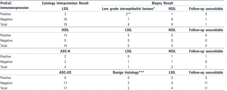
Table 2: Distribution of cases according to ProEx C immunoexpression and correlation of cytology interpretation and biopsy results of the cases
ProExC
immunoexpression Cytology Interpretation ResultLSIL Low grade intraepithelial lesions*Biopsy ResultHSIL Follow-up unavailable
Positive 3 2** 1 0
Negative 16 7 8 1
Total 19 9 9 1
HSIL LSIL HSIL Follow-up unavailable
Positive 14 5 5 4
Negative 0 0 0 0
Total 14 5 5 4
ASC-H LSIL HSIL Follow-up unavailable
Positive 2 0 1 1
Negative 2 1 1 0
Total 4 1 2 1
ASC-US Benign histology*** LSIL Follow-up unavailable
Positive 0 0 0 0
Negative 17 2 4 11
Total 17 2 4 11
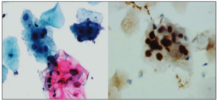
Journal of Cytology Figure 1: Left: A case interpreted as HSIL (Pap, x200), inset: Crowded group of atypical cells (Pap, ×400); Right: Positive ProExC staining in the same case (ProExC, x100) Figure 2: Left: A case interpreted as LSIL (Pap, ×200); Right: Positive ProExC staining in the same case (ProExC, x100)
staining patterns were observed in LSIL [Figure 3] and HSIL lesions [Figure 4].
Overall sensitivity of a ProEx C test for detecting an intraepithelial lesion was 40% whereas specificity was 100%. Because all of the HSIL interpreted Pap-smears resulted as intraepithelial lesions in biopsies, sensitivity of ProEx C for detecting an intraepithelial lesion was 100% for HSIL interpretation category, seperately.
Discussion
All HSIL interpreted cases showed immunoreactivity by ProEx C (100%). This finding is similar to the study of Siddique et al., in which all of the ASC-H patients which were consequtively diagnosed as HSIL on biopsy had positive immunostaining by ProEx C.[18]
The staining pattern was invariably dark brown in color and highly intense without leaving any doubt, and all the stained cells were highly atypical and fulfilled the cytological HSIL criteria. Sixteen percent of the cases which were interpreted as LSIL showed immunoreactivity by ProEx C, and all positive cases
were diagnosed as at least low-grade squamous intraepithelial lesion on biopsy. We used the available residual samples of cervical cytology specimens for immunocytochemistry. During the case selection, some cases with intraepithelial lesion results could not be included in our study because their residual materials were insufficient because of reprocessing and additional Pap-stained slide preperation for diagnostic purposes. Larger series with high number of cases are necessary to reveal the predictive value of ProEx C for the detection of high grade lesions.
Artificial staining in the background of the smear due to mucus and focal mild level of staining in normal endocervical/ metaplastic cell component can be observed. Similar problems have been declared in two other studies based on ProEx C immunocytochemistry.[18,19] Therefore careful investigation
is essential for the detection of true abnormal cells. Hence, ProEx C stained slide should be simultaneously examined with the original Pap-smear preperate. Adequacy of the ProEx C stained slide was a limiting factor for the evaluation because repreperation was made using the remaining vials of the previously prepared smears by the Surepath method. Technical studies to decrease the nonspecific staining could help to improve the specificity of staining.
Figure 3: ProEx C immunoreactivity in lower one‑third of cervical epithelium
Journal of Cytology
All cases which were interpreted as ASC-US were negative by ProEx C. A significant majority of patients (65%) who were interpreted as ASC-US had no consequtive colposcopic biopsy and had lost to follow-up. Because the follow-up data and outcomes of these patients are unavailable, the negative ProEx C score correlation with diagnosis was impossible. Discordances were determined between cytology and biopsy results of the patients. Half of the cytologically interpreted LSIL cases were consecutively diagnosed as HSIL, whereas half of the cytologically interpreted HSIL cases were consequtively diagnosed as LSIL. Sampling errors could be possible indicators for these discordances. A sampling error in cervical brushing could explain how LSIL interpreted smears achieved a final diagnosis of HSIL. Another possible factor could be progression of the lesion (from LSIL to HSIL) while dealing with underdiagnosis of HSIL. All of the HSIL interpreted smears had fulfilled the criteria of HSIL. Therefore, sampling error of colposcopic biopsy could be an indicator for the LSIL biopsy results of the HSIL interpreted smears.
In the study of Shroyer et al., no immunoexpression was determined in 10 pooled samples of NIL smears.[14] Studies
investigating ProEx C immunoexpression status of a large number of smears interpreted as negative for intraepithelial lesion or malignancy (NIL), smears having reactive changes including atypical repair, and atrophic smears could help to evaluate the specificity of this test.
A limitation of our study is the lack of HPV DNA analysis. The reason of this limitation is the inadequacy of vial materials for HPV testing after the repreperations of ProEx C. Investigation of HPV DNA analysis combined with ProEx C expression in cervical smears and biopsies of patients with intraepithelial lesions could provide detailed information regarding cervical carcinogenesis.
Conclusion
Our study shows that ProEx C immunoexpression is evident in cases interpreted as HSIL in cytology. Studies with large number of cases would help to indicate the accurate sensitivity and specificity of this test. Nevertheless, regarding the high mortality rates of cervix carcinoma in developing countries, ProEx C immunocytochemistry could be used in cases suspicious for high grade lesions as a supportive method. Financial support and sponsorship
Nil.
Conflicts of interest
There are no conflicts of interest.
References 1 Parkin DM, Pisani P, Ferlay J. Global cancer statistics. CA Cancer J Clin 1999;49:33‑64. 2 Edwards BK, Brown ML, Wingo PA, Howe HL, Ward E, Ries LA, et al. Annual report to the nation on the status of cancer, 1975‑2002, featuring population‑based trends in cancer treatment. J Natl Cancer Inst 2005;97:1407‑27.
3 Nanda K, McCrory DC, Myers ER, Bastian LA, Hasselblad V, Hickey JD, et al. Accuracy of the Papanicolaou test in screening for and follow‑up of cervical cytologic abnormalities: A systematic review. Ann Intern Med 2000;132:810‑9. 4 Schiffman M, Solomon D. Findings to date from the ASCUS‑LSIL Triage Study (ALTS). Arch Pathol Lab Med 2003;127:946‑9. 5 Baak JP, Kruse AJ, Robboy SJ, Janssen EA, van Diermen B, Skaland I. Dynamic behavioural interpretation of cervical intraepithelial neoplasia with molecular biomarkers. J Clin Pathol 2006;59:1017‑28.
6 Syrjanen KJ. Immunohistochemistry in assessment of molecular pathogenesis of cervical carcinogenesis. Eur J Gynaecol Oncol 2005;26:5‑19.
7 Schiffman M, Khan MJ, Solomon D, Herrero R, Wacholder S, Hildesheim A, et al. A study of the impact of adding HPV types to cervical cancer screening and triage tests. J Natl Cancer Inst 2005;97:147‑50. 8 Kelly D, Kincaid E, Fansler Z, Rosenthal DL, Clark DP. Detection of cervical high‑grade squamous intraepithelial lesions from cytologic samples using a novel immunocytochemical assay (ProEx C). Cancer 2006;108:494‑500. 9 Malinowski DP. Molecular diagnostic assays for cervical neoplasia: Emerging markers for the detection of high‑grade cervical disease. Biotechniques 2005;Suppl: 17‑23
10 Murphy N, Ring M, Heffron CC, King, B, Killalea, AG, Hughes C, et al. p16INK4A, CDC6, and MCM5: Predictive biomarkers in cervical preinvasive neoplasia and cervical cancer. J Clin Pathol 2005;58:525‑34.
11 Chen Y, Miller C, Mosher R Zhao X, Deeds J, Morrissey M, et al. Identification of cervical cancer markers by cDNA and tissue microarrays. Cancer Res 2003;63:1927‑35. 12 Santin AD, Zhan F, Bignotti E, Siegel E, Cane S, Bellone S, et al. Gene expression profiles of primary HPV16‑ and HPV18‑infected early stage cervical cancers and normal cervical epithelium: Identification of novel candidate molecular markers for cervical cancer diagnosis and therapy. Virology 2005;331:269‑91. 13 Champoux JJ. DNA topoisomerases: Structure, function, and mechanism. Annu Rev Biochem 2001;70:369‑413.
14 Shroyer KR, Homer P, Heinz D, Singh M. Validation of a novel immunocytochemical assay for topoisomerase II‑alpha and minichromosome maintenance protein 2 expression in cervical cytology. Cancer 2006;108:324‑30.
15 Bhandarkar ND, Sims HS, David O. ProEx C stain analysis in recurrent respiratory papillomatosis. Ann Otol Rhinol Laryngol 2010;119:99‑104. 16 Wang WC, Wu TT, Chandan VS, Lohse CM, Zhang L. Ki‑67 and ProExC
are useful immunohistochemical markers in esophageal squamous intraepithelial neoplasia. Hum Pathol 2011;42:1430‑7. 17 Yan S, Coffing BN, Li Z, Xie H, Brennick JB, Beg HA, Froehlich HM, Wells WA. Diagnostic and Prognostic Value of ProEx C and GLUT1 in Melanocytic Lesions. Anticancer Res 2016;36:2871‑80. 18 Siddiqui MT, Cohen C, Nassar A. Detecting high‑grade cervical disease on ASC‑H cytology: Role of BD ProEx C and Digene Hybrid Capture II HPV DNA testing. Am J Clin Pathol 2008;130:765‑70.
19 Depuydt CE, Makar AP, Ruymbeke MJ, Benoy IH, Vereecken AJ, Bogers JJ. BD‑ProExC as adjunct molecular marker for improved detection of CIN2+ after HPV primary screening. Cancer Epidemiol Biomarkers Prev 2011;20:628‑37.