Summary
In this study, the effects of total aflatoxin (AF) given orally on silver-staining nucleolus organizer regions (AgNORs) activity were studied in glomerulus and tubular epithelial cells of kidney in Merino rams. In addition, this study was conducted in order to evaluate the efficacy of an esterified glucomannan (EG) for protection against to aflatoxicosis. As materials, 1 year-old 32 Merino rams were used. Rams were fed through the 92 days. Control group (C) fed with the commercial feed. AF group fed with commercial feed added 250 µg/day of total AF. EG group fed with commercial feed added 2 g/day of EG daily. AF+EG group fed with commercial feed added 250 µg/day of total AF and 2 g/day of EG. At the end of the 92nd day the animals were sacrificed, and tissue samples were taken from the kidneys. Whereas ratio of nuclear area of the AgNOR area of cells in examinated regions of kidney was found decreased significantly (P<0.05) in the AF group compared to the control group, AF+EG group was found similar to control group (P>0.05). In conclusion, the adverse effects causing by aflatoxicosis on the kidney AgNOR activity could be ameliorated by adding EG to the ration.
Keywords: Aflatoxin, AgNOR, Glucomannan, Kidney, Ram
Koçlarda Aflatoksinin Böbreğin Farklı Bölgelerindeki Hücrelerin
AgNOR Aktivileri Üzerine Etkileri ve Glukomannanın
Koruyucu Etkinliği
Özet
Bu çalışmada Merinos ırkı koçlara oral yolla verilen total aflatoksinin (AF) glomerulus ve böbrek tubuler epitel hücrelerindeki gümüşleme metoduyla çekirdekçik organizatör bölgelerinin (AgNORs) aktiviteleri üzerine olan etkileri ve AF ile birlikte verilen glukomannanın (EG) aflatoksikozun neden olduğu etkileri azaltıcı/engelleyici etkinliğinin belirlenmesi amaçlandı. Hayvan materyali olarak, 32 adet 1 yaşlı Merinos ırkı koç kullanıldı. Beslemeye 92 gün süreyle devam edildi. Kontrol (K) grubuna ticari yem, AF grubuna ticari yem ile günlük 250 µg AF, EG grubuna ticari yem ile günlük 2 g EG, AF+EG grubuna ise ticari yemle birlikte günlük 250 µg AF ve 2 g EG verildi. 92 günlük besleme periyodunu takiben hayvanlar kesildi. Böbreğin incelenen bölgelerindeki hücrelerin AgNOR alanının çekirdek alanına oranları AF grubunda kontrol grubuna göre önemli oranda düşük (P<0.05); AF+EG grubunda ise kontrol grubuna benzer (P>0.05) bulunmuştur. Sonuç olarak; elde edilen veriler, yemle birlikte alınan AF’ın böbrek AgNOR aktiviteleri üzerinde olumsuz etki oluşturabileceğini, bu etkilerin yeme EG ilave edilerek önemli oranda önlenebileceğini ortaya koymuştur.
Anahtar sözcükler: Aflatoksin, AgNOR, Böbrek, Glukomannan, Koç
Effects of Aflatoxin on AgNOR Activity of Cells in Different
Regions of Kidney, and Protective Effectiveness of Esterified
Glucomannan in Ram
[1] [2]Fatma COLAKOGLU
1Hasan Huseyin DONMEZ
1
[1] [2] 1
This study forms part of the PhD Thesis of Fatma KAYIKCI COLAKOGLU, for which we thank the “Selcuk University Scientific Research Projects (BAP) Coordinating Office”, Project no: 09202023, and TUBITAK, Project No: 107O866, for financial support We are grateful to Prof. Dr. Mustafa ORTATATLI for contributions
Department of Histology and Embryology, Faculty of Veterinary Medicine, University of Selcuk, TR-42075 Konya - TURKEY
Makale Kodu (Article Code): KVFD-2012-8176
One of the most important problems in human and
animal nutrition is contamination of human foods and animal feeds with molds and mycotoxins
1. Aflatoxins are a
group of mycotoxins produced by the strains of Aspergillus
INTRODUCTION
İletişim (Correspondence)
+90 332 2233619
[email protected]flavus and A. parasiticus 2. They are produced on cereal
grains during growth, harvest, storage, or transportation 3.
It is well known AFs are teratogenic 4, immuno-
suppression 5, mutagenic, and carcinogenic 6. There is
evidence that it has harmful effects on the kidneys, though not as much as on the liver 7.
Prevention of feed and feedstuffs from possible mold growth and AF contamination is very important. Practical and cost-effective methods for detoxification of AF containing feed and feedstuff are in great demand 8. Since the early
1990s, the adsorbent-based several studies have been performed to detoxify AF in contaminated food and foodstuffs and to minimize the deleterious effects of AF 9.
An approach to the problem has been the usage of non-nutritive and inert adsorbents in the diet to bind AF and reduce the absorption of AF from the gastrointestinal tract 10.
The non-nutritive clays such as aluminosilicates, zeolites, bentonites, and clinoptilolite were preferred by the researchers 5,11. Recent years, researchers suggested that
the best approach for decontamination would be biological degradation 12. Live yeast (Saccharomyces cerevisiae) initially
used as a performance promoter in the early 1990s, was found to have beneficial effects on aflatoxicosis 13. Esterified
glucomannan (EG) showed considerably high binding ability (80-97%) with AF 14, and it has been preferred for
detoxification of AF in poultry animals 6.
Nucleolar-organizer regions (NORs) are loops of DNA containing ribosomal RNA genes 15. These regions can be
easily stained with silver methods to appear as black dots (AgNORs) in the cell nucleus since they are argyrophilic. NORs are used by cytogeneticists for studying chromosomal disorders. This staining technique is very simple and does not require any special instruments or costly reagents 16.
Additionally, the size, number and dispersion of the silver deposits on the NOR reflect the degree of transcriptional, nucleolar and proliferative activity of the cells 17,18.
The aim of the study was to determine the effects of total AF given orally on AgNOR activity of cells in different regions of kidney in Merino rams. In addition, this study was conducted in order to evaluate the efficacy of EG for protection against to aflatoxicosis.
MATERIAL and METHODS
Animals and Diet
Approval for the present study was obtained from the Animal Ethics Committee of the Faculty of Veterinary Medicine of the Selcuk University (2008/061). Thirty-two Merino rams were approximately purchased 1-year-old (12- 14 months old). Animals were examined for general health. Antiparasitic ivermectin injection (Avromec-F, 1 ml/50 kg) and oksifendazol (oxa-F, 1 tablet/50 kg) were performed. In addition, enterotoxemia (Pluritoxiven-8, 1 ml) and smallpox
vaccines were performed. For adaptation to the environ-ment and the impleenviron-mentation of a new 15-day training program was applied to feeding. Individually weighted rams were divided into four equal groups. Experimental feeding was continued throughout ninety-two days. The rams were fed a commercial food (Table 1). Water and alfalfa were given ad libitum.
Experimental Design
The experimental design consisted of four dietary treatments. Control group (C) fed with the commercial feed. AF group fed with commercial feed added 250 µg/day of total AF. EG group fed with commercial feed added 2 g/day of EG (Alltech, Turkey). AF+EG group fed with commercial feed added 250 µg/day of total AF and 2 g/day of EG. AF dose was determined by the views of the researches 19-21
who were articles on the effects of AF and the pharmaco-logists who were studies on AF in our faculty. EG dose was also determined according to the dose in the prospectus (Alltech, Turkey). To make sure feed consumed, AF and EG that were mixed of 250 g commercial feed were given to animals before morning feeding and then morning feeding was continued.
Aflatoxin
The AF was produced from Aspergillus parasiticus NRLL 2999 culture (USA, Agricultural Research Service, Peoria, IL) via fermentation of rice by the method of Shotwell et al.22 with minor modifications by Demet et al.23. Fermented
rice was sterilized in autoclave, dried at 70ºC, and ground to a fine powder. According to the method reported by Vicam 24 extraction and cleaning of AF in fermented rice
was used immunoaffinity column (Down Test®; Vicam). The amount of AF carried out by high performance liquid chromatography (HPLC) according to the method reported by Stroka et al.25. The amount of total AF in the fermented
rice was found 73.96 ppm. The AF within the rice consisted of 84.15% AFB1, 6.29% AFB2, 9.13% AFG1 and 4.25% AFG2 (rate of return method 97.4%; sensitivity 0.4 ppb).
Collection and Processing of Tissue Samples
At the end of the 92nd day, after sacrificed of the all animals
Table 1. Composition of the commercial feed Tablo 1. Ticari yemin içeriği
Chemical
Composition Percentage Rate Chemical Composition Percentage Rate
Dry matter %88 Na %0.1-0.4
Crude protein %12 NaCl %1.0
Crude Celluluos %12 Metabolic energy 2750 kcal/kg
Crude ash %9 Vit A 7000 IU-kg
İnsoluble ash in HCL %1.0 Vit D3 700 IU-kg
Ca %0.6-1.6 Vit E 25 mg/kg
tissue samples were taken from the kidney in 10% neutral-buffered formalin solution. The tissues were processed and paraffin sections (6 µm) were stained with a solution containing one volume of 2% gelatin in 1% aqueous formic acid and two volumes of 50% silver nitrate (Merck). The staining was performed at 37ºC in the dark for 20-30 min 26.
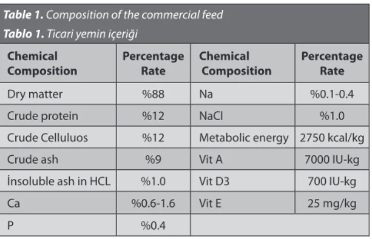
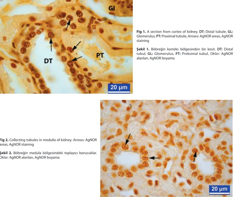
The histological preparations were examined with a light microscope (Leica DM-2500 attached to a DFC-320 digital camera). In different regions of the kidney (glomerulus, proximal tubules, distal tubules and collecting tubules) of each animal, 25 cells having nuclei were evaluated. The nuclear area and the AgNOR area (Fig. 1 and 2) were analyzed with an image analysis program (IM-50). Also, the percentage of the AgNOR area relative to the nuclear area was calculated.
Statistical Analysis
The obtained results were statistically analyzed using Duncan test in SPSS software [version 17; SPSS Inc., Chicago, IL, USA]. The level of significance was P<0.05.
RESULTS
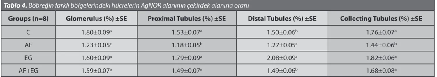
We obtained the nuclear area (Table 2), the AgNOR area (Table 3), and the ratio of the nuclear of the AgNOR area (Table 4) of cells in different regions of the kidney. Nuclear areas and ratio of nuclear area of the AgNOR area of glomerulus and tubular epithelial cells of kidney decreased significantly (P<0.05) in the AF group compared to the other groups. Furthermore, AF group was lowest (P<0.05) AgNOR area.
DISCUSSION
The liver and kidney are considered the main target organ for aflatoxicosis 7,27-29. Lakkawar et al.30 reported that
liver and kidney were the most affected organs in rabbits which fed an AFB1 contaminated diet. The effects of AFs on histopathological changes are directly correlated with the concentration of AF and the duration of the exposure 31.
Fig 1. A section from cortex of kidney. DT: Distal tubule, GL:
Glomerulus, PT: Proximal tubule, Arrows: AgNOR areas, AgNOR staining
Şekil 1. Böbreğin korteks bölgesinden bir kesit. DT: Distal
tubul, GL: Glomerulus, PT: Proksimal tubul, Oklar: AgNOR alanları, AgNOR boyama
Fig 2. Collecting tubules in medulla of kidney. Arrows: AgNOR
areas, AgNOR staining
Şekil 2. Böbreğin medula bölgesindeki toplayıcı borucuklar.
Nuclear areas of glomerulus and tubular epithelial cells were significantly (P<0.05) decreased in all regions of AF group compared to the C group. Measurements in glomerulus, distal and collecting tubule regions of AF+EG group were similar compared with the C group. But, it was observed that measurements in proximal tubule regions in AF+EG group were significantly (P<0.05) different compared with the C group. It was known that AFs have toxic effect on p53 gene which is a protective effect against DNA damage in cells 32,33. The findings of the study were showed
that AF was significantly decreased nuclear areas of cells and formed negative impact on metabolic activity of cells. AgNOR areas of glomerulus and tubular epithelial cells of kidney were similar with C and EG groups. We were observed AF group was lowest (P<0.05) AgNOR area. AgNOR area of AF+EG group was found statistically higher (P<0.05) than that of AF group. These data were in agreement with findings of the researches 34,35 who reported that AFs
caused genetic disorders in cells. In this study, we found the lowest AgNOR area of the AF group since AF caused to modification 36 and reduce number of ribosome. Ultimately,
this situation causes to reduce synthesis of proteins 37,38. On
the other hand, AgNOR area of AF+EG group to be higher than those of AF group, it shows aflatoxin is relatively eliminated pressure on protein synthesis by EG.
The ratio of nuclear area of the AgNOR area of glomerulus and tubular epithelial cells of kidney decreased significantly (P<0.05) in the AF group compared to the other groups. This result reveals that AFs which are caused DNA damage 28,33,39
reduce activity of cell’s protein synthesis 37,38. Zaczek et al.17
have reported that AgNOR parameters associated with the proliferation activity of the epithelium.
As a conclusion, reason of decline in protein synthesis activity of the cells, we can be said AF is partly eliminated the negative impact on the cells by used EG. We were concluded EG is an agent which can be used successfully to prevent aflatoxicosis. Obtained data are showed that there are important changes in the AgNOR parameters. Therefore, we think that AgNOR parameters will also utilize taking into account besides other histopathological assessments for future similar studies.
Table 2. Nuclear area of cells in different regions of kidney Tablo 2. Böbreğin farklı bölgelerindeki hücrelerin çekirdek alanı
Groups (n=8) Glomerulus (μm²)±SE Proximal Tubules (μm²)±SE Distal Tubules (μm²)±SE Collecting Tubules (μm²)±SE
C 15.75±0.34a 26.02±0.46a 23.11±0.32a 28.95±0.48a
AF 13.13±0.28c 22.20±0.29b 20.32±0.32c 25.33±0.30c
EG 15.17±0.32ab 25.30±0.39ab 22.29±0.33ab 28.24±0.35abc
AF+EG 14.33±0.31b 22.33±0.36b 21.65±0.30b 27.00±0.29b
C: Control, AF: Aflatoxin, EG: Glucomannan, AF+EG: Aflatoxin+glucomannan; a-c Values within a column with no common superscripts are significantly
(P<0.05) different
Table 3. AgNOR area of cells in different regions of kidney Tablo 3. Böbreğin farklı bölgelerindeki hücrelerin AgNOR alanı
Groups (n=8) Glomerulus (μm²)±SE Proximal Tubules (μm²)±SE Distal Tubules (μm²)±SE Collecting Tubules (μm²)±SE
C 0.23±0.06a 0.43±0.01a 0.41±0.01a 0.51±0.01a
AF 0.17±0.00c 0.27±0.01c 0.26±0.01c 0.38±0.01b
EG 0.22±0.01a 0.41±0.01a 0.38±0.01a 0.49±0.01a
AF+EG 0.19±0.01b 0.33±0.01b 0.29±0.01b 0.39±0.01b
C: Control, AF: Aflatoxin, EG: Glucomannan, AF+EG: Aflatoxin+glucomannan; a-c Values within a column with no common superscripts are significantly
(P<0.05) different
Table 4. The ratio of the nuclear of the AgNOR area of cells in different regions of kidney Tablo 4. Böbreğin farklı bölgelerindeki hücrelerin AgNOR alanının çekirdek alanına oranı
Groups (n=8) Glomerulus (%) ±SE Proximal Tubules (%) ±SE Distal Tubules (%) ±SE Collecting Tubules (%) ±SE
C 1.80±0.09a 1.53±0.07a 1.50±0.06b 1.76±0.07a
AF 1.23±0.05c 1.18±0.05b 1.27±0.05c 1.44±0.06b
EG 1.60±0.09a 1.79±0.09a 2.08±0.09a 1.82±0.06a
AF+EG 1.59±0.07a 1.49±0.07a 1.49±0.06b 1.68±0.08a
C: Control, AF: Aflatoxin, EG: Glucomannan, AF+EG: Aflatoxin+glucomannan; a-c Values within a column with no common superscripts are significantly
REFERENCES
1. Devegowda G, Raju MVLN, Afzali N, Swamy HVLN: Mycotoxins
picture: worldwide, novel solutions for their counteraction. In, Lyons TP, Jacques KA (Eds): Biotechnology in the Feed Industry. Proceedings of
Alltech’s 14th Annual Symposium. Nottingham, 24 February 1998, UK, pp
241-255, 1998.
2. Bennett JW, Klich M: Mycotoxins. Clinical Microbiology Reviews, 16,
497-516, 2003.
3. CAST: Council for Agricultural Science and Technology Task Force,
Mycotoxins: Economic and Health Risks. Ames, IA, US, Rep No 116, 1989.
4. Sur E, Celik I: Effects of AFB1 on the development of the bursa of
Fabricius and blood lymphocyte acid phosphatase of the chicken. Brit
Poult Sci, 44, 558-566, 2003.
5. Oguz H, Hadimli HH, Kurtoglu V, Erganis O: Evaluation of humoral
immunity of broilers during chronic aflatoxin (50 and 100 ppb) and clinoptilolite exposure. Rev Med Vet, 154, 483-486, 2003.
6. Basmacıoglu H, Oguz H, Ergul M, Col R, Birdane YO: Effect of dietary
esterified glucomannan on performance, serum biochemistry and haematology in broilers exposed to aflatoxin. Czech J Anim Sci, 50 (1): 31-39, 2005.
7. Eraslan G, Karaoz E, Bilgili A, Akdogan M, Oncu M, Essiz D: The
effects of aflatoxin on kidney function in broiler chicks. Turk J Vet Anim Sci, 27, 741-749, 2003.
8. Peraica M, Domijan A, Jurjevic Z, Cvjetkovic B: Prevention of exposure
to mycotoxins from food and feed. Arch Hig Rada Toksikol, 53 (3): 229-237, 2002.
9. Ibrahim IK, Shareef AM, Al-Joubry KMT: Ameliorative effects of SB
on phagocytosis and Newcastle disease antibody formation in broiler chickens during aflatoxicosis. Res Vet Sci, 69, 119-122, 2000.
10. Rosa CAR, Miazzo R, Magnoli C, Salvano M, Chiacchiera SM, Ferrero S, Saenz M, Carvalho EC, Dalcero A: Evaluation of the efficacy of bentonite
from the south of Argentina to ameliorate the toxic effects of AF in broilers. Poult Sci, 80, 139-144, 2001.
11. Kececi T, Oguz H, Kurtoglu V, Demet O: Effects of
polyvinylpoly-pyrrolidone, synthetic zeolite and bentonite on serum biochemical and haemetological characters of broiler chickens during aflatoxicosis. Br
Poult Sci, 39 (3): 452-458, 1998.
12. Bata A, Lasztity R: Detoxification of mycotoxin contaminated
food and feed by microorganisms. Trends Food Sci Technol, 10, 223-228, 1999.
13. Stanley VG, Ojo R, Woldensenbet S, Hutchinson DH: The use of
Saccharomyces cerevisiae, to suppres the effect of aflatoxicosis in broiler
chicks. Poult Sci, 72, 1867-1872, 1993.
14. Diaz DE, Hagler WM, Hopkins BA, Whitlow LW: Aflatoxin binders
I: in vitro binding assay for aflatoxin B1 by several potential sequestering
agents. Mycopathologia, 156, 223-226, 2002.
15. Robert-Fortel I, Junera HR, Geraud G, Hernandez-Verdun D:
Three-dimensional organization of the ribosomal genes and Ag-NOR proteins during interphase and mitosis in PtK1 cells studied by confocal microscopy. Chromosoma,102, 146-157, 1993.
16. Khanna AK, Yadav SK, Dixit VK, Kumar M: AgNOR count and subjective
AgNOR pattern assessment (SAPA) score in carcinoma of the pancreatic
head including tumors. J Pancreas, 6 (6): 575-580, 2005.
17. Zaczek M, Macie jowski J, Gil K, Szot W, Chlap Z: Silver-binding nucleolar
organiser regions (AgNORs) in the normal epithelium of different parts of the digestive tract in rats. Acta Pathol Jpn, 42 (8): 573-578, 1992.
18. Bukhari MH, Niazi S, Khan SA, Hashmi I, Perveen S, Qureshi SS:
Modified method of AgNOR staining for tissue and interpretation in histopathology. Int J Exp Path, 88, 47-53, 2007.
19. Abrams L: Mycotoxicosis. J S Afr Vet Med Assoc, 36, 5-13, 1965. 20. Lewis G, Markson LM, Allcroft R: The effect of feeding toxic groundnut
meal to sheep over a period of five years. Vet Rec, 80, 312-314, 1967.
21. Edrington TS, Harvey RB, Kubena LF: Effect of aflatoxin in growing
lambs fed ruminally degredable or escape protein sources. J Anim Sci, 72, 1274-1281, 1994.
22. Shotwell OL, Hasseltine CW, Stubblefield RD, Sorenson WG:
Production of aflatoxin on rice. Appl Microbiol, 5, 425-429, 1966.
23. Demet O, Oguz H, Celik I, Nizamlıoglu F: Pirinçte aflatoksin üretilmesi.
Vet Bil Derg, 11 (1): 19-23, 1995.
24. Vicam LP: Fluorometer USDA-FGIS procedure for corn, corn meal,
corn/soy blend, milled rice, popcorn, sorghum and soybeans. In, Vicam LP (Ed): Afla Test Instruction Manuel. pp 36-38, Vicam, Watertown, USA, 1999.
25. Stroka J, Anklam E, Otterdijk R: Immunoaffinity columnclean-up
prior to thin layer chromatography for determination of aflatoxins in various food matrices. J Chromat A, 904, 251-256, 2000.
26. Aydın MF, Celik I: Determination of the distribution of silver-stained
nucleolar organizing regions (AgNORs) in the livers broilers and laying hens. Vet Bil Derg, 21 (1-2): 91-99, 2005.
27. Yildirim E, Yalcinkaya I, Kanbur M, Cinar M, Oruc E: Effects of
yeast glucomannan on performance, some biochemical parameters anf pathological changes in experimental aflatoxicosis in broilers chickens.
Revue Med Vet, 162 (8-9): 413-420, 2011.
28. Ozen H, Karaman M, Cigremis Y, Tuzcu M, Ozcan K, Erdag D:
Effectiveness of melatonin on aflatoxicosis in chicks. Res Vet Sci, 86, 485-489, 2009.
29. O’Brien E, Dietrich DR: Mycotoxins Affecting the Kidney. In, Hook JB,
Tarloff JB, Lash LH (Eds): Toxicology of Kidney. First ed., pp. 895-936, Boca Raton, CRC Pr, FL, 2004.
30. Lakkawar AW, Chattopadhyay SK, Johri TS: Experimental aflatoxin
B1 toxicosis in young rabbits-a clinical and patho-anatomical study. Slov
Vet Res, 41 (2): 73-81, 2004.
31. Boonyaratpalin M, Supamattaya K, Verakunpiriya V, Suprasert D: Effects of aflatoxin B1 on growth performance, blood components,
immune function and histopathological changes in black tiger shrimp
(Penaeus monodon fabricius). Aquacult Res, 32, 388-398, 2001.
32. Gerbes AL, Caselmann WH: Point mutations of the p53 gene, human
hepatocellular carcinoma and aflatoxins. J Hepatol, 19 (2): 312-315, 1993.
33. Ozturk M: P53 mutation in hepatocellular carcinoma after aflatoxin
exposure. Lancet, 338, 1356-1359, 1991.
34. Shi CY, Phang TW, Lin Y, Wee A, Li B, Lee HP, Ong CN: Codon 249
mutation of the p53 gene is a rare event in hepatocellular carcinomas from ethnic Chinese in Singapore. Br J Cancer, 72, 146-149, 1995.
35. Sengstag C: The molecular mechanism of aflatoxin B1- induced liver
cancer: is mitotic recombination involved. Mol Carcinog, 19, 147-152, 1997.
36. Lyman BA, Erki L, Biedrzycka DW, Devlin TM, Ch’ih JJ: Modification
of protein synthetic components by aflatoxin B1. Biochem Pharmacol, 37
(8): 1481-1486, 1988.
37. Ergun E, Ergun L, Essiz D: Light and electron microscopic studies on
liver histology in chicks fed aflatoxin. Dtsch Tierarztl Wschr, 49, 243-247, 2006.
38. Sehu S, Ergun L, Cakır S, Ergun E, Cantekin Z, Sahin T, Essiz D, Sareyyupoglu B, Gurel Y, Yigit Y: Hydrated sodium calcium aluminosilicate
for reduction of aflatoxin in quails (Coturnix coturnix japonica). Dtsch Tierarztl
Wschr, 114, 252-259, 2007.
39. Salem MH, Kamel KI, Yousef MI, Hassan GA, EL-Nouty FD: Protective
role of ascorbic acid to enhance semen quality of rabbits treated with sublethal doses of aflatoxin B1. Toxicology, 162, 209-218, 2001.