Original Article
Diagnosis and treatment of intestinal intussusception in
adults: a rare experience for surgeons
Fatih Ciftci
Vocational School of Health Services, Istanbul Gelisim University, Avcılar, Istanbul, Turkey
Received March 21, 2015; Accepted June 3, 2015; Epub June 15, 2015; Published June 30, 2015
Abstract: Aim: We evaluated the aetiological factors, diagnoses and treatment outcomes of adult cases of invagina-tion of the intestine, or intussuscepinvagina-tion. We elucidated the role of ultrasonography (USG), computed tomography (CT) and other techniques in the diagnosis of such cases. Patients and Methods: Six patients with sufficient medi-cal data, all of whom were followed-up and treated for intussusception at Safa Hospital, General Surgery Clinic, Istanbul, Turkey, between July 2008 and December 2013, were enrolled. Results: The site of intussusception was the small bowel in five cases. The aetiopathology was benign in 5 (83.3%) cases and included inflammatory fibroid polyp (IFP) and adhesion. The malignant case was adenocarcinoma. The bowel segments involved were ileoileal in five cases and ileocolic in one case. Conclusions: USG and CT aided with the diagnosis. Although more rare in adults than in children, cases of ileus presenting with acute abdominal disorder, particularly those with an uncertain diagnosis, should be considered possible intussusception cases.
Keywords: Adult, invagination (intussusception), diagnosis
Introduction
The prolapse of one segment of the gastroin-testinal tract into the lumen of an immediately adjoining segment is referred to as intussus-ception [1]. At a paediatric age, this is the most frequent cause of acute abdominal disorder second to acute appendicitis. However, the dis-order is rare in adults, comprising 0.02-0.03% of all cases reported in the hospital and 1-3% of all cases of surgical intestinal obstructions [1, 2]. In contrast to the situation with children, the aetiological factors in adults are known in 80-90% of cases. In the present study, we eval-uated the aetiological factors, diagnoses and treatment outcomes of adult cases of intussus-ception at our clinic over 4 years between July 2009 and December 2013.
Patients and methods
Six patients with sufficient medical data, all of whom were followed-up and treated for invagi-nation at BagcilarSafa Hospital, General Surgery Clinic, Istanbul, Turkey, between July 2008 and December 2013, were enrolled. The complaints and physical findings of the patients were recorded.
A midline incision was employed in five cases, whereas a Pfannenstiel incision was preferred in one patient who was pregnant. Surgical de-intussusception was attempted in one case with idiopathic small bowel intussusception due to adhesion, but it was not successful. In one case of destructed vascular perfusion, resection of the involved segments was accom-plished. Stomas were established in cases with excessive dilation and oedema of the proximal segment of the bowel, and anastomosis was accomplished in those with sufficient perfusion and tolerable oedema and dilation.
Results
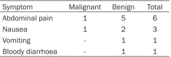
Of the six patients included in our study, three (50%) were males, and three (50%) were females. The patients’ median age was 40 years (range: 15-69 years). The median time between reporting to the emergency clinic and surgery was 2.33 days (1-3 days). All six patients had complaints for less than 7 days, and a median duration of 2.33 days (1-3 days). The most frequent complaints were abdominal pain, nausea and vomiting (Table 1). On physical examination, abdominal tenderness and perito-neal irritation were frequent. The median white
blood cell count was 15.37 ± 3.3/mm3.
Ultrasonography of the abdomen disclosed intussusception in two (33%) cases, and oede-ma and dilation were described in another case. Computed tomography (CT) of the abdo-men revealed intussusception in three (75%) of four patients. Prior to surgery, the definite diag-nosis rate was 50% (n = 3).
The site of intussusception was the small bowel in five cases. The aetiopathology was benign in five (83.3%) cases and included inflammatory fibroid polyp (IFP) and adhesion. The malignant case was adenocarcinoma. Idiopathic intussus-ception was diagnosed in one patient (16.6%) in whom no cause was ascertained. One of the cases that had IFP was 16 weeks pregnant. The bowel segments involved were ileoileal in five cases and ileocolic in one case. Segmentary small bowel resection and end-to-end anasto-mosis were accomplished in five cases. In one case, right hemicolectomy and ileotransversos-tomy were accomplished. The median length of the resected bowel was 28.1 ± 19.1 cm. Wound site infection occurred in one patient. The median length of stay in the hospital was 5.3 days (3-9 days). There was no mortality in our series.
Discussion
Intussusception occurs as a result of unequal motility between adjacent segments of the intestine. The proximal segment is referred to as the intussusceptum, and the distal segment is referred to as the intussuscipiens. The aetio-pathology of intussusception is not well known; however, the predisposing factors of trauma, allergic reaction, genetic tendency, and physi-cal, bacterial, metabolic and chemical stimuli have been suggested as precursors of the pro-cess, leading to lesions on the bowel wall and irritation caused by abnormal peristaltism.
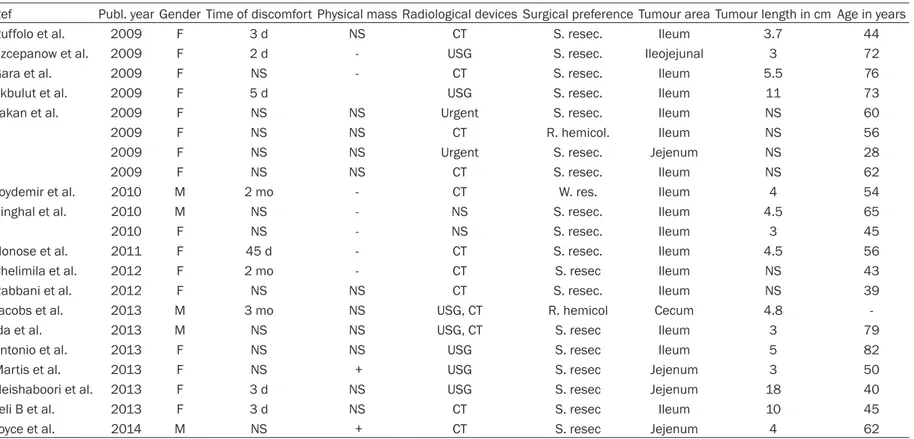
due to IFP presented between 2009 and 2014 (Table 2). However, most frequently, anatomi-cal forms arise that include the gastric antral (65-75%), ileocolic, colocolic (3-8%), enteroen-teral (17-21%), duodenal (1%), oesophageal (1%), gallbladder (1%) and jejunogastric regions [1, 3].
The cause of intussusception in children can be established in only 5% of cases, and the major-ity remain idiopathic. In adults, the aetiology is established in 80-90% of cases; in previous studies, the causes were a benign or malignant neoplasm in 65% of established cases and a non-neoplastic cause in 15-25% of cases [4, 5]. In the present study, the percentage of cases in which the cause of intussusception was estab-lished was 83.3%, in line with previous studies. Intussusception involving the small intestine is more frequent, and 90% of the causes involve polypoid lesions such as haematoma, IFP, ade-noma, leiomyoma and Peutz-Jeghers syndrome; tuberculosis and Meckel’s diverticulum are other causes. Malignant causes of small bowel intussusception are rare and include primary (particularly adenocarcinoma) or metastatic tumours. The aetiology of colocolic intussus-ception is malignancy in 50-60% of all cases [5, 6]. In the present study, most cases involved the small bowel, and the pathological causes were predominantly benign.
Evaluation of previous studies has shown that the median age for intussusception varies between 45 and 57.5 years [6-9]. Gender distri-bution appears to be equal between males and females. In the present study, gender distribu-tion was in agreement with previous data; how-ever, the median age was younger.
The rate of emergent laparotomy for intussus-ception varies from 20% to 61.5% in previous studies [3, 6, 10, 11]. Intussusception fre-quently presents with acute abdominal pain, nausea, vomiting and high WBC count. However, the presentation may be subacute or chronic in the case of partial obstruction. Most of our cases reported to the emergency clinic within 1-3 days of the onset of symptoms and were admitted for emergency surgery with a clinical diagnosis of an acute abdomen disorder or intestinal obstruction.
Table 1. Symptoms of our patients
Symptom Malignant Benign Total
Abdominal pain 1 5 6
Nausea 1 2 3
Vomiting - 1 1
Table 2. Clinicopathological features of 21 cases of intussusception due to IFP presented between 2009 and 2014 in the literature
Ref Publ. year Gender Time of discomfort Physical mass Radiological devices Surgical preference Tumour area Tumour length in cm Age in years
Ruffolo et al. 2009 F 3 d NS CT S. resec. Ileum 3.7 44
Szcepanow et al. 2009 F 2 d - USG S. resec. Ileojejunal 3 72
Gara et al. 2009 F NS - CT S. resec. Ileum 5.5 76
Akbulut et al. 2009 F 5 d USG S. resec. Ileum 11 73
Yakan et al. 2009 F NS NS Urgent S. resec. Ileum NS 60
2009 F NS NS CT R. hemicol. Ileum NS 56
2009 F NS NS Urgent S. resec. Jejenum NS 28
2009 F NS NS CT S. resec. Ileum NS 62
Toydemir et al. 2010 M 2 mo - CT W. res. Ileum 4 54
Singhal et al. 2010 M NS - NS S. resec. Ileum 4.5 65
2010 F NS - NS S. resec. Ileum 3 45
Nonose et al. 2011 F 45 d - CT S. resec. Ileum 4.5 56
Chelimila et al. 2012 F 2 mo - CT S. resec Ileum NS 43
Rabbani et al. 2012 F NS NS CT S. resec. Ileum NS 39
Jacobs et al. 2013 M 3 mo NS USG, CT R. hemicol Cecum 4.8
-Ida et al. 2013 M NS NS USG, CT S. resec Ileum 3 79
Antonio et al. 2013 F NS NS USG S. resec Ileum 5 82
Martis et al. 2013 F NS + USG S. resec Jejenum 3 50
Neishaboori et al. 2013 F 3 d NS USG S. resec Jejenum 18 40
Teli B et al. 2013 F 3 d NS CT S. resec Ileum 10 45
Joyce et al. 2014 M NS + CT S. resec Jejenum 4 62
M: male; F: female; d: days; mo: months; S. resec: segmental resection; NP: not performed; NS: not stated; UA: unavailable; R. hemicol: right hemicolectomy; Ecl: enteroclysis; USG: ultrasonography; DL: diagnostic laparoscopy; CT: computed tomography; MRI: magnetic resonance imaging; Lp: laparoscopy; W. resec: wedge resection.
corporeal examination and exclusive imaging techniques, such as X-ray, ultrasonography (USG), CT, enteroclysis, endoscopic practice, angiography and capsule endoscopy. Parti- cularly, abdominal X-ray is the primary diagnos-tic used because obstructive signs possess clinical characteristics in many cases. Nowa- days, enteroclysis is seldom used for the identi-fication of intussusception. Barium enema was the preferred choice for the identification of intussusception until 1981 when studies showed that USG could be used to precisely diagnose the condition. Although obstructive signs are contraindicated for capsule endosco-py, this new practice for interpreting the small bowel could be useful in cases with a negative outcome on radiological identification. Colo- noscopy is helpful only in cases in which colon-ic involvement is strongly suspected, and lesions can be identified and biopsied [12-15]. USG is frequently employed for the diagnosis of intussusception. Despite its high sensitivity and specificity, a pseudo-kidney sign in the sag-ittal plane is not diagnostic. Target and ox eye signs in the transverse plane favour the diagno-sis of intussusception [15-19]. However, bowel wall oedema and sigmoid volvulus may also manifest the sign [5, 10, 20]. A large amount of faeces in the colon, a perforated Meckel’s diverticulum, malrotation and intraabdominal haematomas are other causes of misdiagnos-es [13, 22]. Moreover, acute obstructive symp-toms and air levels decrease the reliability of USG [5, 23]. The rate of a correct diagnosis using USG was 30-35% in previous studies [7, 23]. In the present study, it was 33% (n = 2) in agreement with this. CT is the gold standard for the diagnosis of intussusception. However, it comprises 50-80% of the diagnostic data [2]. A sausage sign and target mass on CT are not pathognomonic for intussusception [3, 24, 25]. CT may provide sufficient information about metastasis, lymphadenopathy, free liquid and proximal bowel dilation [14, 26-28]. In the pres-ent study, the diagnostic value of CT was in agreement with previous data. However, our population was very small. The limited use of CT may be due to patients undergoing emer-gency surgery. Magnetic resonance imaging is not used routinely for the diagnosis of intussus-ception in either adults or children.
susception may provide successful treatment in cases where bowel perfusion is not disrupt-ed, particularly when intussusception is due to adhesions subsequent to surgery. However, the de-intussusception procedure may lead to per-foration in cases of inflamed, ischemic and colonic intussusceptions. Hence, the proce-dure is not recommended in such cases. Particularly, colonic intussusception may also cause the spread of tumour cells. In that situa-tion, segmental resection or wedge resection of the involved bowel segment should be pre-ferred [3].
Conclusion
The characteristics of adult patients with intus-susceptions in our study agree with those in previous studies. However, the mean age of our cases was older. Intussusception is rare in adults; however, this diagnosis should be con-sidered in cases presenting with suitable mani-festation. In addition, USG and CT aid in the diagnosis. Although more rare in adults than in children, cases of ileus presenting with acute abdominal disorder, particularly those with an uncertain diagnosis, should be considered pos-sible intussusception cases. In addition to cases in which surgery is preferred, resection including the invaginated segment should be accomplished.
Acknowledgements
The authors express their gratitude to all of the participating patients and clinical staff.
Address correpondence to: Fatih Ciftci, Vocational School of Health Services, Istanbul Gelisim University, Basaksehir Mah., Erciyes Sok. No: 15, Daire 24, Basaksehir, Avcılar, Istanbul 34306, Turkey. Tel: +90 505 616 4248; Fax: +90 212 462 7056; E-mail: oprdrfatihciftci@gmail.com
References
[1] Akbulut S. Intussusception due to inflammato-ry fibroid polyp: A case report and comprehen-sive literature review. World J Gastroenterol 2012; 18: 5745-5752.
[2] Azar T, Berger DL. Adult intussusception. Ann Surg 1997; 226: 134-138.
[4] Ahn JH, Choi SC, Lee KJ, Jung YS. A clinical overview of a retrospective study about adult intussusceptions: focusing on discrepancies among previous studies. Dig Dis Sci 2009; 54: 2643-2649.
[5] Ghaderi H, Jafarian A, Aminian A, Mirjafari-Daryasari SA. Clinical presentations, diagnosis and treatment of adult intussusception, a 20 years survey. Int J Surg 2010; 8: 318-320. [6] Wang N, Cui XY, Liu Y, Long J, Xu YH, Guo RX,
Guo KJ. Adult intussusception: a retrospective review of 41 cases. World J Gastroenterol 2009; 15: 3303-3308.
[7] Yakan S, Caliskan C, Makay O, Denecli AG, Kor-kut MA. Intussusception in adults: clinical characteristics, diagnosis and operative strat-egies. World J Gastroenterol 2009; 15: 1985-1989.
[8] Marinis A, Yiallourou A, Samanides L, Dafnios N, AnastasopoulosG, Vassiliou I, Theodoso-poulos T. Intussusception of the bowel in adults: a review. World J Gastroenterol 2009; 15: 407-411.
[9] Theodoropoulos GE, Linardoutsos D, Tsamis D, Stamopoulos P, Giannopoulos D, Zagouri F, Mi-chalopoulos NV. Gastrointestinal stromal tu-mor causing small bowel intussusception in a patient with Crohn’s disease. World J Gastro-enterol 2009; 15: 5224-5227.
[10] Akbulut S, Sevinc MM, Cakabay B, Bakir S, Senol A. Giant inflammatory fibroid polyp of il-eum causing intussusception: a case report. Cases J 2009; 2: 8616.
[11] Gara N, Falzarano JS, Limm WM, Namiki TS, Tom LK. Ileal inflammatory fibroid polyp caus-ing chronic ileocolic intussusception and mim-icking cecal carcinoma. World J Gastrointes-tOncol 2009; 1: 89-92.
[12] Toydemir T. Inflammatory fibroid polyp of the ileum presenting with small bowel obstruction in an adult patient: a case report. J Med Case Rep 2010; 4: 291.
[13] Szczepanowski A, Kaluzniacki P, Mazurek A, Golyski J, Rudzki S. Inflammatory fibroid polyp of the small bowel causing intestinal obstruc-tion due to intussuscepobstruc-tion-a case report. JPCCR 2009; 3: 122-123.
[14] Nonose R, Valenciano JS, da Silva CM, de Sou-za CA, Martinez CA. Ileal Intussusception Caused by Vanek’s Tumor: A Case Report. Case Rep Gastroenterol 2011; 5: 110-116. [15] Singhal M, Singh P, Misra V, Dhingra V, Bhatia
R. Inflammatory fıbroıd polyp of small intes-tine: Report of two cases wıth revıew of litera-ture. JCDR 2010; 4: 3241-3244.
[16] Morales-Fuentes GA, de Ariño-Suárez M, Zárate-Osorno A, Rodríguez-Jerkov J, Terrazas-Espitia F, Pérez-Manauta J. Vanek’s polyp or inflammatory fibroid polyp. Case report and re-view of the literature. Cir Cir 2011; 79: 242-245.
[17] Ruffolo C, Scarpa M, Bassi D, Angriman I. In-flammatory fibroid polyp causing intestinal ob-struction following restorative proctocolectomy for ulcerative colitis. Dig Surg 2009; 26: 285-286.
[18] Joyce KM, Waters PS, Waldron RM, Khan I, Orosz ZS, Nemeth T, Barry K. Recurrent adult jejuno-jejunal intussusception due to inflam-matory fibroid polyp–Vanek’s tumour: a case report. Diagn Pathol 2014; 9: 127.
[19] Tartaglia D, Bertolucci A, Palmeri M, Kauff-mann EF, Napoli N, Galatioto C, Lippolis PV, Zocco G, Seccia M. The role of laparoscopy in adult bowel obstruction caused by intussus-ception. Ann Ital Chir 2014; 85: 328-331. [20] Teli B, Cp M, SS, Mv S. Ileo-ileal
Intussuscep-tion in an Adult Caused by Vanek’s Tumour: A Rare Case Report. Clin Diagn Res 2013; 7: 2994-2995.
[21] Rabbani K, Narjis Y, Jgounni R, Semlani Z, Di-faa A, Benelkhaiat R, Louzi A, Finech B. Adult intussusception caused by an inflammatory fi-broid ileal polyp. Acta Chir Belg 2012; 112: 157-159.
[22] Neishaboori H, Maleki I, Emadian O. Jejunal intussusception caused by a huge Vanek’stu- mor: a case report. Gastroenterol Hepatol Bed Bench 2013; 6: 210-3.
[23] Martis JJ, Rajeshwara KV, Murulya KS, Raghav-endra BK, Alex KM. A rare cause of jejunojeju-nal intussusception in an adult. Indian J Surg 2013; 75: 18-20.
[24] Maya AM, Gallo A, Castelli M, Paz L, Espinosa JC, Giunippero A. [Small bowel intussusception and Vanek’stumor in an elderly woman]. Me-dicina (B Aires) 2013; 73: 461-463.
[25] Ida S, Matsuzaki H, Kawashima S, Watanabe M, Akiyama Y, Baba H. Adult intestinal intus-susception caused by an inflammatory myo- fibroblastictumor. Case Rep Gastroenterol 2013; 7: 224-228.
[26] Jacobs TM, Lambrianides AL. Inflammatory fi-broid polyp presenting as intussusception. J Surg Case Rep 2013; 2: 263-267.
[27] Chelimilla H, Ihimoyan A, Carvajal S, Bhavna B. Ileoileal intussusception secondary to an ileal fibroma. Case Rep Gastroenterol 2012; 6: 734-740.

