1Department of Medicine, Marc and Ruti Bell Program for Vascular Biology and Disease, Leon H. Charney Division of Cardiology, New York University School of Medicine, New York, NY, USA. 2Department of Medicine, Heart Institute & Department of Biomedical Sciences, Cedars Sinai Medical Center, Los Angeles, CA, USA. 3Department of Molecular Biology and Genetics, Bilkent University, Ankara, Turkey. 4National Nanotechnology Center, Bilkent University, Ankara, Turkey. *e-mail: [email protected]
M
acrophages are essential components of mammalian tissues and are strategically positioned throughout the body to maintain tissue homeostasis and act as immune sentinels. Macrophages can be either embryonically seeded in tissues, where they are maintained through self-renewal, or derived from mono-cyte precursors, which infiltrate tissues and differentiate in response to their microenvironment. Regardless of their origin, macrophages are remarkable in their plasticity: they can change their functional phenotype in response to local environmental cues and conse-quently execute a diverse set of functions essential to host defense and tissue repair. Such functions include phagocytosis of apoptotic cells and pathogens, elaboration of immune-effector molecules and growth factors, and remodeling of the extracellular matrix. To fulfill these effector functions, macrophages must coordinate a plethora of intrinsic cellular processes to meet environmental demand. It is becoming increasingly appreciated that the rewiring of meta-bolic pathways and the repurposing of metameta-bolic intermediates to facilitate appropriate extrinsic cellular responses are central to this intrinsic coordination.Early studies of metabolic reprogramming in cancer cells pro-vided a window into the understanding of how metabolism can regulate cell fate. Elevated uptake of glucose and secretion of lactate are metabolic hallmarks of highly proliferative tumor cells, which rely heavily on glycolysis even when sufficient oxygen is present to support mitochondrial oxidative phosphorylation (OXPHOS)—so called ‘aerobic glycolysis’ or the ‘Warburg effect’1. Whereas aerobic glycolysis is much less efficient than OXPHOS at generating ATP, this metabolic pathway is nonetheless well suited for prolifera-tion because it allows metabolic intermediates to be siphoned off for biosynthesis of nucleotides, lipids, and proteins needed in the dividing cell. Similar metabolic alterations have been identified in some immune cells, including macrophages, as they transition from rested to activated states1 to meet cellular demands, such as phago-cytosis, proliferation, and cytokine production2,3. This metabolic modulation can result in energy generation; production of build-ing blocks for cell maintenance and proliferation; and modulation
of cellular signaling4. Studies of macrophages polarized in vitro to either a classically activated (by using lipopolysaccharide (LPS) and the cytokine IFNγ ) or an alternatively activated (by using the cyto-kine IL-4) state have shown that metabolic reprogramming of mac-rophages not only is critical for energy homeostasis but also directly influences cell functions. Although these in vitro phenotypes have been useful models to study, relatively little is known about how macrophage metabolism changes in vivo, when cells encounter a multitude of signals in the tissue microenvironment during either pathogen challenge or sterile inflammatory responses.
During atherosclerosis, sterile inflammation is set in motion by the intramural retention of cholesterol-rich lipoproteins in large- and medium-sized arteries (reviewed in ref. 5). Imbalances in cellular and systemic cholesterol homeostasis promote the artery-wall deposition of lipoproteins containing the apolipopro-tein apoB, particularly in areas of altered blood flow that disturb the normally quiescent endothelium. These sequestered lipopro-teins are susceptible to various modifications (for example, oxi-dation, enzymatic cleavage, and aggregation) that render them proinflammatory; the modified lipoproteins then trigger the recruitment of monocyte-derived cells into the subendothelial space. There, monocytes differentiate into macrophages that clear the accumulated lipoproteins, a process that, although initially beneficial, ultimately results in the accumulation of cholesterol-laden macrophage foam cells that promote plaque formation. These macrophage foam cells have a diminished migratory capacity6,7 and elicit production of proinflammatory cytokines and chemokines that amplify the immune response by recruit-ing reinforcements, includrecruit-ing additional monocytes, T cells, and neutrophils (reviewed in ref. 8). For reasons that remain poorly understood, the resulting low-grade inflammation is nonresolv-ing, and plaque progression can continue over decades of life9. As plaques advance, macrophages proliferate10, secrete inflammatory mediators (for example, cytokines and proteases), and ultimately die from lipid and metabolic stresses11, thereby releasing their lipid contents and other inflammatory debris that contributes
Regulation of macrophage immunometabolism
in atherosclerosis
Graeme J. Koelwyn
1, Emma M. Corr
1, Ebru Erbay
2,3,4and Kathryn J. Moore
1*
After activation, cells of the myeloid lineage undergo robust metabolic transitions, as well as discrete epigenetic changes, that can dictate both ongoing and future inflammatory responses. In atherosclerosis, in which macrophages play central roles in the initiation, growth, and ultimately rupture of arterial plaques, altered metabolism is a key feature that dictates macrophage function and subsequent disease progression. This Review explores how factors central to the plaque microenvironment (for example, altered cholesterol metabolism, oxidative stress, hypoxia, apoptotic and necrotic cells, and hyperglycemia) shape the metabolic rewiring of macrophages in atherosclerosis as well as how these metabolic shifts in turn alter macrophage immune-effector and tissue-reparative functions. Finally, this overview offers insight into the challenges and opportunities of harness-ing metabolism to modulate aberrant macrophage responses in disease.
to the formation of a necrotic core. In plaques, macrophages are exposed to a variety of proinflammatory cytokines, oxidized lip-ids, cholesterol crystals, oxidative stress, and danger-associated molecular patterns derived from dying cells; these biomolecules together create a complex microenvironment that perpetuates macrophage inflammation5.
Even after optimal medical treatment for atherosclerotic risk factors with current antihypertensive and cholesterol-lowering drugs (for example, statins), substantial residual cardiovascular disease risk remains12. There is thus high interest in targeting the inflammatory component of atherosclerosis and developing strate-gies to alter the inflammatory phenotype of plaque macrophages8,13. The encouraging finding, from a recent large clinical trial, that anti-body-mediated inhibition of the proinflammatory cytokine IL-1β can decrease cardiovascular events in high-risk populations14 has provided optimism that such a strategy may work. This possibility has fueled interest in understanding the immunometabolic path-ways that influence macrophage inflammatory responses during atherosclerosis and whether these pathways in turn might repre-sent new therapeutic targets for intervention. Recent studies have begun to elucidate how macrophage metabolism of glucose, choles-terol, fatty acids, and amino acids is rewired in atherosclerosis, and how these changes shape both ongoing and future inflammatory responses. It is already clear that the complex plaque microenvi-ronment results in a variety of activation modes in macrophages and consequently in a more complex and heterogeneous rewiring of metabolic pathways than seen in classically or alternatively acti-vated macrophage phenotypes.
This Review provides a framework for understanding the metabolic programs that influence macrophage activation and inflammatory phenotypes; how these programs are affected by microenvironmental factors in the atherosclerotic plaque; the role of metabolism in the epigenetic programming of future inflamma-tory responses; and whether these various pathways can be targeted for therapeutic intervention in atherosclerosis.
an overview of cellular metabolic programs
Metabolic pathways play a major role in immune-cell function. The intertwined pathways of glycolysis, the tricarboxylic acid (TCA) cycle, the pentose-phosphate pathway (PPP), fatty acid oxidation (FAO), fatty acid synthesis, and amino acid metabo-lism serve to generate energy, produce building blocks necessary for cellular maintenance and proliferation, and modulate cel-lular signaling (in-depth review in ref. 15). Below, we provide a brief description of these pathways, how they are interconnected, and how they intersect in regulating macrophage activation and effector function.
In macrophages, the glucose transporter GLUT-1 initiates uptake of glucose, which is then phosphorylated by hexokinase to glucose 6-phosphate, which in turn is metabolized in glycolysis or in the PPP. During glycolysis, processing of glucose in the cytosol yields two net ATPs as well as pyruvate, which either is converted by lactate dehydrogenase to lactate or enters the TCA cycle. Glycolysis yields two molecules of ATP per molecule of glucose and also pro-vides biosynthetic intermediates that fuel the PPP and fatty acid synthesis. The final glycolytic enzyme, pyruvate kinase (PKM1 or PKM2), regulates glycolytic flux by alternating between catalytically active tetrameric or slow dimeric states, thereby regulating funnel-ing of earlier glycolytic intermediates into the PPP or into serine-biosynthesis pathways16.
The PPP is an anabolic pathway that runs parallel to glycolysis and generates ribose 5-phosphate for synthesis of nucleotides and NADPH, an essential cofactor in lipid biosynthesis and the pro-duction of antioxidants, nitric oxide, and reactive oxygen species (ROS). Macrophages require high levels of PPP flux and NADPH during respiratory bursts both to eliminate extracellular bacteria
and to synthesize antioxidants such as glutathione and thioredoxin, which limit oxidative damage to cells. In contrast, PPP flux is limited in alternatively activated macrophages17, which perform tissue-reparative functions and produce less ROS. PPP-derived NADPH is also used for de novo fatty acid synthesis to expand the endoplasmic reticulum (ER) and Golgi to support enhanced cyto-kine secretion.
OXPHOS diverges from glycolysis after the production of pyru-vate, which is shuttled to the mitochondria and enters the TCA cycle. The TCA cycle converts pyruvate or fatty acids into acetyl coenzyme A (acetyl-CoA), which is condensed with oxaloacetate, thereby forming citrate. This important intermediate is then either further oxidized by the TCA cycle or hydrolyzed by ATP-citrate lyase to generate oxaloacetate and cytosolic acetyl-CoA, the lat-ter of which contributes to protein acetylation and the synthesis of cholesterol and fatty acids needed for new membranes. The TCA cycle produces NADH and flavin adenine dinucleotide (FADH2),
which transfer electrons to the electron-transport chain to support OXPHOS yielding ATP. The TCA cycle and OXPHOS are often associated with quiescent or nonproliferative cells.
FAO is the most fruitful producer of cellular ATP, wherein a single fatty acid molecule such as palmitate can yield over 100 mol-ecules of ATP15. Short-chain fatty acids can diffuse passively into the mitochondria for oxidation, whereas medium- and long-chain fatty acids are imported into the mitochondria through conjugation to carnitine palmitoyltransferase 1. FAO yields a number of prod-ucts including acetyl-CoA, NADH, and FADH2, which can used
by the TCA cycle and electron-transport chain to generate ATP. In contrast, fatty acid synthesis utilizes products derived from other metabolic pathways, including glycolysis, the TCA cycle, and PPP, to provide lipids necessary for differentiation and proliferation18,19. Fatty acid synthesis involves the activity of enzymes including ste-rol regulatory element–binding protein, fatty acid synthase, and acetyl-CoA carboxylase to convert metabolic intermediates, such as TCA-cycle-derived citrate or glycolysis-derived glycerol, into triacylglycerols and phospholipids, which are key components of cellular structures.
Metabolism of amino acids can have important roles in cell effec-tor functions. For example, glutamine feeds the TCA cycle, in which it can be used either for ATP production or as a source of citrate for fatty acid synthesis, whereas arginine and tryptophan can be metab-olized to support cellular proliferation and anabolic growth. Indeed, metabolism of arginine by either inducible nitric oxide synthase or arginase-1 was initially used to define classically and alternatively activated macrophage subsets, respectively20.
the metabolic phenotypes of acutely activated
macrophages
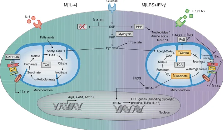
The aforementioned metabolic pathways are intricately dependent on one another during macrophage activation to synergistically ful-fill cell effector functions. For many years, in vitro and in vivo acti-vation of macrophages, and the study of their metabolic and effector responses, focused on two activation phenotypes: ‘M1’ macrophages classically activated by LPS and IFNγ stimulation, and alternatively activated ’M2’ macrophages activated by IL-4 stimulation (hereafter referred to as M[LPS+ IFNγ ] and M[IL-4], respectively). Although it has become clear that these macrophage phenotypes represent extremes, their mechanistic exploration provides a starting point for understanding how macrophage activation and metabolism inter-sect in dictating cellular responses (Fig. 1).
LPS induces a profound metabolic rewiring of macrophages and dendritic cells, which is characterized by increased glucose uptake and enhanced aerobic glycolysis, and is coincident with impaired OXPHOS via the TCA cycle21,22, a response resembling the Warburg effect in highly proliferative tumor cells1. In those cells, pyruvate produced by the glycolytic pathway is metabolized to lactate and
secreted, while TCA cycle intermediates accumulate, thus resulting in decreased OXPHOS and direct effects on immune responses22,23. For example, succinate accumulation leads to stabilization of the transcription factor HIF-1α and upregulation of enzymes involved in glycolysis and of the proinflammatory cytokine IL-1β (ref. 22). Citrate is shuttled to the cytosol, where it is used to generate fatty acids for membrane biogenesis and prostaglandin production24, and to produce the antimicrobial metabolite itaconic acid25. Other TCA-cycle intermediates, such as fumarate, contribute to macrophage ‘innate immune memory’ by modifying epigenetic remodeling by KDM5 demethylases26 and consequently influences future immune responses, as discussed later in this Review.
The PPP is enhanced in M[LPS+ IFNγ ] macrophages through increased flux of glucose intermediates through the pathway and downregulation of the kinase CARKL17, thus allowing for increased NADPH synthesis. NADPH availability from the PPP is essential for cholesterol metabolism and fatty acid synthesis27,28, which supports phagocytosis18,19, as well as for the expansion of the Golgi and ER that is required for production of inflammatory cytokines29,30. M[LPS+ IFNγ ] macrophages also exhibit elevated lipid accumulation via enhanced fatty acid uptake via the scav-enger receptor CD36 and diminished triglyceride lipolysis28. In addition, amino acid metabolism modulates immune-cell func-tions in M[LPS+ IFNγ ] macrophages: glutamine is required for LPS-induced IL-1β secretion31, whereas arginine is metabolized by inducible nitric oxide synthase, thus producing nitric oxide,
which acts both as an antimicrobial agent and as a signal for vaso-dilation, angiogenesis, and insulin secretion. The extent to which other metabolites can alter macrophage functional states is only beginning to be appreciated.
Compared with M[LPS+ IFNγ ] macrophages, M[IL-4] macro-phages have a less well-understood metabolic phenotype. M[IL-4] macrophages exhibit a precipitous increase in FAO and OXPHOS that is thought to contribute to anti-inflammatory responses23. CD36-mediated uptake of triglycerides and subsequent lipolysis via lysosomal lipase feeds the TCA cycle and increases OXPHOS, thereby sustaining M[IL-4] polarization32. However, the specific role of FAO in M[IL-4] macrophages has recently been questioned in studies showing that etomoxir-mediated inhibition of FAO or deficiency in CPT2, an enzyme required for fatty acid import, does not alter the M[IL-4] phenotype33,34. Notably, M[IL-4] macrophages do depend on glucose to drive OXPHOS23, because inhibition of glycolysis suppresses IL-4-induced gene expression35; however, the downregulation of the PPP limits glucose flux into this pathway17. In terms of amino acid metabolism, glutamine flux into both the TCA cycle and hexosamine pathway promotes M[IL-4] macrophage polarization23. Furthermore, arginine metabolism by arginase-1 produces l-ornithine, which is broken down into polyamines and l-proline to support macrophage proliferation and for collagen pro-duction used in tissue repair36,37.
Recent studies have revealed that the metabolic rewiring of M[LPS+ IFNγ ] and M[IL-4] macrophages does not represent
Arg1, Cdh1, Mrc1,2 HRE genes (encoding glycolytic
proteins, TLRs, IL-1β) HIF-1α HIF-1α IL-4 G6P Glycolysis PPP Fatty acids Acetyl-CoA OAA Citrate Isocitrate Succinate Fumarate Malate TCA OXPHOS cc ar M c a M c a M Su Fuma OXPHOS CI CII CIII CIV α-Ketoglutarate ↑CARKL ↑↑Lactate ↑Succinate ↑ROS ↑ROS Etc. ↑Membrane potential TCA Isocitrate α-Ketoglutarate ↑Nucleotides Amino acids NADPH iNOS Acetyl-CoA OAA Malate Fumarate FAS ↑NO ↑↑ATP Pyruvate Mitochondrion Mitochondrion Nucleus Glucose M[LPS+IFNγ] M[IL-4] LPS/IFNγ F6P PPP ↑Citrate et ↑ROS Is to Etc.
↑↑MembraM aneanee otentialalalaall pote l socitrate e oglutarate CI CII CIII CIV
Fig. 1 | Metabolic pathways controlling macrophage activation states. Glycolysis involves the conversion of glucose molecules into various metabolic
by-products, culminating in the end product pyruvate as well as two net ATPs. In macrophages treated with IL-4 (M[IL-4], left side of the cell) pyruvate (and fatty acids) enters the intact TCA cycle as acetyl-CoA, thus resulting in sustained ATP production via OXPHOS and leading to the upregulation of genes associated with tissue repair. In contrast, in macrophages treated with LPS and IFNγ (M[LPS+ IFNγ ], right side of the cell), most pyruvate is converted into lactate and secreted. Furthermore, the enzyme CARKL is downregulated, and as a result, glycolysis also feeds the PPP, thereby generating nucleotides, amino acids, and NADPH. The TCA cycle is broken in two places in M[LPS+ IFNγ ] macrophages, thus resulting in the accumulation of citrate, which in turn is used to drive fatty acid synthesis (FAS) and succinate, which stabilizes the transcription factor HIF-1α . HIF-1α enters the nucleus and promotes the expression of hypoxia response element (HRE)-containing genes, which encode both glycolytic and proinflammatory proteins such as IL-1β ). The disturbed TCA cycle and decreased OXPHOS also result in increased levels of ROS, which further stabilize HIF-1α , allowing it to drive glycolysis and inflammatory-gene expression. G6P, glucose 6-phosphate; F6P, fructose 6-phosphate; OAA, oxaloacetate. Credit: Kim Caesar/Springer Nature
a metabolic blueprint for all activated macrophages. Different microbial stimuli, endogenous danger ligands, and tissue micro-environmental signals lead to specific and complex metabolic rewiring of macrophages. As these unique metabolic signa-tures of macrophages have been unraveled, it has become clear that the type and fate of nutrients used are linked not only to the macrophage’s need for energy but also to the generation of metabolites that act as signaling molecules and building blocks (for example, lipids and nucleic acids) needed for cellular main-tenance or proliferation.
Studies using agonists of Toll-like receptors (TLRs) other than TLR4 or using whole microbial lysates suggest that the Warburg metabolism of M[LPS+ IFNγ ] macrophages is not a universal response to pathogenic stimuli2. In vitro stimulation of human CD14+ monocytes with ligands of TLR2 (with its ligands Pam3Cys
or P3C) or TLR3 (with its ligand poly(I:C)), or with whole patho-gen lysates from Escherichia coli, Staphylococcus aureus, or
Mycobacterium tuberculosis, induces increases in glycolysis and
OXPHOS2. These metabolic responses are connected to specific functional activities; blocking glycolysis with 2-deoxyglucose restricts production of IL1β in response to LPS and P3C, whereas blocking OXPHOS with rotenone lowers IL1β production and phagocytosis only in P3C-stimulated cells.
Cytokine stimulation of monocytes and macrophages also induces a distinct metabolic rewiring that is only beginning to be understood. As described for IL-4, stimulation with the cytokine IL-13, which also polarizes macrophages toward alternative activa-tion, increases mitochondrial biogenesis, FAO and OXPHOS38,39. Stimulation of macrophages with the myeloid growth factors M-CSF or GM-CSF increases [3H]2-deoxyglucose uptake and
oxy-gen consumption rate. Interestingly in mouse peritoneal macro-phages, M-CSF is a stronger inducer of the glycolytic extracellular acidification rate and lactate production than GM-CSF, whereas the opposite is true in human monocyte-derived macrophages40 and mouse bone marrow–derived macrophages41. The functional conse-quences of M-CSF- and GM-CSF-induced macrophage metabolism on cytokine production, phagocytosis, and cell migration remain to be determined. Interestingly, in human monocytes, IFNγ alone can enhance glycolysis and lactate production42, yet stimulation of mouse peritoneal macrophages with IFNγ in combination with other cytokines (for example, TNF39 or TNF plus IL-1β (ref. 43)) does not alter [3H]2-deoxyglucose uptake. Additionally, IL-10
pro-motes anti-inflammatory effects by suppressing the kinase mTOR44, regulating GLUT-1 translocation to inhibit glycolytic flux, and pro-moting mitophagy of dysfunctional mitochondria to help preserve respiratory capacity.
The divergent macrophage effector responses to in vitro TLR, pathogen, and cytokine stimuli described above highlight that mac-rophage cellular responses to environmental cues probably require different functional outputs accomplished through specific meta-bolic rewiring. Although these studies of macrophage metameta-bolic responses in vitro have led to an appreciation of the versatility and extent to which metabolic rewiring regulates the inflammatory function of these cells, macrophages in vivo are exposed to multiple stimuli simultaneously and must mount a coordinated response to balance a multitude of cellular demands, each dependent on spe-cific energy and metabolic-intermediate requirements. In the next section, we discuss how macrophage metabolism is dynamically regulated within the multifaceted microenvironment of atheroscle-rotic plaques. Specifically, we consider how priming of macrophage precursors by the systemic atherosclerotic milieu may alter cell function before recruitment to plaques, and how plaque microen-vironmental stimuli, such as lipoproteins, cholesterol, hypoxia and oxidative stress, apoptotic and necrotic cell death, and hypergly-cemia, may regulate macrophage metabolism within this dynamic microenvironment (Fig. 2).
Monocyte priming in hematopoietic reservoirs and the
circulation in atherosclerosis
Myeloid cells experience complex signals in vivo as they transit from hematopoietic reservoirs into different tissues, where they undergo differentiation into macrophages in unique microenviron-ments. Monocytes, which are derived from reservoir populations in the bone marrow and spleen, then released into the circulation, are the major precursors of the atherosclerotic-plaque macrophage population (reviewed in ref. 5). Before their arrival in the artery wall, these monocytes are exposed to a variety of pathologic stimuli within the systemic milieu. Evidence from studies of monocytes isolated from people with atherosclerosis and from mouse models of atherosclerosis indicates that this exposure modulates both cel-lular metabolism and function, thus priming monocytes toward an inflammatory phenotype well before their differentiation within the plaque microenvironment.
Levels of monocytes in the circulation, particularly those of the CD14+ subpopulation in humans and the Ly6Chi subpopulation
in mice, are strongly correlated with atherosclerosis progression8. Multiple factors in the atherosclerotic systemic milieu have been shown to drive an increase in myelopoiesis, including cholesterol homeostasis45, chronic inflammation, activation of the sympathetic nervous system46,47, and defective cholesterol efflux48. Enhanced glu-cose uptake by monocytes and hematopoietic precursor cells has been observed in mouse models of atherosclerosis and is thought to contribute to enhanced myelopoiesis. Inhibition of GLUT-1 directly, or by increasing high-density lipoprotein (HDL) levels, decreases myleoproliferation in mouse models49. Using a trans-plant approach in which atherosclerosis-susceptible Apoe–/– mice
were reconstituted with either wild-type or Apoe–/– bone marrow,
Sarrazy et al.50 observed enhanced uptake of [14C]2-deoxyglucose
in the bone marrow, spleen, and plaques of Apoe–/– bone marrow–
transplanted mice. Within the bone marrow compartment, the mice with Apoe–/– bone marrow exhibited elevated glucose uptake by
multipotent progenitors, abundance of granulocytic-macrophage progenitors (GMPs), and increased GLUT-1 expression and uptake of the fluorescent d-glucose analog 2-NBDG by hematopoietic stem and progenitor cells (HSPCs). These alterations in glucose uti-lization were accompanied by enhanced oxygen consumption and increased mitochondrial membrane potential. Mechanistically, the upregulation of GLUT-1 was found to be dependent on signaling by the interleukin receptor IL-3Rβ , which drove subsequent glycolytic substrate utilization by the mitochondria and accelerated myelopoi-esis in Apoe–/– mice.
These metabolic alterations also extend to monocytes isolated from atherosclerotic patients. In a study comparing human mono-cytes isolated from healthy individuals or individuals with athero-sclerosis, Shirai et al.51 have mechanistically explored the concept of ‘monocyte priming’ in relation to cellular metabolic status. After ex vivo stimulation with LPS and IFNγ , monocytes isolated from the atherosclerotic patients produced more IL-6 and IL-1β than did monocytes from control individuals, and this primed pheno-type was maintained after their in vitro differentiation into mac-rophages. Together with these differences, LPS+ IFNγ -stimulated monocytes from atherosclerotic patients exhibited a higher oxygen consumption rate and respiratory reserve capacity, as well as higher glycolytic extracellular acidification rate and glycolytic flux, than did monocytes from healthy individuals. Notably, the enhanced glucose uptake of atherosclerotic monocytes was proportional to mitochondrial ROS production, oxidative stress, and subse-quent inflammatory signaling. Molecular-level investigation of the underlying mechanisms identified a glucose–ROS–PKM2–STAT3 pathway through which glucose utilization led to unbalanced ROS generation from the mitochondrial respiratory chain, which in turn induced the redox-sensitive enzyme PKM2 (one of the isoforms of the enzyme that catalyzes the final step of glycolysis and converts
phosphoenolpyruvate to pyruvate) to translocate from the cyto-plasm to the nucleus, thus resulting in activation of the transcrip-tion factor STAT3 and transcriptranscrip-tion of IL6 and IL1B.
Cumulatively, the preclinical and clinical evidence to date pro-vides direct evidence that the atherosclerotic systemic milieu primes both reservoir and circulating hematopoietic myeloid populations before recruitment to the plaque microenvironment. Specifically, these cells utilize more glucose, have enhanced rates of glycoly-sis and OXPHOS, and are hyperinflammatory before recruitment to plaques.
Microenvironmental cues shape macrophage metabolism
and function in plaques
In atherosclerotic vessels, as in other tissue sites, distinct microen-vironmental factors influence macrophage metabolism and phe-notype, and these forces change as plaques evolve and progress. As described below, the environment of atherosclerotic plaques is influenced by hyperlipidemia, oxidative stress, hypoxia, and cell death. In addition, in people with diabetes, hyperglycemia, a comorbid condition that accelerates atherosclerosis, is also a major factor influencing macrophage metabolism. Together, these micro-environmental signals are important drivers of the metabolic repro-gramming of macrophages and of their inflammatory responses, both of which ultimately affect disease progression and plaque stability. Below, we summarize in vivo evidence, and support from in vitro mechanistic work, of plaque microenvironmental features
that drive changes in macrophage function alongside metabolic adaptations and consequently facilitate cellular effector responses in atherosclerosis.
Macrophage uptake of modified lipoproteins and resulting phe-notypes. Monocyte-derived and tissue-resident macrophages lead
the response to lipoproteins that are retained and modified in the artery wall. These macrophages attempt to clear the inflammatory lipids and are transformed into ‘foamy’ macrophages as they store the excess cholesterol and triglyceride in cytoplasmic lipid droplets. Cholesterol loading of macrophages diminishes their migratory capacity, and, for reasons that are still unclear, these macrophage foam cells persist in the artery wall, where they foster chronic inflammation. In vitro studies mimicking macrophage foam cell formation by using oxidized low-density lipoprotein (oxLDL) have shown that oxLDL is a potent inducer of macrophage gly-colysis52, inflammation53, and mitochondrial oxidative damage54. Both oxLDL and its associated oxidized phospholipids have been shown to induce inflammatory signaling via TLR4 homodimers or TLR4–TLR6 heterodimers, thus leading to cytokine and chemo-kine expression as well as changes in the actin cytoskeleton55,56. In addition, the unregulated uptake of oxLDL via CD36 results in the nucleation of intracellular cholesterol crystals that destabilize lyso-somes, thereby impairing cholesterol and fatty acid metabolism, and activating the NLRP3 inflammasome and production of IL-1β (refs 57,58). Numerous cellular metabolic disturbances, all present in
↑Glycolysis ↓OXPHOS ↑ROS ↑Glut-1, HIF-1α ↑Inflammatory cytokines ↑Mitchondrial dysfunction ↓Mitchondrial fission ↓Efferocytosis ↓Glycolysis ↑OXPHOS ↓Inflammatory cytokines ↑Cholesterol efflux ↑Tissue repair ↑Glycolysis ↑Glucose consumption ↑Inflammatory cytokines ↑OXPHOS ↑Myelopoiesis
↑Glutathione- and regulation ↓Phagocytosis ↓Migration ↑Glycolysis ↑Glut-1 ↑Myelopoiesis ↑↑Glycolysis ↑↑Glut-1 ↓OXPHOS ↓OXPHOS Hypoxia Necrotic core Hyperglycemia Monocyte priming oxLDL LDL ↑HIF-1α ↑Inflammatory-cytokine production ↑↑Glycolysis ↑↑Glut-1 ↑↑Glycolysis ↑OXPHOS M(ox) Lumen Intima SMC Media EC ↑↑Glut-1 ↑NRF2 M[LPS+ IFNγ]-like ↑OXPHOS ↓Glycolysis M[IL-4] ↑PPARγ
LDL oxLDL Inflammatory cytokine
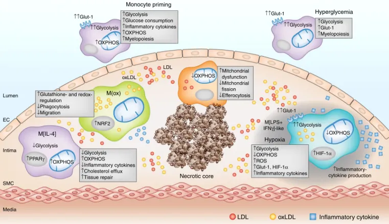
Fig. 2 | Macrophage metabolic reprogramming in atherosclerotic plaques. In the hypercholesterolemic systemic environment, monocytes exhibit a
‘primed’ inflammatory phenotype characterized by enhanced inflammatory responses that correspond to increases in both glycolysis and OXPHOS. After entering into the plaque microenvironment, a variety of stimuli including lipids, cytokines, cell apoptosis and necrosis, hypoxia, and hyperglycemia can influence macrophage metabolic reprogramming and subsequent function. Distinct macrophage phenotypes have been identified within plaques, including M[IL-4]-like, M(ox), M[LPS+ IFNγ ]-like, and hypoxia-associated macrophages. These different types of macrophages are associated with distinct metabolic signatures and inflammatory functions. PPARγ , peroxisome proliferator–activated receptor gamma; NRF2, nuclear factor 2. Credit: Kim Caesar/ Springer Nature
plaque macrophages, have been linked to NLRP3-inflammasome activation, including the release of lysosomal cathepsins, mitochon-drial ROS, and extracellular ATP59. Furthermore, free-cholesterol enrichment of macrophage foam cell membranes can enhance inflammatory signaling from lipid rafts, particularly via activation of TLR4 signaling and the transcription factor NF-κ B, indepen-dently of ligand stimulation60–62. In addition, oxLDL can activate HIF-1α , which fuels increased glucose uptake, glycolysis, and mac-rophage inflammatory responses52,63. Thus, macrophage foam cells generated in vitro exhibit multiple characteristics of M[LPS+ IFNγ ] macrophages, including elevated glycolysis, TLR4 activation, and IL-1β production.
Attempts to characterize the plaque macrophage inflammatory phenotype in vivo have revealed multiple subsets of macrophages, which are dynamically regulated during the progression and regres-sion of disease. Macrophages with general markers of the M[LPS+ IFNγ ] phenotype are present in early atherosclerotic lesions, and their proportion increases as plaques progress to more complex inflammatory lesions64. These M[LPS+ IFNγ ]-like macrophages are enriched in lipid, in agreement with an increase in glycolysis, and they populate different regions within plaques than macrophages with markers of the M[IL-4] phenotype65. During atherosclero-sis regression, the balance of M[LPS+ IFNγ ]-like and M[IL-4]-like macrophages switches, such that M[IL-4]-M[IL-4]-like macrophages become more prominent66–68. Plaque macrophages resembling the M[IL-4] phenotype show little lipid accumulation, in agreement with a high level of FAO, and these cells are thought to be atheropro-tective because of their anti-inflammatory and profibrotic proper-ties. Indeed, treatment of Ldlr−/− mice with the M[IL-4]-polarizing
cytokine IL-13 inhibits atherosclerosis progression69. In addition, a third macrophage phenotype, termed M(ox), has been identified in atherosclerotic plaques and found to be induced in vitro in response to oxidized phospholipids70. M(ox) macrophages are characterized by high expression of genes whose transcription is dependent on the transcription factor NRF2 (including Hmox1, Srxn1, and Txnrd1), and by a glutathione- and redox-regulating phenotype. These mac-rophages have lower phagocytic and migratory capacity than do macrophages stimulated with M[IL-4] or M[LPS+ IFNγ ], character-istics that may potentially contribute to their persistence in plaques. Attempts to quantify these macrophage populations in advanced plaques of Ldlr–/– mice have indicated a distribution of ~40%
clas-sically activated (CD86hi), ~20% alternatively activated (CD206+),
and ~30% M(ox) macrophages.
To date, metabolic phenotyping of plaque macrophages beyond cell-surface markers has been limited, not only because of the chal-lenges of isolating macrophages from plaques (typically through aortic digestion, which can alter the cellular metabolic state) but also because of previous technological limitations in understand-ing cellular phenotypes at the sunderstand-ingle-cell level. Within plaques, it is likely that discrete phenotypic populations and/or a phenotypic spectrum of atherosclerosis-induced macrophage activation states exist, which are driven by the various plaque microenvironmental signals encountered in a spatiotemporal fashion. Indeed, emerging and established single-cell and imaging technologies hold promise in identifying, characterizing, and understanding these spatiotem-poral activation states within plaques, thus enabling subsequent targeting of either distinct phenotypic populations or shifting the phenotypic spectrum for therapeutic benefit.
Macrophage cholesterol storage and efflux. Under homeostatic
conditions, lipoproteins taken up by macrophages are transported to late endolysosomal compartments, where cholesterol esters are hydrolyzed. The endolysosomal proteins NPC1 and NPC2 func-tion together to transport free cholesterol to the cytoplasm, where it is trafficked to the cell membrane for export or delivered to the ER for reesterification and storage in lipid droplets. Macrophages
in advanced plaques show evidence of massive free-cholesterol accumulation, thus suggesting a breakdown in these processes that maintain cholesterol homeostasis. Excess free-cholesterol accumu-lation is damaging to membranes and causes dysreguaccumu-lation of meta-bolic processes that occur in the ER and mitochondria.
Cholesterol efflux via the lipid transporters ABCA1 and ABCG1 is key to maintaining cholesterol homeostasis in plaque macro-phages and promoting reverse cholesterol transport to the liver for excretion71. This process relies on proper mitochondrial function. A sustained supply of ATP is required to fuel the activity of ABC transporters to move their substrates (free cholesterol and phos-pholipids) across the membrane to an acceptor, and treatment of macrophages with oligomycin, which blocks the production of ATP from OXPHOS, markedly decreases cholesterol efflux to apoA1 (ref. 72). Furthermore, macrophages from mice lacking PGC1α , a master regulator of mitochondrial biogenesis, have impaired cho-lesterol efflux72, and accordingly Ldlr–/– mice deficient in
hema-topoietic Ppargc1a expression exhibit accelerated progression of atherosclerosis73. Mitochondria are also an important source of oxy-sterol ligands for the liver X receptor transcription factors, which coordinate the expression of genes whose products are involved in the response to cholesterol excess, including ABCA1 and ABCG1. The mitochondrial sterol 27-hydroxylase CYP27A1 converts excess cholesterol to 27-hydroxycholesterol, and mitochondrial transport of cholesterol from the outer to inner membrane is rate limiting in CYP27A1’s generation of oxysterols.
Studies of hypercholesterolemic mouse models suggest that mitochondrial metabolism is disturbed in atherosclerotic-plaque macrophages. In the REVERSA mouse model of genetically revers-ible hypercholesterolemia, mitochondrial genes have been found to be significantly downregulated in the aorta during progression of atherosclerosis, coincident with rapid lesion expansion and elevated CD68+ macrophage content within plaques74. Functional enrich-ment analysis indicated an overrepresentation of mitochondrial and ROS homeostasis pathways, and markedly diminished expression of genes involved in mitochondrial biogenesis and the antioxidant response (for example, Sod2, Ucp1, and Ucp3). Notably, after genetic reversal of hypercholesterolemia, more than 200 genes were found to be upregulated in the aorta, 30% of which were nuclear-encoded mitochondrial genes. Of those mitochondrial cholesterol-respon-sive genes, 70% corresponded to genes downregulated during hypercholesterolemia and lesion expansion, thus suggesting that this effect can be reversed. Although this study did not examine isolated macrophages, given their prominence in mouse atheroscle-rotic plaques, macrophage mitochondrial function may be specu-lated to drive this response.
Strategies that increase mitochondrial biogenesis or function have been shown to enhance the export of cholesterol from macro-phages. In atherosclerotic-plaque macrophages, microRNA (miR)-33 appears to be a nexus for repressing mitochondrial function, cholesterol efflux, and FAO. miR-33a and miR-33b are upregulated in human and mouse atherosclerotic plaques68,72, and these noncod-ing RNAs post-transcriptionally inhibit genes in networks regu-lating mitochondrial biogenesis and function (Ppargc1a, Slc25a3,
Pdk4, and Nrf1)72, intracellular cholesterol transport and efflux
(NPC1 (in humans only), OSBPL6, ABCA1, and Abcg1 (in mice only))75,76, autophagy and lipid-droplet catabolism (ATG5, ATG12,
LC3B, and LAMP1)77,78, and FAO (HADHB, CPT1A, CROT, and
PRKAA1)79. Antagonism of miR-33 in mouse models of
atheroscle-rosis has been shown to be atheroprotective through raising plasma levels of HDL cholesterol, promoting macrophage cholesterol efflux and reverse cholesterol transport, enhancing mitochondrial func-tion, and decreasing macrophage inflammatory polarization68,72,80,81.
Oxidative stress and hypoxia. Oxidative stress and
cycle that fuels atherosclerotic-plaque progression. During ath-erosclerosis, macrophage ROS are generated through the actions of mitochondrial oxidative metabolism, NADPH oxidases, peroxi-dases, nitric oxide synthases, cyclooxygenases, and lipoxygenases. The macrophage antioxidant response is key to decreasing cellular levels of ROS and protecting mitochondria and other organelles, proteins, and nucleic acids from oxidative damage; however, the transcription of antioxidant genes and mitochondrial transport of the antioxidant glutathione are suppressed in plaque macrophages, thus magnifying inflammation in the artery wall. For example, NADPH oxidase–derived ROS from macrophages promotes oxida-tion of LDL in the artery wall, amplifying macrophage foam cell formation82. Furthermore, excessive mitochondrial ROS generated as a by-product of electron-transport-chain reactions can dam-age mitochondrial DNA, proteins, and lipids, and these processes increase during atherosclerosis in mice83. The oxidation or release of mitochondrial DNA can also serve as a danger-associated molec-ular pattern that activates multiple innate immunological signal-ing pathways in macrophages, includsignal-ing the CpG DNA receptor TLR9, the NLRP3 inflammasome84,85, and the cyclic GMP–AMP synthase–stimulator of interferon genes pathway86–88. Decreasing mitochondrial oxidative stress in macrophages can prevent inflam-mation in plaques and decrease atherosclerotic burden in mice54.
In addition to cellular oxidative stress, the imbalanced demand and supply of oxygen in the plaque microenvironment leads to hypoxia in human and mouse atherosclerotic plaques and is a key contributor to atherogenesis89. Local hypoxia within the plaque environment arises because of a combination of increased cel-lular metabolic demand coupled with impaired oxygen delivery due to increased diffusion distance within plaques90. In humans, hypoxic regions in the plaque microenvironment are rich in HIF-1α -expressing macrophages and foam cells91, which are associated with angiogenesis and plaque hemorrhage90. In atherosclerotic mice, a decrease in plaque hypoxia after administration of hyperoxic car-bogen gas (95% O2, 5% CO2) results in favorable changes in plaque
characteristics, including an increase in M[IL-4]-like macrophages and a decrease in both IL-6 production and necrotic-core size92.
ROS and hypoxia are potent drivers of macrophage metabolic reprogramming that results in enhanced reliance on glycolysis. Critical to this metabolic switch is activation of the HIF-1α tran-scription factor, which induces the expression of GLUT-1 and glycolytic enzymes (for example, HK, PFK, and PFKB3), and lim-its OXPHOS93. Hypoxia response element–containing genes also encode a variety of inflammatory proteins, such TLR2, TLR4, IL-1β , and CXCR4, thus resulting in a coordinated inflammatory response. As such, activation of the HIF-1α pathway, particularly in mac-rophages, is critically involved in modulating both the metabolic and inflammatory effector responses. Increased glucose uptake associated with HIF-pathway activation has been demonstrated in mouse and human atherosclerotic lesions. In human plaques, expression of HIF-1α colocalizes with high expression of GLUT1 (official symbol SLC2A1), GLUT3 (official symbol SLC2A3), HK1, and HK2 in macrophage-rich regions94, thus suggesting enhanced glycolysis in these cells. Furthermore, expression of the HIF-1α -regulated cytokine IL-1β is also elevated within macrophage-rich hypoxic plaque regions94, and hypoxia potently induces NLRP3 activation and IL-1β expression in human macrophages. Similarly, in the plaques of Ldlr–/– and Apoe–/– mice, there is an abundance
of hypoxic HIF-1α -expressing macrophages90,95, and laser capture of HIF-1α -positive macrophages has revealed significantly greater
Glut1 (Slc2a1) expression than that in macrophages from normoxic
regions. In vitro studies of macrophage foam cells support these in vivo observations. Hypoxia markedly potentiates macrophage gly-colytic flux in the presence of proatherogenic mediators such as oxLDL and proinflammatory cytokines (IL-1β , IFNγ , and TNF)63, and this effect is dependent on HIF-1α and PFKFB3. Interestingly,
the glycolytic shift in response to hypoxia relies on mitochondria for appropriate oxygen sensing, because macrophages that lack mitochondria do not accumulate HIF-1α in response to hypoxia96. HIF-1α also increases macrophage sterol and triglyceride content by stimulating sterol synthesis and suppressing cholesterol efflux via ABCA1 (ref. 95), and consequently further exacerbates cholesterol accumulation in plaque macrophages. Macrophages deficient in HIF-1α exhibit low expression of inflammatory genes (for example, those encoding monocyte chemoattractant protein-1 and osteopon-tin), as well as apoptosis, and hematopoietic deficiency of HIF-1α (ref. 90) or GLUT-1 (ref. 50) in mice decreases atherosclerotic burden. Cumulatively, these studies implicate hypoxia as a driver of macro-phage phenotype in atherosclerosis progression, highlighting that HIF-1α activation and increased glycolysis are central to this patho-logic response.
Macrophage death and efferocytosis. Prolonged defects in
cho-lesterol efflux and/or free-chocho-lesterol esterification by the ER can ultimately lead to macrophage death in plaques. ER stress has been documented in human and mouse atherosclerotic-plaque macro-phages, particularly in the advanced stages of disease, and decreas-ing ER stress can mitigate atherosclerosis97,98. ER stress leads to the induction of the unfolded-protein response, which reestablishes homeostasis in the ER, thereby maintaining functions in folding and secretion of proteins, calcium storage, and lipid synthesis for membrane biogenesis or energy storage. However, if prolonged, ER stress can trigger apoptosis through the ER-resident protein IRE1 or downstream effectors such as CHOP5. In advanced plaques, clear-ance of apoptotic cells by surrounding macrophages can become compromised as these cells undergo metabolic dysfunction. Efficient clearance of apoptotic cells by macrophages requires intact lysosomal function and lipid metabolism to cope with the dramatic increase in lipid, protein, nucleotide, and carbohydrate content that occurs after the ingestion of apoptotic bodies. Thus, the combina-tion of defective macrophage cholesterol efflux, enhanced apoptotic cell death, and defective efferocytosis by plaque macrophages can result in secondary necrosis and the release of cellular components into the plaque milieu, effects that in turn are thought to contribute to the formation of the lipid-rich, acellular necrotic core that char-acterizes vulnerable plaques.
Efferocytosis is dependent on a coordinated metabolic cellular response. In vitro, FAO is enhanced during efferocytosis, and alter-ing the mitochondrial membrane potential by glucose deprivation or providing excess glucose can increase and decrease efferocyto-sis, respectively99. Thus, the mitochondrial membrane potential within macrophages is a critical determinant of phagocytic capacity and continued apoptotic cell uptake99. Mitochondrial morphology exists in dynamic flux, changing rapidly through fission or fusion in response to external stimuli and cellular metabolic needs. In set-tings of nutrient deprivation, increased demand for mitochondrial respiration leads to mitochondrial fusion, which is associated with increased ATP production and resistance to autophagy100. In athero-sclerosis, mitochondrial fission is required in response to apoptotic cell uptake for effective efferocytosis by macrophages in the ath-erosclerotic plaque101. Interestingly, M[IL-4] macrophages, which have high OXPHOS, have a greater efferocytosis capacity than do M[LPS+ IFNγ ] macrophages, thus suggesting that this difference in metabolism may determine macrophage efferocytosis ability in ath-erosclerotic plaques. In agreement with this possibility, factors asso-ciated with M[LPS+ IFNγ ] macrophages, such as oxidative stress and hypoxia, inhibit efferocytosis in atherosclerotic plaques92,102.
Hyperglycemia. Hyperglycemia associated with type 1 or type 2
diabetes mellitus is an independent risk factor for atherosclerosis and cardiovascular events103, and is associated with elevated inflam-matory monocyte and macrophage activation and hematopoiesis104.
Type 1 diabetes–induced hyperglycemia results in proliferation and expansion of bone marrow myeloid cell progenitors and release of inflammatory monocytes into the circulation, coinciding with impairment of atherosclerosis resolution, which is mitigated with treatment for hyperglycemia104. Interestingly, overexpres-sion of the glucose transporter GLUT1 in myeloid cells has been found to increase glycolysis and flux through the PPP, coupled with a compensatory decrease in FAO; however, this overexpres-sion alone does not induce cytokine production105. Furthermore, in
Apoe–/– mice, myeloid-specific overexpression of GLUT-1 does not
promote atherosclerosis. Thus, macrophage glycolytic flux alone is not sufficient to drive atherosclerosis, and the atheropromoting macrophage response to a hyperglycemic systemic milieu is driven by factors beyond increased rates of glycolysis. The metabolic changes in myeloid cells in the diabetic setting remain an area of active investigation.
Epigenetic and metabolic memory shape macrophage
responses
Although metabolic modulation is integral to the macrophage activation response, recent discoveries have also unveiled its role in ‘learned’ innate immune functional responses. Macrophages exposed to acute or chronic stimuli, both in vitro and in vivo, can remodel their chromatin to mount a ‘learned’ response to subse-quent stimulation with the same or different stimuli, in a process termed innate immune memory. These memory responses are char-acterized phenotypically by cellular inflammatory responses that are partly regulated by changes in metabolic machinery. Although these effects have predominately been defined in the context of pathogen response, emerging evidence suggests that innate immune memory also regulates the metabolic and effector response to sterile inflam-matory disease states, such as atherosclerosis.
Two categories of innate immune memory responses to patho-genic stimuli have been described: ‘trained’ or ‘tolerant’ effector responses. Innate immune training, which results in a potentiated inflammatory response of monocytes and macrophages, occurs in response to pathogenic stimuli such as bacillus Calmette–Guéri vaccination and β -glucan (Fig. 3). Innate immune tolerance, which results in a dampened response to secondary stimuli, occurs after exposure to LPS. The divergent responses of training and toler-ance are facilitated in part by metabolic cellular adaptations. For example, β -glucan-induced training is accompanied by a meta-bolic switch from OXPHOS to aerobic glycolysis via activation of the mTOR–HIF1α pathway. Epigenetic analysis of cells after β -glucan stimulation has revealed an increase in trimethylated his-tone H3K4 and acetylated hishis-tone H3K27 in the promoter region of EIF4EBP1, the main target of mTOR, and highly increased expression of genes involved in the mTOR signaling and glyco-lytic pathways106. The increase in acetylated histone H3K27 in β -glucan-trained monocytes has been linked to a decrease in the NAD+-dependent class III histone deacetylase sirtuin-1, and early
treatment with the sirtuin-1 activator resveratrol during β -glucan training partially decreases IL-6 production106. Integration of the transcriptome and metabolome of β -glucan-trained monocytes and macrophages has also revealed upregulation of metabolic pathways, including glucose metabolism, glutaminolysis, and cho-lesterol synthesis26,106. In contrast, macrophages undergoing innate immune tolerance after exposure to LPS exhibit a metabolic switch from glycolysis to OXPHOS and an increased NAD+/NADH ratio,
thus resulting in activation of the histone deacetylases sirtuin-1 and sirtuin-6 and consequent inhibition inflammatory-gene tran-scription107. Collectively, these findings have established the para-digm that the innate immune memory of macrophages is linked to their metabolic programs and can influence their subsequent inflammatory response to microbial and probably endogenous inflammatory ligands.
Innate immune memory occurs not only in monocytes and mac-rophages but also within myeloid progenitor populations in the bone marrow. β -glucan-treated mice, up to 28 d after treatment, show increased HSPC populations, including long-term hema-topoietic stem cells108. RNA-seq of long-term hematopoietic stem cells 7 d after β -glucan injection has identified glycolysis, cholesterol biosynthesis, and the mevalonate pathway as functions overrepre-sented among significantly upregulated genes, whereas the expres-sion of genes involved in cholesterol efflux is decreased. Within the bone marrow, IL-1β is increased, and increased numbers of CD131+
(the GM-CSF receptor) HSPCs have been observed. Treatment with an IL-1-receptor antagonist or blockade of GM-CSF restricted the β -glucan-induced expansion of HSPCs, as did inhibition of glycoly-sis with 2DG or inhibition of the cholesterol-biosyntheglycoly-sis pathway with statin therapy.
In monocytes, activation of the cholesterol-biosynthesis pathway, but not cholesterol synthesis itself, has been found to be essential for the induction of trained immunity109. Statins prevent training induced by β -glucan and bacillus Calmette–Guéri, thus blunting both upregulation of cytokine production after secondary LPS stimulation and increased glycolysis. The addition of exogenous mevalonate, the metabolite directly downstream of HMG-CoA reductase (the target of the statin class of drugs), is sufficient to res-cue the β -glucan innate immune training phenotype in the presence of a statin, and mevalonate alone induces a trained immunity phe-notype through epigenetic reprogramming, enhanced glycolysis, and mTOR signaling. The ability of mevalonate to induce innate immune training indicates that this response is not restricted to microbial stimuli and that factors associated with sterile inflamma-tory insults, such as those that occur in atherosclerosis and other chronic inflammatory conditions, are likely to induce similar effec-tor and metabolic responses that heighten inflammation.
Recent studies have implicated two microenvironmental fac-tors of atherosclerosis, namely oxLDL and hypercholesterolemia, as sterile drivers of innate immune training. Human monocytes exposed to oxLDL53 and then re-stimulated with TLR2 (Pam3Cys) or TLR4 (LPS) agonists exhibit increased expression of inflam-matory cytokines (IL-6, TNFα , IL-8, and MCP-1), and this effect is reversed by treatment with a methylation inhibitor, thus impli-cating epigenetic reprogramming in this response. In an indepen-dent study, human monocytes trained with oxLDL for 5 d and then stimulated with LPS have also been found to exhibit enhanced IL-6 and TNF-α responses110, which are abrogated by treatment with a recombinant IL-1R antagonist. Notably, statin treatment of oxLDL-trained monocytes also blunts the immune-training phenotype by mitigating the enhanced glycolytic and proinflammatory-cytokine response to LPS re-stimulation109. In vivo, a hypercholesterolemic environment has been found to drive innate immune training in mice110. Monocytes isolated from Ldlr–/– mice fed a western diet
(WD) for 4 weeks showed enhanced cytokine and chemokine production when stimulated ex vivo with various TLR agonists. Furthermore, in vivo treatment of mice fed a WD for 4 weeks with LPS resulted in higher levels of serum inflammatory cytokines than were observed in chow-diet-fed mice. In agreement with findings from previous studies, the WD increased myelopoiesis, including levels of the myeloid progenitor GMP population, which showed priming of inflammatory genes and a downregulation of choles-terol-biosynthesis pathways in RNA-seq analysis. Notably, switch-ing the WD-fed mice back to chow diet for 4 weeks normalized the systemic inflammatory milieu but did not normalize the enhanced monocyte response to ex vivo TLR stimulation. RNA-sequencing and assay for transposase-accessible chromatin using sequencing (ATAC-sequencing, which identifies changes in open and acces-sible chromatin) revealed that the WD effectively reprogrammed GMPs at the transcriptional and epigenetic levels. Notably, treat-ment of mice with an IL-1R antagonist, or deficiency of the NLRP3
inflammasome, which regulates IL-1β production, abrogated the enhanced cytokine response of WD-trained monocytes.
In clinical studies, circulating monocytes from patients with ath-erosclerosis have also been found to have an enhanced inflamma-tory signature that is associated with epigenetic remodeling, which may also be connected to metabolic alterations. For example, an enhanced inflammatory-cytokine response to TLR agonists also occurs in patients with either high lipoprotein(a) levels111 or estab-lished atherosclerosis112. Monocytes isolated from patients with symptomatic atherosclerosis elicit a stronger cytokine response to ex vivo challenge with LPS and P3C than do monocytes from controls. A subsequent analysis of mRNA from whole-blood circulating cells has revealed higher expression of glycolytic (HK2, PFKFB3, PKM, and PDHA1), TCA-cycle (ASL and MDH1), and PPP (TALDO1) genes, as compared with expression in controls112. Collectively, these studies in mice and humans have unveiled the potent effects of innate immune training on circulating monocytes and hematopoi-etic progenitors that may contribute to atherogenesis. Furthermore, the role of IL-1β in macrophage innate immune training in response to a hypercholesterolemic environment110 is noteworthy, given the recent findings from the CANTOS trial that therapeutic targeting of IL-1β with the monoclonal antibody canakinumab decreases the rate of recurrent cardiovascular events in patients with previ-ous myocardial infarction14, as well as lung cancer incidence and mortality113. These findings raise the possibility that part of the ben-eficial effects of blocking IL-1β signaling in cardiovascular disease may derive from the ability of this treatment to reverse myeloid cell epigenetic reprogramming during inflammation, as well as subse-quent heightened immune responses.
Summary and future directions
Much work remains to be done to unravel the intertwined and dynamic metabolic and epigenetic landscapes that determine immunologic function in myeloid cells as these cells are exposed to microbial stimuli and unique tissue microenvironments in health and disease. In atherosclerosis, it is clear that macrophages exhibit metabolic rewiring in response to dyslipidemia and environmen-tal factors (hypoxia, dying cells, and cytokines) that lead to meta-bolic heterogeneity among plaque macrophages. These metameta-bolic changes are likely to be dynamic, because the microenvironment evolves during both plaque progression and treatment of disease. Although not discussed in this review, metabolic reprogramming is also ongoing in the other cell types in plaques, including T cells, dendritic cells, endothelial cells, and smooth muscle cells, and these changes in turn affect macrophage metabolic programs through nutrient competition, extracellular accumulation of metabolic by-products, and cell–cell signaling and interactions.
In the field of basic macrophage biology, there is a need for bet-ter delineation of metabolic adaptations and the roles of specific metabolites in regulating distinct macrophage effector responses, such as phagocytosis, cytokine production, cholesterol efflux, autophagy, and extracellular matrix synthesis and degradation. Although these processes are often studied in isolation in cul-tured cells, there is also a need to understand how macrophages coordinate their metabolic needs when they must exert multiple effector responses simultaneously. Furthermore, beyond single environmental stimulatory agents, there is a need to understand how macrophages behave in vivo, where the microenvironment is more complex, and where macrophages experience multiple stimuli Trained macrophage a b Nontrained macrophage ↓OXPHOS ↑Lactate ↑Succinate ↑Inflammatory-cytokine production ↑Citrate ↑Glycolysis ↑H3K4me3 ↑H3K27ac Pyruvate Glut-1 Glucose β-glucan Akt mTOR Acetyl-CoA Histone acetyl transferase ↑HIF-1α Lactate Nucleus Mitochondrion Fumarate ↓TCA ↓NAD+/↑NADH Histone deacetylase sirtuin-1+6 ↓OXPHOS Lactate Acetyl-CoA ↑Glycolysis ↑H3K4me3 ↑H3K27ac Pyruvate Glut-1 Glucose Nucleus Mitochondrion
↑TCA ↑↑NAD+/↓NADH
Histone deacetylase sirtuin-1+6
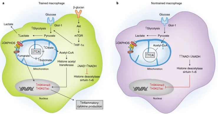
Fig. 3 | Epigenetic alterations that shape the macrophage immune response. Stimuli that activate immune cells and induce metabolic rewiring are also
capable of inducing epigenetic remodeling through histone modifications, thereby resulting in innate immune memory. a, In trained macrophages (e.g.,
those stimulated with bacillus Calmette–Guéri or β -glucan), a broken TCA cycle results in the accumulation of citrate and succinate. These metabolites activate histone-modifying enzymes, such as histone acetyltransferase, thereby increasing K4-trimethylated histone H3 (H3K4me3) and K27-acetylated histone H3 (H3K27ac) at promoter sites of genes encoding inflammatory cytokines and subsequently resulting in an increased inflammatory response. Additionally, a decreased ratio of NAD+ to NADH in trained cells inhibits the activity of NAD+-dependent histone deacetylases sirtuin-1 and sertuin-6, thus supporting the increased inflammatory response. b, This response is absent in nontrained macrophages, which have lower basal levels of H3K4me3 and
H3K27ac, owing to an intact TCA cycle, which prevents the accumulation of citrate and succinate, and an elevated NAD+/NADH ratio, which results in activation of sirtuin-1 and sirtuin-6. Akt; protein kinase B. Credit: Kim Caesar/Springer Nature
concurrently. Further study, particularly at the single-cell level, will be needed to determine how macrophages prioritize the different signals that they are exposed to and adopt metabolic signatures that fulfill these different needs.
From an atherosclerosis perspective, it will be important to bet-ter understand how the metabolic signatures of monocytes and macrophages change during early atherogenesis, and to determine the point at which these programs become maladaptive and disease promoting. Such an understanding will require studying macro-phage metabolism across multiple stages of atherosclerosis, includ-ing early foam cell lesions, advanced plaques, and, importantly, plaques vulnerable to rupture, which present the highest risk for myocardial infarction. To date, metabolic studies of macrophages taken from plaques have been limited because their isolation is chal-lenging, yet these cells offer metabolic clues for understanding the switch from functional to dysfunctional metabolic programs.
There is intense interest in exploiting the plasticity of macro-phage metabolism to rebalance inflammatory responses and restore protective immune functions in atherosclerosis. A better under-standing of the specific metabolic rewiring of activated macro-phages during both progression and regression of atherosclerosis will be of fundamental importance in identifying novel therapeutic approaches. Evidence from mouse models of atherosclerosis regres-sion has already indicated that lowering apoB-lipoprotein produc-tion or raising circulating HDL levels shifts the macrophage plaque phenotype toward that of M[IL-4]-like tissue-reparative macro-phages66–68 and that this shift requires ongoing recruitment of Ly6chi
monocytes into plaques114. The identification of drugs targeting metabolic intermediates or pathways that could reorient macro-phage effector functions to restore homeostasis to the artery wall are needed. In addition, as more evidence emerges that epigenetic memory and metabolic memory are interconnected, drugs that alter epigenetic marks on chromatin are being explored as potential tools to manipulate macrophage inflammatory responses. The increasing availability of small molecules capable of manipulating metabolic routes and epigenetic marks is opening new avenues for the dis-covery and potential therapeutic application of small molecules in atherosclerosis and beyond.
Received: 30 March 2018; Accepted: 18 April 2018; Published online: 18 May 2018
References
1. Andrejeva, G. & Rathmell, J. C. Similarities and distinctions of cancer and immune metabolism in inflammation and tumors. Cell Metab. 26, 49–70 (2017).
2. Lachmandas, E. et al. Microbial stimulation of different Toll-like receptor signalling pathways induces diverse metabolic programmes in human monocytes. Nat. Microbiol. 2, 16246 (2016).
3. Liu, L. et al. Proinflammatory signal suppresses proliferation and shifts macrophage metabolism from Myc-dependent to HIF1α -dependent. Proc. Natl Acad. Sci. USA 113, 1564–1569 (2016).
4. Stienstra, R., Netea-Maier, R. T., Riksen, N. P., Joosten, L. A. B. & Netea, M. G. Specific and complex reprogramming of cellular metabolism in myeloid cells during innate immune responses. Cell Metab. 26, 142–156 (2017). 5. Moore, K. J., Sheedy, F. J. & Fisher, E. A. Macrophages in atherosclerosis: a
dynamic balance. Nat. Rev. Immunol. 13, 709–721 (2013).
6. Park, Y. M., Febbraio, M. & Silverstein, R. L. CD36 modulates migration of mouse and human macrophages in response to oxidized LDL and may contribute to macrophage trapping in the arterial intima. J. Clin. Invest.
119, 136–145 (2009).
7. van Gils, J. M. et al. The neuroimmune guidance cue netrin-1 promotes atherosclerosis by inhibiting the emigration of macrophages from plaques. Nat. Immunol. 13, 136–143 (2012).
8. Tabas, I. & Lichtman, A. H. Monocyte-macrophages and T cells in atherosclerosis. Immunity 47, 621–634 (2017).
9. Tabas, I. & Glass, C. K. Anti-inflammatory therapy in chronic disease: challenges and opportunities. Science 339, 166–172 (2013).
10. Robbins, C. S. et al. Local proliferation dominates lesional macrophage accumulation in atherosclerosis. Nat. Med. 19, 1166–1172 (2013).
11. Tabas, I. Macrophage death and defective inflammation resolution in atherosclerosis. Nat. Rev. Immunol. 10, 36–46 (2010).
12. Grundy, S. M. et al. Implications of recent clinical trials for the National Cholesterol Education Program Adult Treatment Panel III guidelines. Arterioscler. Thromb. Vasc. Biol. 24, e149–e161 (2004).
13. Libby, P., Ridker, P. M. & Hansson, G. K. Progress and challenges in translating the biology of atherosclerosis. Nature 473, 317–325 (2011). 14. Ridker, P. M. et al. Antiinflammatory therapy with canakinumab for
atherosclerotic disease. N. Engl. J. Med. 377, 1119–1131 (2017).
15. O’Neill, L. A., Kishton, R. J. & Rathmell, J. A guide to immunometabolism for immunologists. Nat. Rev. Immunol. 16, 553–565 (2016).
16. Christofk, H. R. et al. The M2 splice isoform of pyruvate kinase is important for cancer metabolism and tumour growth. Nature 452, 230–233 (2008).
17. Haschemi, A. et al. The sedoheptulose kinase CARKL directs macrophage polarization through control of glucose metabolism. Cell Metab. 15, 813–826 (2012).
18. Ecker, J. et al. Induction of fatty acid synthesis is a key requirement for phagocytic differentiation of human monocytes. Proc. Natl Acad. Sci. USA
107, 7817–7822 (2010).
19. Ménégaut, L., Thomas, C., Lagrost, L. & Masson, D. Fatty acid metabolism in macrophages: a target in cardio-metabolic diseases. Curr. Opin. Lipidol.
28, 19–26 (2017).
20. Van den Bossche, J., O’Neill, L. A. & Menon, D. Macrophage immunometabolism: where are we (going)? Trends Immunol. 38, 395–406 (2017).
21. Palsson-McDermott, E. M. et al. Pyruvate kinase M2 regulates Hif-1α activity and IL-1β induction and is a critical determinant of the warburg effect in LPS-activated macrophages. Cell Metab. 21, 65–80 (2015). 22. Tannahill, G. M. et al. Succinate is an inflammatory signal that induces
IL-1β through HIF-1α . Nature 496, 238–242 (2013). 23. Jha, A. K. et al. Network integration of parallel metabolic and
transcriptional data reveals metabolic modules that regulate macrophage polarization. Immunity 42, 419–430 (2015).
24. Infantino, V. et al. The mitochondrial citrate carrier: a new player in inflammation. Biochem. J. 438, 433–436 (2011).
25. Tallam, A. et al. Gene regulatory network inference of immunoresponsive gene 1 (IRG1) identifies interferon regulatory factor 1 (IRF1) as its transcriptional regulator in mammalian macrophages. PLoS One 11, e0149050 (2016).
26. Arts, R. J. et al. Glutaminolysis and fumarate accumulation integrate immunometabolic and epigenetic programs in trained immunity. Cell Metab. 24, 807–819 (2016).
27. Posokhova, E. N., Khoshchenko, O. M., Chasovskikh, M. I., Pivovarova, E. N. & Dushkin, M. I. Lipid synthesis in macrophages during inflammation in vivo: effect of agonists of peroxisome proliferator activated receptors alpha and gamma and of retinoid X receptors. Biochemistry (Mosc.) 73, 296–304 (2008).
28. Feingold, K. R. et al. Mechanisms of triglyceride accumulation in activated macrophages. J. Leukoc. Biol. 92, 829–839 (2012).
29. Everts, B. et al. TLR-driven early glycolytic reprogramming via the kinases TBK1-IKKɛ supports the anabolic demands of dendritic cell activation. Nat. Immunol. 15, 323–332 (2014).
30. Ham, M. et al. Macrophage glucose-6-phosphate dehydrogenase stimulates proinflammatory responses with oxidative stress. Mol. Cell. Biol. 33, 2425–2435 (2013).
31. Wallace, C. & Keast, D. Glutamine and macrophage function. Metabolism
41, 1016–1020 (1992).
32. Huang, S. C. et al. Cell-intrinsic lysosomal lipolysis is essential for alternative activation of macrophages. Nat. Immunol. 15, 846–855 (2014). 33. Tan, Z. et al. Pyruvate dehydrogenase kinase 1 participates in macrophage
polarization via regulating glucose metabolism. J. Immunol. 194, 6082–6089 (2015).
34. Nomura, M. et al. Fatty acid oxidation in macrophage polarization. Nat. Immunol. 17, 216–217 (2016).
35. Van den Bossche, J. et al. Mitochondrial dysfunction prevents repolarization of inflammatory macrophages. Cell Rep. 17, 684–696 (2016).
36. Pesce, J. T. et al. Arginase-1-expressing macrophages suppress Th2 cytokine-driven inflammation and fibrosis. PLoS Pathog. 5, e1000371 (2009). 37. Boutens, L. et al. Unique metabolic activation of adipose tissue
macrophages in obesity promotes inflammatory responses. Diabetologia 61, 942–953 (2018).
38. Huang, S. C. et al. Metabolic reprogramming mediated by the mTORC2-IRF4 signaling axis is essential for macrophage alternative activation. Immunity 45, 817–830 (2016).
39. Tavakoli, S., Zamora, D., Ullevig, S. & Asmis, R. Bioenergetic profiles diverge during macrophage polarization: implications for the interpretation of 18F-FDG PET imaging of atherosclerosis. J. Nucl. Med. 54,