www.mbs-journal.de
Dentin Phosphoprotein Mimetic Peptide Nanofibers
Promote Biomineralization
Gulcihan Gulseren, Gulistan Tansik, Ruslan Garifullin, Ayse B. Tekinay,*
and Mustafa O. Guler*
Dr. G. Gulseren, Dr. G. Tansik, Dr. R. Garifullin, Prof. A. B. Tekinay Institute of Materials Science and Nanotechnology
National Nanotechnology Research Center (UNAM) Bilkent University
Ankara 06800, Turkey
E-mail: [email protected] Dr. G. Tansik
Department of Biomedical Engineering Department of Pathology
DJTMF Biomedical Nanotechnology Institute University of Miami
Coral Gables, FL 33136, USA Dr. R. Garifullin
Institute of Fundamental Medicine and Biology Kazan Federal University
Kazan 420021, Russia Prof. M. O. Guler
Institute for Molecular Engineering University of Chicago
Chicago, IL 60637, USA E-mail: [email protected]
The ORCID identification number(s) for the author(s) of this article can be found under https://doi.org/10.1002/mabi.201800080. DOI: 10.1002/mabi.201800080
hydroxyapatite within a collagenous matrix.[1–3] Understanding the
mecha-nisms of mineral deposition on collagen is vital for the development of treatments for mineralization-related diseases, and may have key importance for the design of bioinspired materials for hard tissue repair.[1,4–9] Both collagen and
noncolla-genous proteins modulate the nucleation, growth, and inhibition of hydroxyapatite during hard tissue formation.[2] Among
these noncollagenous proteins, dentin sialophosphoprotein (DSPP) is a member of the small integrin-binding ligand, N-linked glycoprotein (SIBLING) family and was originally thought to be dentin-specific.[10] Several studies showed
expres-sion of DSPP in bone,[11] cementum,[12]
and certain nonmineralized tissues.[13,14]
Human and mouse studies demonstrated that DSPP gene mutations and ablations lead to mineralization defects in dentin and bone, which indi-cates the importance of DSPP in biomineralization.[15–18] As a
proprotein, DSPP is activated through cleavage into three dis-tinct proteins; dental phosphoprotein (DPP), dentin sialopro-tein (DSP), and dentin glycoprosialopro-tein (DGP).[19]
DPP is abundantly present in the dentin matrix and play crucial roles in the initiation and maturation phases of dentin formation, respectively.[20,21] It has exceptionally high degree
of phosphorylation and contains numerous Ser-Asp, Asp-Ser-Ser, and Asp-Ser repeats, in which the serine residues are potential phosphorylation sites.[22,23] This unique structure
allows DPP to facilitate mineral deposition by attracting cal-cium ions and promoting the formation of hydroxyapatite crys-tals that form the bulk of the dentin structure.[24] While the
hydroxyapatite-binding and mineralization-inducing capacities of DPP have been replicated in a short, serine-rich, nonphos-phorylated peptide sequence,[25] the potential effects of serine
phosphorylation and dephosphorylation have not been previ-ously studied in biomaterials.
Peptide nanofibers have been widely used since they pre-sent various advantages for producing tailored protein inspired materials.[26–29] In this work, we designed a peptide amphiphile
molecule (SpDSp-PA) that is capable of emulating the struc-ture and function of DSPP in general and DPP in particular, and studied its capacity to support deposition of hydroxyapa-tite and mediate the survival and osteogenic differentiation of Saos-2 cells.[30] Like DPP, the SpDSp-PA nanofibers contain Osteogenic Differentiation
Dentin phosphoprotein (DPP) is a major component of the dentin matrix playing crucial role in hydroxyapatite deposition during bone mineralization, making it a prime candidate for the design of novel materials for bone and tooth regeneration. The bioactivity of DPP-derived proteins is controlled by the phosphorylation and dephosphorylation of the serine residues. Here an enzyme-responsive peptide nanofiber system inducing biomineralization is demon-strated. It closely emulates the structural and functional properties of DPP and facilitates apatite-like mineral deposition. The DPP-mimetic peptide molecules self-assemble through dephosphorylation by alkaline phosphatase (ALP), an enzyme participating in tooth and bone matrix minera lization. Nanofiber net-work formation is also induced through addition of calcium ions. The gelation process following nanofiber formation produces a mineralized extracellular matrix like material, where scaffold properties and phosphate groups promote mineralization. It is demonstrated that the DPP-mimetic peptide nanofiber net-works can be used for apatite-like mineral deposition for bone regeneration.
1. Introduction
Mineralization of hard connective tissues such as bone and dentin is a complex process that involves deposition of
numerous copies of Ser(phos)-Asp-Ser(phos) sequence in order to effectively induce mineralization and subsequent cellular dif-ferentiation. Self-assembled nanofiber formation was induced with two different triggers: through removal of phosphate groups on the SpDSp-PA by ALP; or through neutralization of the peptide with cation addition while a nonphosphoryl-ated control molecule (SDS-PA) was used to study the effect of phosphate groups produced by dephosphorylation process on biomineralization. The SpDSp-PA nanofibers exhibited a strong potential for biomineralization, especially after dephos-phorylation by ALP, while soluble peptides were ineffective. In contrast to dephosphorylated SpDSp-PA, nonphosphorylated SDS-PA network failed to facilitate mineral deposition despite their nanofibrous structure, suggesting that phosphate resi-dues and scaffold formation are both critical for the formation of hydroxyapatite like material.[31] The phosphorylated DPP
has been demonstrated to display a greater binding affinity to hydroxyapatite due to greater resistance to enzymatic degrada-tion and a greater capacity for calcium deposidegrada-tion.[31–34] Due
to their ability to support the survival and osteogenic differen-tiation of Saos-2 cells, these materials can be used for tissue biomineralization through combination of cellular and mineral components. In addition, we demo nstrate the effects of dif-ferent scaffold formation on biomineralization and osteogenic differentiation. These results are important for designing new complex orthopedic and dental support materials and under-standing scaffold effects in regenerative medicine efforts.
2. Experimental Section
2.1. Materials
4-(2′,4′-Dimethoxyphenyl-Fmoc-aminmethyl)-phenoxyacet-amido-methylbenzhydryl amine resin (Rink amide MBHA resin), Val-OH, Ala-OH, Gly-OH, Fmoc-Ser(PO(OBzl)OH)-OH, Fmoc-Ser(tBu)-OH, Fmoc-Asp(OtBu)-OH, 2-(1H-benzotriazol-1-yl)-1,1,3,3-tetramethyluronium hexafluorophosphate (HBTU), N,N-diisopropylethylamine (DIEA), triisopropylsilane (TIS), trifluoroacetic acid (TFA),
N,N-dimethylformamide (DMF), dichloromethane (DCM), and
diethyl ether were purchased from Merck and Fisher Scientific. Calcein-AM and other cell culture materials were obtained from Invitrogen, Thermo-Fisher and Abcam.
2.2. Synthesis and Purification of Peptide Amphiphile Molecules
Lauryl-VVAGS(Phos)DS(Phos)G-Am and Lauryl-VVAGSDSG-Am peptides were synthesized on Rink amide MBHA resin at 0.25 mmol scale. Amino acid couplings were done with 2 equivalents of Fmoc-protected amino acid, 1.95 equivalents of HBTU, and 3 equivalents of DIEA for 2 h. Fmoc removal was performed with 20% piperidine solution in DMF for 20 min. Peptides were cleaved from the resin with a mix-ture of TFA:TIS:H2O in the ratio of 95:2.5:2.5 for 3 h. Excess
TFA was removed by rotary evaporation, and the remaining viscous peptide solution was triturated with ice-cold ether. The resulting white product was completely separated from
the ether by centrifugation; the centrifugate was dissolved in water, frozen at −80 °C and finally freeze-dried. Peptide mol-ecules were characterized by liquid chromatography–mass spectrometry (LC–MS). An Agilent Technologies 6530 Accu-rate-Mass Q-TOF LC–MS equipped with a Zorbax SB-C18 column was used for LC–MS analysis. The concentration of the sample was 0.5 mg mL−1 and samples were analyzed in mobile phase which was water (0.1% NH4OH) and
acetoni-trile (0.1% NH4OH).
2.3. Secondary Structure Analysis of DPP-mimetic PAs
Secondary structure analysis was carried out following the syn-thesis and purification of DPP-mimetic peptide amphiphile molecules. All peptides were dissolved in water at a concentra-tion of 1 × 10⁻³m, and ALP or calcium chloride solution (10-fold
excess, 10 × 10−3m) were used for gel formation. Peptide/ALP
and peptide/Ca2+ mixtures were incubated until gel equilib-rium was reached, and subsequently diluted to a final concen-tration of 5 × 10⁻⁵m for CD analysis. CD spectra were recorded
on JASCO J-815 spectrophotometer with 3 accumulations and scan speed 100 nm min−1.
2.4. TEM Imaging of DPP-Mimetic PAs
1 × 10⁻³m peptide solution was prepared and gelled with ALP
or 10 × 10−3 m CaCl
2 solution and incubated for 6 h. Peptide
gels were then diluted with water and a small amount of solu-tions was dropped to carbon coated copper grids. Peptide fibers were stained with 2% w/v uranyl acetate solution for contrast enhancement: the carbon grids were air-dried prior to TEM measurements. A FEI Tecnai G2 F30 TEM was used for the imaging of peptide amphiphile nanofibers.
2.5. Enzyme Induced Gel Formation
300 µL (2 × 10⁻³ m) SpDSp-PA and SDS-PA solutions were
prepared, and 1 µL ALP (30 U mg−1) was added. Enzyme
con-taining peptide solutions and only peptide solutions were incu-bated in humidified incubators with constant 5% CO2 at 37 °C
for 2 h. Resulting solution was analyzed with Q-TOF LC–MS system equipped with a reverse phase analytical column. The concentration of the sample was 0.5 mg mL−1. The mobile
phase was a gradient of water (0.1% NH4OH) and acetonitrile
(0.1% NH4OH).
2.6. Oscillatory Rheology
Oscillatory rheology measurements were performed to deter-mine viscoeslastic properties of Ca2+-triggered DPP-like peptide
gels with Anton Paar Physica RM301. 25 mm parallel plate con-figuration was used for rheometer operating at 25 °C. 125 µL of each sample (peptide) mixed with 125 µL 10-fold excess cal-cium chloride solution with a final peptide concentration of 1 wt% was carefully loaded on the center of the lower plate and
incubated for 15 min before measuring. After equilibration, the upper plate was lowered to a gap distance of 0.5 mm. Storage modulus (G′) and loss modulus (G″) values were swept with 10 rad s−1 angular frequency and 0.1% shear strain. The linear
viscoelastic region data for each experimental group were com-pared after 1 h time sweep measurement.
2.7. Surface Mineralization Assay and Energy-Dispersive X-Ray (EDX) Analysis
Coverslip surfaces were coated with 2 × 10⁻³m peptide
solu-tions and supplied with 1.5× simulated body fluid (SBF). After incubation for 12 h, the solution was discarded and the cov-erslips were washed repeatedly with water, coated with 3 nm Au−Pd and imaged under scanning electron microscope oper-ated at 10−15 keV.
EDX spectra analysis was performed to prove presence of calcium (Ca) and phosphate (P) minerals on pPA peptide nanofiber-coated surfaces (Figure S8, Supporting Information). Peaks were collected from n = 3 coatings per sample and peak intensities collected from each replica were compared to obtain quantitative comparison of mineral deposition among experi-mental groups.
2.8. X-Ray Diffractometry Analysis
The crystallographic structure of calcium phosphate crystals was evaluated with a PAN analytical X’Pert X-ray diffractometer using Cu Kα radiation. Peptide-coated glass surfaces were pre-pared as described and measured without further modification. The rotation time was 16 s, scan range was from 20° to 50°, and step size was 0.013°. The planes observed were (002), (210), (211), (202), (310), (113), (222), (312), and (320) for all of the crystals formed on surfaces.
2.9. Cell Culture and Maintenance
Saos-2 human osteosarcoma cells (ATCCHTB-85) were used in viability, immunocytochemistry, and gene expression experi-ments. All cells were cultured in 75 cm2 cell culture flasks using
Dulbecco’s modified Eagle medium (DMEM) supplemented with 10% fetal bovine serum (FBS), 1% penicillin/streptomycin, and 2 × 10⁻³ml-glutamine. The cells were kept at 37 °C in a
humidi-fied chamber supplied with 5% CO2. Cell passage was carried
out at cell confluency between 80% and 90% using trypsin/ ethylenediaminetetraacetic acid (EDTA) chemistry. The culture medium was changed every 3−4 days. The osteogenic differenti-ation experiments (immunocytochemistry stainings and quanti-tative reverse transcription-polymerase chain reaction (qRT-PCR) analysis) were conducted in two groups: osteogenic medium group [osteogenic medium is a combination of the mainte-nance medium with 10 × 10−3m β-glycerophosphate, 50 µg mL−1
ascorbic acid, and 10 × 10−9m dexamethasone] and maintenance
medium group [Dulbecco’s modified Eagle’s medium (DMEM, Gibco), containing 10% fetal bovine serum, 100 µg mL−1
strepto-mycin and 100 U mL−1 penicillin/streptomycin].
2.10. Viability of Saos-2 Cells on Peptide Networks
The viability of Saos-2 cells incubated on peptide nanofibers and uncoated tissue culture plates (TCP) for 24 h was studied by Live/Dead Assay (Invitrogen). Briefly, cells were seeded on peptide coated nanofibers and uncoated 96 well tissue culture plates at a density of 5 × 10³ cells per well for 24 h. After
incuba-tion under standard condiincuba-tions for 24 h, cells were stained with 2 × 10⁻⁶m Calcein-AM and 4 × 10⁻⁶m ethidium homodimer I
(EthD-1) in PBS (1×) for 30 min at room temperature. After incubation, images were taken at three random points per well with a fluorescent microscope at 10× magnification. Viable cells were imaged under fluorescence microscope. All samples were studied in triplicate. Live and dead cells were counted by using ImageJ and number of live cells was calculated for each sample.
2.11. Immunocytochemistry
Before immunocytochemistry staining, differentiated cells were fixed with 4% formaldehyde for 15 min and permeabilized with 0.5% Triton-X for 10 min at room temperature. 3 wt % BSA/PBS was used for blocking for 1 h. Rabbit-raised, anti-human, DMP-1 polyclonal antibodies and goat-raised, anti-rabbit, IgG H&L DyLight 488 conjugated secondary antibody (Abcam) (ab103203 and ab96899, respectively) were obtained from Abcam. The cell nuclei were stained with TO-PRO-3 iodide. The samples were visualized with the Zeiss LSM 510 confocal microscope.
2.12. Gene Expression Analysis
For gene expression studies, Saos-2 cells were seeded on peptide nanofiber-coated and uncoated wells of 6 well plates at a density of 2.5 × 10⁵ cells cm−2. Gene expression profiles
of Runx2, Collagen I, and osteopontin were evaluated by qRT-PCR analysis for investigating the osteogenic differentia-tion process. RNA isoladifferentia-tion from Saos-2 cells seeded on pep-tide nanofiber-coated and bare surfaces was performed after 7 days of incubation by using TRIzol (Invitrogen) according to the manufacturer’s instructions. Yield and purity of extracted RNAs were assessed by Nanodrop 2000 (Thermo Scientific). Samples were diluted to a concentration of 100 ng µL−1 prior
to their use. Primers for PCR amplification of Runx2, Collagen I, osteopontin and GAPDH are shown in Table S1 (Supporting Information). Melting temperatures (Tm) for the primers were
determined as 58 °C for Runx2 and GAPDH, 57.3 °C for osteo-pontin, and 61.4 °C for Collagen I. cDNA synthesis from RNA and qRT-PCR were performed using SuperScript III Platinum SYBR Green One-Step qRT-PCR Kit according to the manu-facturer’s instructions. mRNA levels were calculated and normalized to GAPDH according to comparative Ct method for each target gene.[35]
2.13. Statistical Analysis
All quantitative values are presented as mean ± SEM (standard error of mean), and all experiments were performed with at
least three replicates. One-way analysis of variance (ANOVA) was used for the statistical analysis of viability tests, gene expression studies and Ca2+ deposition quantification analyses. All statistical tests were performed using Graph-pad Prism v5.0.
3. Results and Discussion
3.1. Design and Characterization of DPP-Mimetic Peptide Amphiphile Molecules
Both phosphorylated and nonphosphorylated peptide amphiph-iles were designed with the Lauryl-VVAGSDSG-Am sequence to mimic the Ser-Asp-Ser motifs present in large numbers in DSPP and DPP. The hydrophobic lauryl group acts in con-junction with the β-sheet-forming VVAG motif to enhance the self-assembly of the peptide into nanofibers, while the SDSG sequence presents phosphorylated or nonphosphorylated serine residues to facilitate the deposition of hydroxyapatite on the nanofiber matrix. This sequence was synthesized with and without phosphorylated serine residues through the inclusion
of the appropriate amino acid during the synthesis, and mole-cules were named as SDS-PA (for nonphosphorylated PA) and SpDSp-PA (for phosphorylated PA) (Figure 1a). Following purification and identification of the peptides by LC and MS (Figure S1, Supporting Information), the physicochemical char-acteristics of SDS-PA, SpDSp-PA, and their gels were charac-terized by circular dichroism (CD) spectroscopy, oscillatory rheology, scanning electron microscopy (SEM), and transmis-sion electron microscopy (TEM).
Self-assembly of SpDSp-PA molecules was triggered both by ALP-mediated dephosphorylation and calcium ion addition (Figure S2, Supporting Information). Dephosphorylation of SpDSp-PA resulted in cleavage of the negatively charged phos-phate groups that normally prevent the self-assembly of the peptide, while calcium ion addition promoted the nanofiber for-mation through charge screening and electrostatic crosslinking by divalent metal ion. Both ALP and Ca2+ treated solutions were observed to rapidly form gels at room temperature and exhib-ited CD spectra consistent with a β-sheet structure (Figures 1b,d and 3a,b, and Figure S4, Supporting Information). In contrast, SpDSp-PA did not exhibit any β-sheet formation in water due
Figure 1. Enzymatic cleavage and gel formation of DPP-like peptides. a) Enzymatic dephosphorylation process of SpDSp-PA with ALP. b) TEM Images of peptide nanofibers formed after ALP treatment. c) Circular dichroism data showing β-sheet formation before and after enzymatic treatment, and control SDS-PA. d) Illustration of enzyme induced gel formation and e) LC–MS analysis of enzymatic cleavage process.
to its strong negative charge preventing self-assembly, while SDS-PA had a CD signal similar to that of the ALP-treated SpDSp-PA.
The enzymatic dephosphorylation of SpDSp-PA was moni-tored in real time through LC-MS and CD measurements. Unmodified SpDSp-PA eluted at 8 min in water/acetonitrile gradient due to its strong negative charge, while the ALP-treated group was observed later (at ≈12 min) due to removal of the phosphate groups (Figure 1e). Completely dephosphoryl-ated SpDSp-PA had mass spectrum similar to that of synthetic SDS-PA, confirming successful dephosphorylation (Figure S3, Supporting Information). In addition, time-lapse CD measure-ments demonstrated that a negative signal appears at 221 nm in the CD spectrum of SpDSp-PA following ALP-treatment, and gradually increases with time (Figure 2a). This signal is associated with the β-sheet formation and corresponds to the self-assembly of the SpDSp-PA, concurrent with its dephos-phorylation by ALP. The 221 nm signal reached a plateau at 1 h suggesting that dephosphorylation and self-assembly are complete (Figure 2b). Kinetics of the self-assembly was moni-tored by nanofiber formation with half-life of 7.37 min. Then, the system slowly reached equilibrium in accordance with decreasing precursor. The rate of catalytic self-assembly could be tuned simply by changing the amount of enzyme present in the system. Concentration-dependent CD measurements also suggested that SpDSp-PA can form β-sheet structures at concentrations as low as ≈5 × 10⁻⁶m (Figure S4, Supporting
Information).
Calcium ion-induced gel formation was also studied by TEM, CD, and oscillatory rheology. While SDS-PA did not substan-tially react to the addition of calcium ion in CD measurements, SpDSp-PA exhibited a substantial increase in the intensity of β-sheet-associated CD signal at 221 nm, as the negatively charged phosphoserine residues in its structure allowed it to form self-assembled nanostructures following the addition of calcium ions (Figure 3a). In contrast, SDS-PA did not require a neutralizing agent for its self-assembly, and consequently maintained a β-sheet structure with or without the inclusion of a cation. However, it should be noted that cation inclusion sup-ports self-assembly and nanofiber stability due to the negative charge on the peptides. It is notable that the storage modulus of Ca2+ treated SpDSp-PA was substantially higher than that of
Ca2+ treated SDS-PA, suggesting that ion-mediated assemblies
of phosphorylated peptides are mechanically more rigid. Phos-phate groups on the nanostructure improve cation mediated self-assembly (Figure 3c and Figure S5, Supporting Informa-tion). Nevertheless, both hydrogel forms exhibited a nanofi-brous structure similar to many peptide amphiphile assemblies (Figures 3b,d).
3.2. Mineral Deposition Capacity of DSPP-Mimetic PAs under Enzymatic and Ionic Self-Assembly
Following the material characterization of SpDSp-PA, SDS-PA, and their self-assembled forms, an in vitro biomineralization assay was performed to study the formation of a mineralized matrix. Interestingly, the ALP-treated SpDSp-PA group exhib-ited a very high rate of biomineralization, heavily coating the glass coverslip with calcium depositions matching the Ca:P ratio of hydroxyapatite (Figure 4 and Figure S6, Supporting Informa-tion). Ca:P ratio of all groups was demonstrated to understand the structure of crystalline content on each experimental group (Figure 4). Ca2+-treated SpDSp-PA also supported biominer-alization, although with lower crystalline content (Figure S7, Supporting Information). Nontreated SpDSp-PA and SDS-PA were both largely ineffective in facilitating the mineralization of the glass surface. Effective hydroxyapatite deposition in enzyme treated group is in contrast to prior studies suggesting that dephosphorylated DPP loses a portion of its affinity for hydroxyapatite, resulting in lower rates of biomineralization (or even the inhibition of mineral deposition) compared to its phosphorylated form.[36,37] It should be noted that free
SpDSp-PA is in soluble form while the ALP-treated peptide self-assem-bles into a nanofibrous matrix that may assist in the deposition of hydroxyapatite by presenting a well-ordered series of amino acid residues. We therefore suggest that the presence of a well-organized nanofiber scaffold can support or substitute for the strongly negative charge of naturally occurring DPP, allowing for efficient mineralization even in the absence of bound phosphoryl groups. In addition, the phosphoryl functional groups removed by ALP may be available for the formation of hydroxyapatite or even remain in association with the peptide matrix to better facilitate the mineral deposition process.
Some of these effects may also apply to the in vivo develop-ment of dentin and enamel. For example, the DPP and DSP
Figure 2. In-depth monitoring of network formation by dephosphorylated SpDSp-PA. The β-sheet formation rate was monitored by biocatalytic con-version of the SpDSp-PA. a) Time-course circular dichroism measurement of ALP-treated SpDSp-PA showing gradual formation of β-sheet signal. b) Quantification of the 221 nm signal of β-sheet structure showing that dephosphorylation is complete in 1 h.
in teeth are partially dephosphorylated with age,[38] which
results in a lowered capacity for biomineralization that may be mitigated through the presence of a well-established scaffold matrix. In addition, the phosphorylation and dephosphoryla-tion of DPP is constantly regulated by enzymatic activity, and a balance between the two processes is necessary for modulating the mineral content in hard tissues by controlling the con-centration of calcium and phosphorus in free and HA-bound forms.[24] As SpDSp-PA can be dephosphorylated, and retains
function to act as a bioactive matrix in its dephosphorylated form, we suggest that the present system can serve as a model for further investigation of enzyme activity and its effects on biomineralization. In order to better demonstrate the efficacy of the SpDSp-PA system as a mimic of the native DPP matrix, we further studied osteogenic differentiation of Saos-2 cells on these networks.
3.3. Cell Viability of Saos-2 Cells on Peptide Nanofibers
Biocompatibility of the DPP-mimetic PAs was tested with live/ dead assay for Saos-2 cells. Cells were viable on all surfaces over a 24 h period. No significant difference was observed between the viability of cells on different peptide nanofiber scaffolds and bare tissue culture plate (TCP) suggesting that the peptide nanofibers provided a biocompatible environment for cellular survival (Figures S9 and S10, Supporting Information).
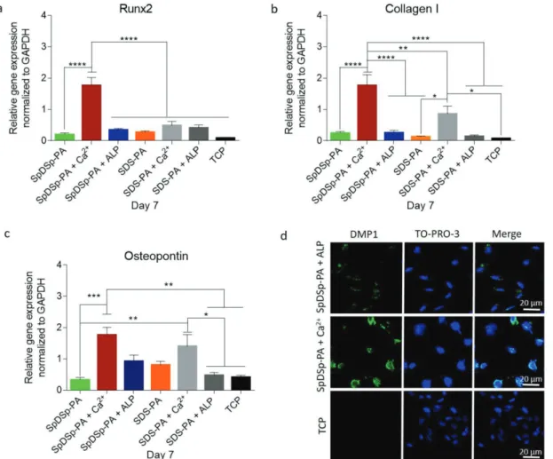
3.4. Molecular Analysis of Osteogenic Differentiation
The differentiation of progenitor cells into the osteogenic line age is regulated by the expression of specific marker genes such as transcription factors, adhesion molecules and proteins of the extracellular matrix.[39] Here, expressions of the
osteo-genic markers Runt-related transcription factor 2 (Runx2), type I collagen (Collagen I) and osteopontin were studied on day 7 in both osteogenic medium and maintenance medium groups (Figures 5 and 6). Runx2 is often referred to as the key regulator of osteogenic differentiation[40,41] and its expression is typically
analyzed during the early phases. In the osteogenic medium group, the expression of Runx2 was upregulated when cells were cultured on ALP-treated SpDSp-PA nanofibers compared to cells on nontreated SpDSp-PA peptide nanofibers, nontreated SDS-PA nanofibers, Ca2+-treated SDS-PA peptide nanofibers,
and bare TCP surfaces by ≈2.34, ≈8, ≈2.3, and ≈8.3 folds, respec-tively. The expression of Runx2 was also enhanced by ≈2.37 folds on ALP-treated SpDSp-PA nanofibers compared to the Ca2+-treated SpDSp-PA group (Figure 5a). Early stage mineral
deposition on collagen matrix acts as a template for minerali-zation and serves as a marker for osteogenic differentiation.[42]
The expression of Collagen I was significantly upregulated when Saos-2 cells were cultured on ALP-treated SpDSp-PA nanofibers compared to cells on nontreated SpDSp-PA nanofibers, non-treated SDS-PA nanofibers, Ca2+-treated SDS-PA nanofibers,
and bare TCP surfaces by ≈7.6, ≈16, ≈5, and ≈34 folds,
Figure 3. Calcium ion induced gel formation of DPP-like peptides. In-depth monitoring of network formation by calcium-treated SpDSp-PA and SDS-PA. a) Circular dichroism spectra showing Ca2+-triggered self-assembly of SpDSp-PA, and self-assembly of SDS-PA with and without cation pres-ence. b,d) TEM images of SDS-PA + Ca2+ and SpDSp-PA + Ca2+ nanofiber networks. c) Oscillatory rheology of SpDSp-PA + Ca2+ and SDS-PA + Ca2+
respectively (Figure 5b). The expression of Collagen I was also enhanced by ≈3 folds on ALP-treated SpDSp-PA nanofibers compared to Ca2+-treated SpDSp-PA group.
In addition to Collagen I, several noncollagenous proteins have crucial roles in the formation and maturation of miner-alized tissues. Osteopontin is one of the most notable among these proteins and is a highly phosphorylated sialoprotein that occurs as a prominent component of the mineralized extracel-lular matrices of bones.[43] The osteopontin expression of cells
on ALP-treated SpDSp-PA nanofibers was also significantly higher compared to cells on nontreated SpDSp-PA and SDS-PA nanofibers, and bare TCP surfaces by ≈3.8, ≈5.8, and ≈9.6 folds, respectively (Figure 5c). The expression of osteopontin was like-wise enhanced by ≈1.4 and ≈2 folds on ALP-treated SpDSp-PA nanofibers compared to Ca2+ treated SpDSp-PA group and Ca2+
-treated SDS-PA group, respectively (Figure 5c). Dentin matrix protein 1 (DMP1) is an acidic phosphoprotein that is also pre-sent in mineralized tissues and plays an important role in both the intra- and extracellular biomineralization process of osteo-blasts and odontoosteo-blasts.[44–46] DMP1 expression was also
exam-ined on day 14, and immunocytochemistry stainings showed that cells on ALP treated SpDSp-PA nanofibers and Ca2+ treated SpDSp-PA group showed DMP1 staining (Figure 5d). Similar to gene expression analyses, however, the ALP-treated SpDSp-PA group was considerably more effective in eliciting DMP1 deposition than the Ca2+ treated SpDSp-PA group.
In contrast to the osteogenic medium results, cells in main-tenance medium exhibited enhanced differentiation on Ca2+ treated PA nanofibers compared to ALP treated SpDSp-PA as well as other groups tested. In maintenance medium, the expression of Runx2 was significantly upregulated on Ca2+
treated SpDSp-PA nanofibers compared to cells on nontreated
SpDSp-PA peptide nanofibers, nontreated SDS-PA nanofibers, Ca2+ treated SDS-PA nanofibers and bare TCP surfaces by ≈9.77, ≈6.76, ≈3.6, and ≈22 folds, respectively. The expression of Runx2 was also significantly enhanced by ≈5.17 on Ca2+
ion-treated SpDSp-PA nanofibers compared to ALP treated SpDSp-PA group (Figure 6a). Similarly, the expression of Collagen I was significantly upregulated when cells were cul-tured on Ca2+ treated SpDSp-PA nanofibers compared to cells on nontreated SpDSp-PA and SDS-PA nanofibers, Ca2+ treated
SDS-PA nanofibers and bare TCP surfaces by ≈7.6, ≈14.6, ≈2, and ≈25.14 folds, respectively, and by ≈7 folds compared to the ALP treated SpDSp-PA group (Figure 6b). The expression of osteopontin was also upregulated when cells were cultured on Ca2+ treated SpDSp-PA nanofibers compared to cells on non-treated SpDSp-PA and SDS-PA peptide nanofibers, Ca2+ treated
nanofibers and bare TCP surfaces by ≈5.6, ≈2.2, ≈1.25, and ≈4.29 folds, respectively, and ≈1.91 folds compared to the ALP-treated SpDSp-PA group (Figure 6c). DMP1 staining results were found to support the gene expression profiles, with cells on Ca2+ treated SpDSp-PA showing enhanced DMP1 expres-sion compared to ALP-treated SpDSp-PA nanofibers, while the TCP group did not show any staining (Figure 6d).
It is interesting to note that Saos-2 cells grown in mainte-nance medium show enhanced osteogenic gene expression on Ca2+ treated PA compared to ALP treated
SpDSp-PA, while the reverse trend holds true for cells in osteogenic culture medium. This phenomenon may be attributed to the effect of β-glycerophosphate, which is a component of the oste-ogenic medium and promotes osteogenesis by donating free phosphate groups following its cleavage by ALP. As phosphate groups act as both structural components for the biominerali-zation process and signaling molecules for crucial osteogenic
Figure 4. Hydroxyapatite deposition on peptide coated surfaces. Mineral deposition on peptide surfaces with and without nanostructure formation. Ca/P ratio was evaluated to reveal mineral composition (above SEM images) of different experimental groups. EDX analysis results of Ca2+ content collected from mineralized peptide surface are shown as qualitative mineralization data.
pathways (such as ERK and cAMP/PKA), the combined effect of phosphate release from β-glycerophosphate and SpDSp-PA may have provided Saos-2 cells with an enhanced stimulus for osteogenic differentiation, especially in the context of enhanced Runx2 activation by dexamethasone and collagen deposition by ascorbic acid (which are also found in the osteogenic culture medium). Although cells in maintenance medium are also sup-plied with a free phosphate source on ALP-treated SpDSp-PA, inorganic phosphate by itself is evidently insufficient to pro-mote the matrix modification effects (e.g., collagen deposi-tion, calcification and attachment) that accompany osteogenic development, as evidenced by considerably lower expressions of osteogenic marker genes (and especially Collagen I) on ALP treated SpDSp-PA in maintenance medium. Consequently, cells cultured in maintenance medium are unable to establish their own mineralized matrix on ALP treated SpDSp-PA, and the presence of an existing calcified scaffold in Ca2+ treated SDS-PA is more conducive for osteogenesis through promo-tion of cell-matrix interacpromo-tions. Indeed, the presence of a stable scaffold structure is a major contributor to osteogenic dif-ferentiation in both osteogenic and normal media, as soluble
SpDSp-PA was relatively ineffective in stimulating osteogenic differentiation under both conditions. Overall, immunostaining and gene expression profiles showed that Saos-2 cells differ-entiate into osteogenic lineage on both Ca2+ treated and ALP
treated SpDSp-PA nanofibers, since enhanced osteogenic dif-ferentiation was observed on these peptide nanofiber surfaces. In addition, we observed that different medium conditions may change the differentiation ability of cells, potentially due to the tendency of Saos-2 cells to establish a native matrix in osteo-genic medium and rely on their interactions with the existing scaffold in maintenance medium.
4. Conclusion
In summary, we showed that DPP-mimetic peptide nanofiber network can mediate biomineralization process through the phosphorylation of its serine residues. DPP-mimetic peptide nanofibers provide a physical scaffold matrix for formation of hyroxyapatite like mineral formation utilizing phosphate and carboxylate groups as a substrate. In contrast, soluble
Figure 5. Molecular analysis of osteogenic differentiation in osteogenic medium. Gene expression analysis of Runx2 a), Collagen I b), and osteopontin c) on day 7 of osteogenic differentiation. The expression level of each gene was normalized to GAPDH. Values represent mean ± SEM (**p < 0.01, *p < 0.05). d) Confocal images of DMP-1 immunostaining on day 14. Green shows DMP-1, and blue shows the nucleus.
SpDSp-PA and nonphosphorylated SDS-PA nanofibers both failed to facilitate mineral deposition. The presence of phos-phate groups and the nanofibrous peptide matrix are both vital for the biomineralization process. In addition, the SpDSp-PA and SDS-SpDSp-PA nanofibers supported the viability of Saos-2 cells. The Ca2+ treated SpDSp-PA and ALP treated SpDSp-PA nanofibers enhanced osteogenic differentiation, and they can be utilized to develop new treatments for assisted repair of hard tissue defects because of structural, biochemical and functional resemblance to the DPP.
Supporting Information
Supporting Information is available from the Wiley Online Library or from the author.
Acknowledgements
G.G. and G.T. were supported by TUBITAK-BIDEB 2211-C PhD fellowship. R.G. acknowledges RFBR for the grant (16-33-60146) for young researchers and the Program of Competitive Growth of Kazan
Federal University. The authors acknowledge A. D. Ozkan for fruitful discussions and thank Mr. M. Guler for help in TEM imaging.
Conflict of Interest
The authors declare no conflict of interest.
Keywords
biomineralization, dentin phosphoprotein, osteogenic differentiation, peptide amphiphile
Received: February 25, 2018 Revised: March 22, 2018 Published online: May 10, 2018
[1] F. Nudelman, A. J. Lausch, N. A. Sommerdijk, E. D. Sone, J. Struct. Biol. 2013, 183, 258.
[2] E. Villarreal-Ramirez, D. Eliezer, R. Garduño-Juarez, A. Gericke, J. M. Perez-Aguilar, A. Boskey, Bone 2017, 95, 65.
Figure 6. Molecular analysis of osteogenic differentiation in maintenance medium. Gene expression analysis of Runx2 a), Collagen I b), and osteopontin c) on day 7 of osteogenic differentiation. The expression level of each gene was normalized to GAPDH. Values represent mean ± SEM (****p < 0.0001, ***p < 0.001, **p < 0.01, *p < 0.05). d) Confocal images of DMP-1 immunostaining on day 14. Green shows DMP-1, and blue shows the nucleus.
[3] G. Tansik, E. Kilic, M. Beter, B. Demiralp, G. Kiziltas Sendur, N. Can, H. Ozkan, E. Ergul, M. O. Guler, A. B. Tekinay, Biomater. Sci. 2016, 4, 1328.
[4] A. S. Deshpande, E. Beniash, Cryst. Growth Des. 2008, 8, 3084. [5] Y. Ha, J. Yang, F. Tao, Q. Wu, Y. Song, H. Wang, X. Zhang, P. Yang,
Adv. Funct. Mater. 2018, 28, 1704476.
[6] E. D. Eren, G. Tansik, A. B. Tekinay, M. O. Guler, ChemNanoMat 2018, https://doi.org/10.1002/cnma.201700354.
[7] W. Zhang, X. Yu, Y. Li, Z. Su, K. D. Jandt, G. Wei, Prog. Polym. Sci. 2017, 80, 94.
[8] G. Wei, Z. Su, N. P. Reynolds, P. Arosio, I. W. Hamley, E. Gazit, R. Mezzenga, Chem. Soc. Rev. 2017, 46, 4661.
[9] J. D. Hartgerink, E. Beniash, S. I. Stupp, Science 2001, 294, 1684. [10] L. Fisher, D. Torchia, B. Fohr, M. Young, N. Fedarko, Biochem.
Bio-phys. Res. Commun. 2001, 280, 460.
[11] C. Qin, J. Brunn, E. Cadena, A. Ridall, H. Tsujigiwa, H. Nagatsuka, N. Nagai, W. Butler, J. Dent. Res. 2002, 81, 392.
[12] O. Baba, C. Qin, J. C. Brunn, J. E. Jones, J. N. Wygant, B. W. McIntyre, W. T. Butler, Eur. J. Oral Sci. 2004, 112, 163.
[13] K. Alvares, Y. S. Kanwar, A. Veis, Dev. Dyn. 2006, 235, 2980. [14] K. U. Ogbureke, L. W. Fisher, J. Histochem. Cytochem. 2007, 55, 403. [15] S. Xiao, C. Yu, X. Chou, W. Yuan, Y. Wang, L. Bu, G. Fu, M. Qian,
J. Yang, Y. Shi, Nat. Genet. 2001, 27, 201.
[16] X. Zhang, J. Zhao, C. Li, S. Gao, C. Qiu, P. Liu, G. Wu, B. Qiang, W. H. Lo, Y. Shen, Nat. Genet. 2001, 27, 129.
[17] T. Sreenath, T. Thyagarajan, B. Hall, G. Longenecker, R. D’Souza, S. Hong, J. T. Wright, M. MacDougall, J. Sauk, A. B. Kulkarni, J. Biol. Chem. 2003, 278, 24874.
[18] K. Verdelis, Y. Ling, T. Sreenath, N. Haruyama, M. MacDougall, M. C. van der Meulen, L. Lukashova, L. Spevak, A. B. Kulkarni, A. L. Boskey, Bone 2008, 43, 983.
[19] Y. Yamakoshi, J. Oral Biosci. 2009, 51, 134.
[20] S. Suzuki, T. Sreenath, N. Haruyama, C. Honeycutt, A. Terse, A. Cho, T. Kohler, R. Müller, M. Goldberg, A. B. Kulkarni, Matrix Biol. 2009, 28, 221.
[21] Y. Yamakoshi, J. C.-C. Hu, M. Fukae, H. Zhang, J. P. Simmer, J. Biol. Chem. 2005, 280, 17472.
[22] A. S. Deshpande, P.-A. Fang, X. Zhang, T. Jayaraman, C. Sfeir, E. Beniash, Biomacromolecules 2011, 12, 2933.
[23] K. Kawasaki, T. Suzuki, K. M. Weiss, Proc. Natl. Acad. Sci. USA 2004, 101, 11356.
[24] S. L. Lee, A. Veis, T. Glonek, Biochemistry 1977, 16, 2971.
[25] Y. S. Choi, J. Y. Lee, J. S. Suh, G. Lee, C. P. Chung, Y. J. Park, J. Biomed. Mater. Res., Part A 2013, 101, 590.
[26] G. Gulseren, M. A. Khalily, A. B. Tekinay, M. O. Guler, J. Mater. Chem. B 2016, 4, 4605.
[27] G. Gulseren, I. C. Yasa, O. Ustahuseyin, E. D. Tekin, A. B. Tekinay, M. O. Guler, Biomacromolecules 2015, 16, 2198.
[28] E. Arslan, I. C. Garip, G. Gulseren, A. B. Tekinay, M. O. Guler, Adv. Healthcare Mater. 2014, 3, 1357.
[29] A. E. Topal, G. Tansik, A. D. Ozkan, M. O. Guler, A. Dana, A. B. Tekinay, Adv. Mater. Interfaces 2017, 4, 1700090.
[30] Y. Sun, Y. Lu, S. Chen, M. Prasad, X. Wang, Q. Zhu, J. Zhang, H. Ball, J. Feng, W. Butler, J. Dent. Res. 2010, 89, 498.
[31] R. Fujisawa, Y. Kuboki, S. Sasaki, Calcif. Tissue Int. 1986, 39, 248. [32] A. M. Milan, R. V. Sugars, G. Embery, R. J. Waddington, Eur. J. Oral
Sci. 2006, 114, 223.
[33] K. Ibaraki, H. Shimokawa, S. Sasaki, Matrix 1991, 11, 115. [34] R. Fujisawa, Y. Kuboki, S. Sasaki, Calcif. Tissue Int. 1987, 41, 44. [35] T. D. Schmittgen, K. J. Livak, Nat. Protoc. 2008, 3, 1101.
[36] M. Wallwork, J. Kirkham, H. Chen, S. Chang, C. Robinson, D. Smith, B. Clarkson, Calcif. Tissue Int. 2002, 71, 249.
[37] C. F. Nawrot, D. J. Campbell, J. K. Schroeder, M. Van Valkenburg, Biochemistry 1976, 15, 3445.
[38] P. A. Cloos, A. L. Jensen, Biogerontology 2000, 1, 341.
[39] W. Huang, S. Yang, J. Shao, Y.-P. Li, Front. Biosci.: A J. Virtual Libr. 2007, 12, 3068.
[40] C. Ge, W. P. Cawthorn, Y. Li, G. Zhao, O. A. MacDougald, R. T. Franceschi, J. Cell. Physiol. 2016, 231, 587.
[41] F. Otto, A. P. Thornell, T. Crompton, A. Denzel, K. C. Gilmour, I. R. Rosewell, G. W. Stamp, R. S. Beddington, S. Mundlos, B. R. Olsen, Cell 1997, 89, 765.
[42] E. Birmingham, G. Niebur, P. McHugh, G. Shaw, F. Barry, L. McNamara, Eur. Cells Mater. 2012, 23, 13.
[43] J. Sodek, B. Ganss, M. McKee, Crit. Rev. Oral Biol. Med. 2000, 11, 279.
[44] J. Feng, H. Huang, Y. Lu, L. Ye, Y. Xie, T. Tsutsui, T. Kunieda, T. Castranio, G. Scott, L. Bonewald, J. Dent. Res. 2003, 82, 776. [45] A. George, R. Silberstein, A. Veis, Connect. Tissue Res. 1995,
33, 67.
[46] R. D’souza, A. Cavender, G. Sunavala, J. Alvarez, T. Ohshima, A. Kulkarni, M. MacDougall, J. Bone Miner. Res. 1997, 12, 2040.