Effect of surgeon’s judgement on the diagnosis of acute
appendicitis
Akut apandisit tanısında cerrah kararının etkisi
Objective: The accuracy of a surgeon’s judgement still remains to be controversial in the diagnosis of acute appendici-tis, which is a diagnosis usually based on laboratory data and imaging tests.
Material and Methods: Patients with a possible diagnosis of acute appendicitis were reviewed retrospectively with regard to demographic variables, laboratory and imaging results, and treatment modalities.
Results: There were 128 patients with a mean age of 31.2±14 years. The mean white blood cell count and the propor-tion of polymorphonuclear leukocytes were 11403±4669/mm3 and 75±11%, respectively. Appendectomy was perfor-med on 66 (51.6%) patients. Conservative management was applied to 62 (48.4%) patients. Statistical analysis showed that patients with appendicitis have a higher white blood cell count (p=0.015) and a higher proportion of polymorp-honuclear leukocytes (p=0.023). Sensitivity, specificity and accuracy rates were 84.6%, 63.7% and 74.3% for ultrasound and 100%, 86.7% and 92.2% for computed tomography, respectively.
Conclusion: Diagnosis based on patients’ laboratory and imaging data, in combination with, the surgeon’s judgement appears to yield the best outcomes in patients with suspicion of acute appendicitis.
Key Words: Acute appendicitis, diagnosis, ultrasound, tomography, appendectomy
Amaç: Sıklıkla laboratuvar verileri ve görüntüleme testlerine göre temellendirilmiş akut apandisit tanısı uygulan-makla birlikte, cerrah kararının akut apandisit için tanısal doğruluktaki etkisi halen tartışmalıdır.
Gereç ve Yöntemler: Olası akut apandisit tanısı olan hastalar geriye dönük olarak demografik değişkenler, laboratu-var ve görüntüleme sonuçları ve tedavi seçenekleri açısından değerlendirildi.
Bulgular: Yaş ortalaması 31,2±14 yıl olan toplam 128 hasta vardı. Ortalama lökosit sayısı ve polimorfonükleer lökosit oranı sırası ile 11,403±4,669/mm3 ve %75±11 olarak bulundu. Apendektomi 66 hastaya (%51,6) uygulandı. Konservatif yaklaşım 62 hastada (%48,4) gerçekleştirildi. Akut apandisit olan hastaların daha yüksek lökosit sayısına (p=0,015) ve daha yüksek oranda polimorfonükleer lökosit oranına sahip olduğu gösterildi (p=0,023). Duyarlılık, özgüllük ve doğruluk oranlarının ultrason için %84,6, %63,7 ve %74,3; bilgisayarlı tomografi için %100, %86,7 ve %92,2 olduğu saptandı.
Sonuç: Laboratuvar verileri ile birlikte ultrason ve bilgisayarlı tomografinin uygun kullanımının yanında cerrahın kararına göre temellendirilmiş akut apandisit tanısı, olası akut apandisit olgularında en iyi sonuçların elde edilme-sine yol açmaktadır.
Anahtar Kelimeler: Akut apandisit, tanı, ultrason, tomografi, apendektomi
INTRODUCTION
Acute appendicitis (AA) is the most frequently suspected disorder in patients presenting with acute abdominal pain and is the most common indication for urgent abdominal surgery. Diagnosis of AA based only on clinical and laboratory data results in high negative appendectomy rates and missed di-agnoses with increased morbidity (1-4). Imaging tests such as graded compression ultrasound (US) with or without color Doppler evaluation and computed tomography (CT) have been used to improve diag-nostic performance for the last several decades (1, 5-8). The lower sensitivity of US compared to CT and the great variability caused by operator dependency may result in a higher number of false negative diagnoses if US is used as the only imaging technique (1). However, CT is associated with considerable ionizing radiation exposure, which discourages its use. Quality assurance for patients with suspected AA should aim to minimize the negative appendectomy rate, without delaying the treatment of perforated AA, by optimal diagnostic use of US and CT (3, 9).
The purpose of this retrospective study was to evaluate the optimal use of laboratory investigation, imaging techniques and surgeon’s judgement to diagnose patients presenting with acute abdominal pain and possible AA.
1Department of General Surgery, Faculty of Medicine, Bezmialem Vakıf University, İstanbul, Turkey 2Department of Radiology, Faculty of Medicine, İstanbul Medipol University, İstanbul, Turkey 3Department of Radiology, 29 Mayıs Hospital, Ankara, Turkey 4Department of Radiology, 29 Mayıs Hospital, İstanbul, Turkey
Address for Correspondence
Yazışma Adresi
Mustafa Hasbahçeci, MD Department of General Surgery, Faculty of Medicine, Bezmialem Vakıf University, İstanbul, Turkey Phone: +90 533 356 01 32 e-mail:
[email protected] Received / Geliş Tarihi: 12.12.2013 Accepted / Kabul Tarihi: 21.12.2013 ©Copyright 2014
by Turkish Surgical Association Available online at www.ulusalcerrahidergisi.org ©Telif Hakkı 2014 Türk Cerrahi Derneği Makale metnine www.ulusalcerrahidergisi.org web sayfasından ulaşılabilir.
22
Mustafa Hasbahçeci
1, Cengiz Erol
2, Mustafa Törü
3, Mehmet Şeker
4Original Investigation / Özgün Araştırma
Ulusal Cer Derg 2014; 30: 22-27MATERIAL AND METHODS
This study was a descriptive study based on retrospectively collected data. Patients who presented with right lower quad-rant pain and possible AA between June 2006 and June 2010 in a private hospital were included. The institutional review board approved the study protocol (Istanbul 29 Mayıs Hospi-tal-04.02.2011/4). A clinical worksheet list was used to collect patient’s demographic information.
On admission (Figure 1), all patients underwent physical ex-amination and blood testing to determine white blood cell (WBC) count and proportion of polymorphonuclear (PMN) leukocytes. For the standard values of our laboratory, the WBC count and proportion of PMN leukocytes were considered normal when lower than 10400/mm3 and 75%, respectively. All patients underwent diagnostic imaging techniques (Figure 1), including US, CT or both depending on the discretion of the on-call surgeon at the general surgical unit. The imaging tech-niques were performed and reviewed during office hours (8 am-6 pm). If patients were admitted out of office hours, their imaging evaluations were performed and reviewed on the next day. Within the first 24 hours after admission, the attend-ing surgeon decided between surgical treatment and conser-vative management based on clinical and laboratory findings and imaging results. Conservative management included pain control, restricting oral intake except fluids and active obser-vation with serial clinical examinations performed by the same attending surgeon on either an in-patient or outpatient basis. During the conservative management, intravenous or oral an-tibiotics were not given. Pregnant patients and patients with-out complete data were excluded from the study.
Diagnosis of AA or perforated AA at the time of operation was based on macroscopic findings. Neither diagnostic
lapa-roscopy nor laparoscopic appendectomy was used. Surgeons with at least 5 years of experience performed all operations. Normal-looking appendices discovered during laparotomy were removed via split-muscle McBurney incision. All excised appendices were microscopically analyzed by pathology us-ing paraffin sections. Histological diagnosis of appendicitis was based on infiltration of the muscularis propria by PMN leukocytes. The proportion of patients with perforated appen-dices and negative appendectomy were identified by patho-logic determination of perforated AA and the proportion of normal-looking appendices noted during surgery, respective-ly. Patients in whom conservative management was initially intended and then, within 48 hours, AA was diagnosed, were considered as missed AA cases.
Imaging Techniques
Senior radiologists performed all US examinations. For pa-tients examined by US (Logiq 9, GE Healthcare, Milwaukee, Wisconsin, USA), both convex and linear probes were used. A routine US examination of the upper abdomen and pelvis using a 3-5-MHz convex transducer was initially performed to rule out alternative abnormalities related to the liver, gall-bladder, pancreas, kidney, or pelvic organs, and the presence of peritoneal fluid. Afterwards, graded compression and color Doppler US of the right lower quadrant, with special empha-sis directed to the site of maximal tenderness, was performed using a linear 5-12-MHz or 4-8-MHz transducer, according to body size. The criteria for diagnosis of AA included the follow-ing direct signs: distended (≥7 mm) and non-compressible appendix, and inflammation of periappendiceal fat. Indirect signs included the presence of appendicoliths and increased flow observed via color Doppler US. All other results, including cases in which the appendix could not be visualized or entirely verified as normal, were considered negative. When a final diagnosis was negative based on US examination, patients additionally underwent CT examination of the abdomen and pelvis, if deemed appropriate by the surgeon.
All abdominal CT examinations reviewed by senior radiologists were performed with a 64-detector CT scanner (Lightspeed VCT, GE Healthcare, Milwaukee, Wisconsin, USA). Images were obtained in the cranio-caudal direction with detector collima-tion of 64×0.625 mm, a voltage of 100-120 kVp, and a tube current of 150-250 mAs. Water or water soluble oral contrast agents (2%) between 750-1500 mL were consumed one hour before each examination. An upper extremity 18-20 gauge IV cannula was used for venous access. 70-100 mL of non-ionic contrast medium with a 300 mg/ml iodine concentration was injected at a flow rate of 2-3 mL/s and followed by a 50 mL saline with the same flow rate. The scan was started after a 70-80 s delay. All images were reconstructed as 2.5 mm axial sections. A diagnosis of acute appendicitis was determined via CT if thickening of the appendix (≥7 mm) and associated inflammation of the periappendiceal fat (fat stranding) were observed. No inconclusive CT assessments were reported. Both US and CT were assessed for their ability to determine the best mode of treatment-surgery or conservative man-agement-compared to reference standards and based on the findings of the operation, follow-up period, and pathological analysis. When evaluating the correlation between treatment modality decided by the attending surgeon and imaging re-Figure 1. Schematic representation of the diagnostic flow
diagram
WBC: white blood cell; PMN: polymorphonuclear; US: ultrasound; CT: computed tomography; S: surgical treatment for acute appen-dicitis; CM: conservative management
+: positive test result for acute appendicitis -: negative test result for acute appendicitis
Physical examination WBC count PMN leukocyte percentage Admission Imaging + (n=57) - (n=20) + (n=5) - (n=19) + (n=13) - (n=14) CM (n=14) CM (n=1) S (n=12) CT (n=24) CM (n=20) + (n=12) S (n=10) CM (n=2) CM (n=12) – (n=12) S (n=44) CM (n=13) US (n=101)
US alone (n=77) First US (n=24) CT alone (n=27)
CT (n=51)
sults, if both US and CT were performed on the same patient, the CT results were regarded as the final diagnosis. Otherwise, whichever technique was performed was accepted as the final diagnosis.
Follow-up information was acquired from all patients over-the-phone or in-person follow-up appointments six months after discharge.
Statistical Analysis
Sensitivity, specificity, positive and negative predictive values, positive and negative likelihood values, and accuracy were calculated for each imaging technique to compare their diag-nostic accuracy rates.
The collected data were entered in an electronic database (Mi-crosoft Excel for Windows, Mi(Mi-crosoft Corporation, Redmond, WA). Statistical calculations were performed using NCSS (Num-ber Cruncher Statistical System, 2007) and PASS Statistical software (Utah, USA, 2008). Normally distributed continuous variables were expressed as mean±standard deviation (SD). Cat-egorical variables were expressed as frequencies and percent-ages of an appropriate denominator. The Student’s t-test was used for analysis of normally distributed, descriptive continuous variables, which were expressed as mean ± SD. The chi-square test and McNemar’s test were used to compare qualitative vari-ables. The sensitivity, specificity, positive and negative predic-tive values, posipredic-tive and negapredic-tive likelihood ratios, and accuracy of diagnostic screening tests were also used to compare the diagnostic efficiency of imaging techniques. Differences were considered statistically significant if the p value was equal to or less than 0.05 with a 95% confidence interval.
RESULTS
One hundred forty three patients were admitted with right lower quadrant pain complaint. Fifteen patients had incom-plete data and were excluded from the study; a total of 128 patients were included. The mean age was 31.2±14 years, with a range of 10 to 83 years. There were 58 (45.3%) female and 70 (57.7%) male patients. Demographic findings and labora-tory parameters are shown in Table 1. Comparative analysis of laboratory parameters revealed that sixty-five patients (51%) had WBC counts above 10400/mm3, and 73 (57%) had PMN leukocyte percentages above 75%.
For diagnostic purposes, 101 US and 51 CT examinations were performed. 77 only underwent a US examination, and 27 only underwent CT; 24 patients required both US and CT examina-tions. US results were positive for appendicitis in 62 patients.
Five of them underwent additional CT at the discretion of the surgeon. Although three of the patients were regarded as positive for AA, appendectomy was performed in only two of them. Fifteen patients with a diagnosis of AA after US, includ-ing the other two patients who underwent additional CT, were managed conservatively. Surgical treatment was performed on 44 patients diagnosed with AA by US. Appendicitis was confirmed by histological examination in 42 patients, and two patients had normal-looking appendices.
Computed tomography was applied to 27 patients without an initial US examination. All 14 of the patients whose CT re-sults were negative for AA were managed conservatively. Of the remaining 13 patients with a diagnosis of AA, one patient was followed in a conservative manner. All of the other pa-tients were operated on, but AA was only confirmed in 11; one patient had a normal-looking appendix. Three perforated ap-pendicitis cases were diagnosed preoperatively using US (one patient) and CT (two patients).
Sixty-six (51.6%) patients diagnosed with AA were operated. Three cases of perforated AA and three normal-looking ap-pendices were confirmed by histological examination after the operation. Therefore, both perforated AA and negative ap-pendectomy occurred at a rate of 4.5%. The actual prevalence of AA was 49% after exclusion of the three negative appen-dectomy cases. Conservative management was applied to 62 (48.4%) patients who recovered without any complaints. Four alternative diagnoses (three cases of ovarian cyst rupture and one case of pyelonephritis) were determined using imaging techniques. During the follow-up period, no patients present-ed with misspresent-ed AA requiring surgical treatment or additional hospitalization.
The treatment modality correlated with the final radiological diagnosis in 87.5% of the cases (112 out of 128 patients); there were three cases of negative appendectomy. The remaining 16 (12.5%) patients (13 US patients and 3 CT patients) did not undergo surgical treatment, although all the patients had a radiological diagnosis of AA. Conservative management with active serial examinations by the attending surgeon was per-formed with no complications.
Patients with and without proven AA diagnoses (n=63 and n=65, respectively) were compared with regards to age, gen-der, WBC count, proportion of PMN leukocytes, and the num-ber of patients with WBC counts and PMN leukocyte percent-ages higher than the cut-off value (Table 2, 3). Patients with proven AA diagnoses had significantly higher WBC counts and Table 1. Demographic characteristics and laboratory findings of the study group, and results of statistical comparison be-tween female and male patients
Parameter Total (n=128)† Female (n=58)† Male (n=70)† p
Age (year)‡ 31.2±14 (30) 33±15 (29) 29.6±13 (30) 0.156
WBC count (/mm3) ‡ 11403±4669 (11000) 10360±3519 (9300) 12266±5312 (11500) 0.017*
Proportion of PMN leukocytes (%)‡ 75±11 (76) 73±12 (75) 77±9 (76) 0.055
†: Number of patients in each group ‡: Mean±SD (median)
*: Statistical significance
24
PMN leukocyte percentages (p<0.05). AA was more frequently seen in patients with WBC counts higher than the cut-off value (p<0.05).
Eighteen of the 62 patients diagnosed with AA using US were not confirmed as having AA (18 false positive results), and eight of the 39 patients not diagnosed with AA were con-firmed as having AA (8 false negative results). Four of the 25 patients diagnosed with AA using CT were not confirmed as having AA (4 false positive results). However, all patients not diagnosed with AA by CT were confirmed as not having AA (no false negative results; Table 4). There was no difference in the diagnostic performance of the imaging techniques for either female or male patients (p>0.05).
DISCUSSION
Although appendectomy is the most common surgical pro-cedure performed throughout the world, diagnoses based on clinical history, physical examination, and routine labora-tory tests are not always accurate. The role of imaging in con-firming the diagnosis of AA and detecting alternative AA-like disorders has been well demonstrated (3). Therefore, imag-ing techniques have been used to increase diagnostic accu-racy and to lower negative appendectomy and perforated AA rates without causing any negative effects (5). As a policy, we performed at least one kind of imaging technique on each
patient presenting with right lower quadrant pain. Many dif-ferent studies have examined the efficacy of diagnosing AA using clinical history, physical examination, laboratory tests, and/or imaging techniques (2, 3, 5-8, 10). After review of these studies, it was clear that guidelines regarding the optimal di-agnostic approach to AA were lacking and that each center should find its own algorithm after consideration of current AA protocols and local factors, such as the availability of imag-ing techniques (11).
Although previous studies had conflicting results regarding the inflammatory parameters of AA, we aimed to use both the WBC count and the proportion of PMN leukocytes in every pa-tient (7, 12-15). In one study, 15.6% of the papa-tients presenting with right lower quadrant pain had normal WBC counts and C-reactive protein levels (12). However, our results showed that a higher WBC count was significantly correlated with the presence of AA (p<0.05). Although the proportion of PMN leu-kocytes was higher in AA patients, the correlation lacked sta-tistical significance. AA was also more frequently seen in pa-tients with WBC counts higher than the cut-off value (p<0.05). Overall, laboratory parameters related to inflammation should be regarded as adjuncts to clinical and imaging findings, and any increase in these parameters should raise suspicion of AA. US and CT were the most commonly used imaging techniques for AA. Previous studies have shown that the two techniques diagnose AA with different accuracies (5). CT is recognized as the most accurate imaging method for the detection of AA in patients with right lower quadrant pain (16). Several factors of imaging techniques must be taken into consideration, includ-ing the operator dependency inherent to US, the length of time required to perform the imaging techniques, the radia-tion dosage during CT examinaradia-tion, and any correlaradia-tion with clinical findings (1-3, 17).
Ultrasound performance has been shown to be more accu-rate during the day, when senior radiologists perform US, than during the night, when on-duty residents are respon-sible for US examinations (3). In accordance with the litera-ture, senior radiologists performed all US examinations in the present study during the day with an acceptable diagnostic accuracy rate. In the present study, sensitivity, specificity, and Table 2. Comparison of continuous parameters with regard
to the presence or absence of AA
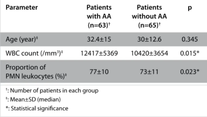
Parameter Patients Patients p
with AA without AA (n=63)† (n=65)† Age (year)‡ 32.4±15 30±12.6 0.345 WBC count (/mm3)‡ 12417±5369 10420±3654 0.015* Proportion of 77±10 73±11 0.023* PMN leukocytes (%)‡
†: Number of patients in each group ‡: Mean±SD (median)
*: Statistical significance
Table 3. Comparison of categorical parameters with regard to the presence or absence of AA
Parameter Patients Patients p
with AA without AA (n=63)† (n=65)† Gender (n (%))‡ Female 28 (44) 30 (46) 0.846 Male 35 (56) 35 (54) Number of patients (n (%))‡ WBC count <10 400 24 (38) 39 (60) 0.013* WBC count ≥10 400 39 (62) 26 (40) Proportion of PMN leukocytes <75 23 (37) 32 (49) 0.146 Proportion of PMN leukocytes ≥75 40 (63) 33 (51) †: Number of patients in each group
‡: Mean±SD (median) *: Statistical significance
Table 4. Statistical analysis of the ability of US and CT to diagnose AA Parameter US CT True positive† 44 21 False negative† 8 0 False positive† 18 4 True negative† 31 26 Sensitivity 0.85 1.0 Specificity 0.64 0.87
Positive predictive value 0.71 0.84
Negative predictive value 0.80 1.0
Accuracy 0.74 0.92
Positive likelihood ratio 2.3 7.5
Negative likelihood ratio 0.24 0
†: number of patients
US: ultrasound; CT: computed tomography
positive and negative predictive values for US were found to be 0.85, 0.64, 0.71, and 0.80, respectively. Although all cases in which the appendix could not be visualized were regard-ed as negative, the results were within the reportregard-ed ranges (3, 5, 7-10, 18, 19).
Previous studies reported several pitfalls and limitations which lowered the diagnostic accuracy of US examinations, such as obesity, excessive bowel gas, unusual appendix location, ap-pendicitis in the tip of the appendix, right lower quadrant ab-scess without visualization of the appendix, and edematous incompressible terminal ileum or cecum (3, 8). In general, very obese patients suspected of having AA are sent directly to CT because of the difficulty to penetrate tissues during US (3, 18). We usually referred our obese patients to CT without US. How-ever, we were not able to give the body mass index values be-cause of the retrospective nature of our study.
Mean sensitivities and specificities are substantially in favor of CT over US. CT has a sensitivity approaching 100%, is not oper-ator dependent, and can be performed on patients on which US is difficult to perform. However, contrast administration, ionizing radiation, and cost are limiting factors to CT, and we used CT as the primary imaging technique for only 27 patients (21%) despite its higher diagnostic accuracy (5, 19, 20). It is known that positive and negative likelihood ratios above 10 and below 0.1, respectively, imply strong effects, whereas a like-lihood ratio of 1 implies no effect. Mean sensitivity and specific-ity for CT were reported as high as 0.91 and 0.90 (5). Given these results, sensitivity, negative predictive value and negative likeli-hood ratio were accepted as powerful indicators of CT effective-ness in this study, especially for patients without AA.
Previous studies demonstrated that routine referral of patients suspected of having acute appendicitis to US, and limited re-ferral to CT based on the US results and clinical judgement, improves diagnostic accuracy and therapeutic management (1-3, 10, 16, 18, 19, 21). This approach reduces the necessity of CT examinations and has been shown to be cost effective (1). However, the use of imaging was largely successful because of multidisciplinary cooperative imaging algorithms involving the emergency department, physicians, surgeons, and radi-ologists. The staged protocol was also successful, especially in children, because of the effective use of US (2, 21). Therefore, in the present study, US were regarded as the primary imaging technique in most of the cases. However, CT was performed as the primary or adjunctive imaging technique in select cases as decided by the attending surgeon.
Current practice has a high negative appendectomy rate of 10%–20% but is regarded as acceptable for preventing a missed appendicitis rate of up to12%, which is considered clinically more important (4, 5, 14). In missed appendicitis cases, perforation and abscess leading to increased morbidi-ty and mortalimorbidi-ty, may occur more frequently (4). However, ac-cording to comparative studies published in the last decade, a mean uniform decrease in the negative appendectomy rate has occurred due to widespread implementation of CT (18, 22). A meta-analysis has similarly demonstrated an overall significant decrease in the negative appendectomy rate due to CT: from 16.7% to 8.6% for all patients, and from 27.3% to
9.6% for female patients, specifically (22). However, CT may cause more appendiceal perforations due to the delay before the scan. While the difference in the appendiceal perforation rate between the CT group and the clinical evaluation group was not statistically significant (CT: 23.4%; clinical evaluation: 16.7%), further studies should be done for clarification (22). Others and we believe that the lower rates of negative ap-pendectomy and perforated AA were due to clinical judge-ment, in addition to the laboratory and imaging results (23). It was also observed that in 16 cases (12.5%), treatment mo-dality was decided based on the clinical judgement of the attending surgeon, although all 16 had a radiological diag-nosis of AA. Our findings also showed that surgical treatment should be avoided for patients with negative CT results for AA. Therefore, it should be kept in mind that clinical judge-ment is especially important in atypical cases with positive imaging findings for AA.
Study Limitations
The present study was limited, as the absence of AA could not be absolutely confirmed in patients who did not undergo sur-gery. The retrospective design and small number of patients were other limiting factors.
CONCLUSION
Laboratory data demonstrating the severity of inflammation should be used for the clinical diagnosis of AA. US should be the primary imaging technique performed on every pa-tient presenting with right lower quadrant pain. However, CT should be chosen as the primary imaging technique in selected cases at the discretion of the attending surgeon. CT should also be performed in addition to US on patients who are highly suspected of having AA but showing negative US results. A good clinical approach including the skillful use of laboratory data, as well as US and CT on selected patients per the surgeon’s judgement, should be considered as part of an effective diagnostic algorithm for AA.
Ethics Committee Approval: Ethics committee approval was received for this study from the Institutional Review Board of Ethical Commit-tee, Istanbul 29 Mayıs Hospital.
Informed Consent: Due to retrospective design of the study, written informed consent could not be taken from the patients. Institutional Review Board from the Ethical Committee of the Hospital has been considered.
Peer-review: Externally peer-reviewed.
Author Contributions: Concept - M.H.; Design - M.H., C.E.; Supervision - M.Ş.; Funding - M.H., C.E., M.T., M.Ş.; Materials - M.H., C.E., M.T., M.Ş.; Data Collection and/or Processing - M.H.; Analysis and/or Interpreta-tion - M.H., C.E., M.T., M.Ş.; Literature Review - M.H.; Writer - M.H., C.E.; Critical Review - M.T., M.Ş.
Conflict of Interest: No conflict of interest was declared by the au-thors.
Financial Disclosure: The authors declared that this study has re-ceived no financial support.
Etik Komite Onayı: Bu çalışma için etik komite onayı İstanbul 29 Mayıs Hastanesi Etik Kurulu’ndan (04.02.2011/4) alınmıştır.
26
dan yazılı onam alınmayıp, Hastane Etik Kurulu’nun verdiği Kurumsal Değerlendirme onamı dikkate alınmıştır.
Hakem değerlendirmesi: Dış bağımsız.
Yazar Katkıları: Fikir - M.H.; Tasarım - M.H., C.E.; Denetleme - M.Ş.; Kaynaklar - M.H., C.E., M.T., M.Ş.; Malzemeler - M.H., C.E., M.T., M.Ş.; Veri toplanması ve/veya işlemesi - M.H.; Analiz ve/veya yorum - M.H., C.E., M.T., M.Ş.; Literatür taraması - M.H.; Yazıyı yazan - M.H., C.E.; Eleştirel İnceleme - M.T., M.Ş.
Çıkar Çatışması: Yazarlar çıkar çatışması bildirmemişlerdir.
Finansal Destek: Yazarlar bu çalışma için finansal destek almadıklarını beyan etmişlerdir.
REFERENCES
1. Krishnamoorthi R, Ramarajan N, Wang NE, Newman B, Rubesova E, Mueller CM, et al. Effectiveness of a staged US and CT proto-col for the diagnosis of pediatric appendicitis: reducing radiation exposure in the age of ALARA. Radiology 2011; 259: 231-239.
[CrossRef]
2. Toorenvliet BR, Wiersma F, Bakker RF, Merkus JW, Breslau PJ, Ham-ming JF. Routine ultrasound and limited computed tomography for the diagnosis of acute appendicitis. World J Surg 2010; 34: 2278-2285. [CrossRef]
3. Gaitini D, Beck-Razi N, Mor-Yosef D, Fischer D, Ben Itzhak O, Krausz MM. Diagnosing acute appendicitis in adults: accuracy of color Doppler sonography and MDCT compared with surgery and clin-ical follow-up. Am J Roentgenol 2008; 190: 1300-1306. [CrossRef]
4. Augustin T, Cagir B, Vandermeer TJ. Characteristics of perforated appendicitis: effect of delay is confounded by age and gender. J Gastrointest Surg 2011; 15: 1223-1231. [CrossRef]
5. van Randen A, Bipat S, Zwinderman AH, Ubbink DT, Stoker J, Boermeester MA. Acute appendicitis: meta-analysis of diagnostic performance of CT and graded compression US related to preva-lence of disease. Radiology 2008; 249: 97-106. [CrossRef]
6. van Randen A, Laméris W, van Es HW, ten Hove W, Bouma WH, van Leeuwen MS. Profiles of US and CT imaging features with a high probability of appendicitis. Eur Radiol 2010; 20: 1657-1666.
[CrossRef]
7. İnan M, Tulay SH, Besim H, Karakaya J. The role of ultrasonogra-phy and Alvarado score in diagnosis of acute appendicitis. Ulusal Cer Derg 2011; 27: 149-53.
8. Eryılmaz R, Baş G, Alimoglu O, Ercan M, Şahin M. The increasing importance of ultrasonography in the differential diagnosis of suspected acute appendicitis. Ulusal Cer Derg 2001; 17: 28-33. 9. Howell JM, Eddy OL, Lukens TW, Thiessen ME, Weingart SD,
Decker WW. Critical issues in the evaluation and management of emergency department patients with suspected appendicitis. Ann Emerg Med 2010; 55: 71-116. [CrossRef]
10. Gamanagatti S, Vashisht S, Kapoor A, Chumber S, Bal S. Compari-son of graded compression ultraCompari-sonography and unenhanced
citis. Singapore Med J 2007; 48: 80-87.
11. Frush DP, Frush KS, Oldham KT. Imaging of acute appendicitis in children: EU versus US. or US versus CT? A North American per-spective. Pediatr Radiol 2009; 39: 500-505. [CrossRef]
12. Monneuse O, Abdalla S, Pilleul F, Hervieu V, Gruner L, Tissot E. Pain as the only consistent sign of acute appendicitis: lack of inflam-matory signs does not exclude the diagnosis. World J Surg 2010; 34: 210-215. [CrossRef]
13. Kim HC, Yang DM, Lee CM, Jin W, Nam DH, Song JY. Acute ap-pendicitis: relationships between CT-determined severities and serum white blood cell counts and C-reactive protein levels. Br J Radiol 2011; 84: 1115-1120. [CrossRef]
14. Ortega-Deballon P, Ruiz de Adana-Belbel JC, Hernández-Matías A, García-Septiem J, Moreno-Azcoita M. Usefulness of laboratory data in the management of right iliac fossa pain in adults. Dis Co-lon Rectum 2008; 51: 1093-1099. [CrossRef]
15. Başak F, Kınacı E, Çalışkan YK, Kaygusuz A, Aren A. Retrospective analysis of 562 cases with a previous diagnosis of acute appendi-citis. İstanbul Tıp Dergisi 2006; 1: 16-20.
16. Poletti PA, Platon A, De Perrot T, Sarasin F, Andereggen E, Rutschmann O. Acute appendicitis: prospective evaluation of a diagnostic algorithm integrating ultrasound and low-dose CT to reduce the need of standard CT. Eur Radiol 2011; 21: 2558-2566.
[CrossRef]
17. Pritchett CV, Levinsky NC, Ha YP, Dembe AE, Steinberg SM. Man-agement of acute appendicitis: the impact of CT scanning on the bottom line. J Am Coll Surg 2010; 210: 699-705. [CrossRef]
18. Poortman P, Oostvogel HJ, Bosma E, Lohle PN, Cuesta MA, de Lange-de Klerk ES. Improving diagnosis of acute appendicitis: results of a diagnostic pathway with standard use of ultrasonog-raphy followed by selective use of CT. J Am Coll Surg 2009; 208: 434-441. [CrossRef]
19. Reich B, Zalut T, Weiner SG. An international evaluation of ultra-sound vs. computed tomography in the diagnosis of appendici-tis. Int J Emerg Med 2011; 4: 68. [CrossRef]
20. Pickhardt PJ, Lawrence EM, Pooler BD, Bruce RJ. Diagnostic per-formance of multidetector computed tomography for suspected acute appendicitis. Ann Intern Med 2011; 154: 789-796. [CrossRef]
21. Ramarajan N, Krishnamoorthi R, Barth R, Ghanouni P, Mueller C, Dannenburg B. An interdisciplinary initiative to reduce radiation exposure: evaluation of appendicitis in a pediatric emergency department with clinical assessment supported by a staged ul-trasound and computed tomography pathway. Acad Emerg Med 2009; 16: 1258-1265. [CrossRef]
22. Krajewski S, Brown J, Phang PT, Raval M, Brown CJ. Impact of computed tomography of the abdomen on clinical outcomes in patients with acute right lower quadrant pain: a meta-analysis. Can J Surg 2011; 54: 43-53. [CrossRef]
23. Andersson RE. Routine ultrasound and limited computed to-mography for the diagnosis of acute appendicitis: a surgeon›s perspective. World J Surg 2011; 35: 295-296. [CrossRef]