DOI: 10.5455/jtomc.2018.04.063 2018;25(2):266-73
Do we damage nucleus pulposus tissue while treating
cerebrovascular ischemic neurological deficits with
nimodipine?
Numan Karaarslan1, Ibrahim Yilmaz2, Duygu Yasar Sirin3, Derya Baykiz4, Aykut Demirkiran4, Ozkan Ates5 1Namik Kemal University Faculty of Medicine, Department of Neurosurgery, Tekirdag, Turkey
2Istanbul Medipol University Faculty of Medicine, Department of Medical Pharmacology, Istanbul, Turkey
3Namik Kemal University, Faculty of Arts and Sciences, Department of Molecular Biology and Genetics, Tekirdag, Turkey 4Republic of Turkey, Ministry of Health, State Hospital, Department of Cardiology, Tekirdag, Turkey
5Istanbul Esenyurt University, Esencan Hospital, Department of Neurosurgery, Istanbul, Turkey Copyright © 2018 by authors and Annals of Medical Resarch Publishing Inc
Abstract
Aim: Nimodipine is used to prevent cerebrovascular-originated ischemic neurological deficits, yet its effects on nucleus pulposus (NP) cells or annulus fibrosus (AF) cells weren’t studied. This study aimed to examine nimodipine’s effects on vitality and proliferation of chondroadherin (CHAD), type II collagen (COL2A1), and hypoxia-inducible factor 1 alpha (HIF 1α) gene expression in human primary NP/AF cells.
Material and Methods: NP/AF cell cultures obtained from 6 patients who underwent microdiscectomy were treated with 100 µMolar nimodipine and analyzed at 0, 24, and 48 h. Data were evaluated using one-way ANOVA and post-hoc Tukey HSD with 95% confidence interval.
Results: We observed suppressed cell proliferation and increased necrosis in nimodipine-treated NP/AF cell cultures, especially degenerated tissue. COL2A1 gene expression wasn’t detected in any experimental groups. CHAD and HIF 1α expression had time-dependent decreases in control. CHAD and HIF 1α expression were found to decrease at 24h, but increased at 48h in degenerated tissue. In nimodipine-applied intact tissues, CHAD expression was stable at 24h but 1.62 times higher than control at 48h. HIF 1α levels were lower than control.
Conclusion: In nimodipine-treated degenerated AF/NP cultures, CHAD and HIF 1α expressions had time-dependent decreases. However, after complete RT-PCR data evaluation, no correlation between nimodipine application and gene expression occurred. Keywords: Annulus Fibrosus; Cytotoxicity; Nimodipine; Nucleus Pulposus; Primary Cell Culture.
Received: 15.04.2018 Accepted: 17.04.2018 Available online: 18.04.2018
Corresponding Author: Numan Karaarslan, Namik Kemal University Faculty of Medicine, Department of Neurosurgery, Tekirdag, Turkey, E-mail: numikara@yahoo.com
INTRODUCTION
Toxicity is a major problem for the drug industry. Predictive research projects are urgently needed to determine drugs with a potential to cause toxicity so that appropriate measures can be taken (1).
With the developments in pharmaceutical technology and regenerative medicine, questions about the toxicity of drugs on a molecular level and how to repair damaged tissues have been raised. Regenerative studies have been carried out in surgical sites and other branches of medicine that make use of tissue engineering for the purpose of preserving skeletal muscles or repairing damaged tissues (2). Simultaneous with these studies are studies examining the toxic effects of drugs on healthy intervertebral disc
tissues. Experimental setups are frequently comprised of tissues obtained from animals (3). However, it has been emphasized in the literature that studies performed on animal tissues may be inapplicable to human tissue because of differences in tissue sensitivity (4).
Commercial cell lines have been used in studies as an alternative to animal tissues (5-7). However, it is known that commercial cell lines contain monotype cells, have a microenvironment such as an extracellular matrix, and that their phenotypic and genotypic characteristics may change. Thus, results from commercial cell line studies may also be unreliable (4,8).
No animal tissues or commercial cell lines were used in the current study of nimodipine effects. Therefore, we are
of the opinion that our results are of particular importance. Nimodipine is used to prevent cerebrovascular-originated ischemic neurological deficits. It is especially used following subarachnoidal bleeding, to inhibit and treat neurologic deficits due to cerebral vasospasm.
In treatments where, cerebrovascular ischemic neurological deficits are prevented, it is known that nimodipine has been used if prevalent cerebral edema in brain tissue, and a corresponding increase in intracranial pressure, existed. Side effects such as headaches, gastrointestinal symptoms, nausea, and flushing are reported. If the initial dose is high, reported side effects include perspiration, decreasing blood pressure, listlessness, nervousness, decreased heart rate, and in rare instances, increased heart rate (1,9,10).
There are studies indicating that there is no deterioration in fertility of next-generation rats when nimodipine is taken at 30 mg/kg/day (11). A cardiotoxicity study was performed on zebrafish that reported “nimodipine treatment resulted in atrial arrest with much slower but regular ventricular heart beating” (1). Pregnant rats were given 10 mg/kg/day during embryogenesis, and no detrimental effects were found (12,13).
Many drugs, whether taken orally, injected, or any other method of delivery, accumulate in tissues, especially in compartments of synovial liquid (4,8), and some research has shown nimodipine accumulation in NP cell tissue (14,15). Nimodipine has some negligible effects on atrioventricular node transmission much the same as other dihydropyridine subgroup drugs. Although verapamil and diltiazem have balanced effects on sinoatrial nodes, atrioventricular nodes, and calcium channels in vascular shapes, nimodipine and other dihydropyridine groups are mainly effective in vascular structures, causing strong peripheral vasodilatation (16). When nimodipine is taken orally, the possibility of hypertension is higher than when taken intravenously (17), and it has been reported that cardiac dysfunction deteriorates in subarachnoidal bleeding cases (18).
With the purpose of preventing ischemic neurologic deficits caused by vasospasm following subarachnoidal bleeding, nimodipine infusion is applied for 5 to 14 days along with 6×60 mg/d posology for 7 days. The treatment consists of 5 days given as an infusion and 7 days given orally, with the total dosage slightly exceeding 2970 mg.
We found no reports in the literature indicating this drug has any neuropharmacological or psychopharmacologic characteristics or cerebral antivasoconstrictive and antiischemic activities on nucleus pulposus (NP) or annulus fibrosus (AF) cells. The current study aimed to examine the effects of a pharmacological agent with nimodipine active ingredients on human primary NP/AF cells at a molecular level.
The present study a pioneering work in the literature as nimodipine was examined in human primary NP/AF cell cultures. At a pharmaco-molecular level, the study aimed to analyze the effects of nimodipine on chondroadherin
(CHAD) (19, 20); NP-specific marker proteins; type II collagen (COL2A1) (21), which is responsible for extracellular matrix development; and hypoxia-inducible factor 1 alpha (HIF 1α) (22), which is induced by hypoxia and is a continuous expression of the NP-specific marker.
MATERIAL and METHODS
The present scientific research project was carried out with the approval of the research ethics committee of the School of Medicine of Namık Kemal University for the purpose of examining tissue materials used for the isolation of NP/ AF cells. The experiments were performed after informed consent had been obtained from the patients. Pure human primary NP/AF cell cultures without nimodipine were used in the control group.
Researchers did not know the dosages of drugs in each of the groups. They were blind to culture environment content, even in the control groups. In order to minimize experimental errors, analyses were performed by the same researchers. All analyses and experiments were repeated 3 times.
Study Design
Human primary NP/AF cell cultures that had been obtained from 2 different anatomic sites were prepared and, afterward, were tested at 0, 24, and 48 h.
The cell cultures were monitored using an inverted microscope. Nimodipine was prepared in appropriate solutions and concentrations. In the well plates, color-coded agents were added, and an extra plate from each treatment was set aside for 3 (4,5 dimethylthiazol 2 yl) 2,5-diphenyltetrazolium bromide (MTT) analysis, acridine orange/propidium iodide (AO/PI) staining, and real-time polymerase chain reaction (RT-PCR) analysis.
Selection Criteria of Tissue Donor Cases
Tissues were obtained from patients who had applied to clinics with complaints of lumbar or leg pain and were pre-diagnosed as having lumbar disc hernia after initial physical and neurologic examinations. Magnetic resonance imaging (MRI) was used in these cases before surgery.
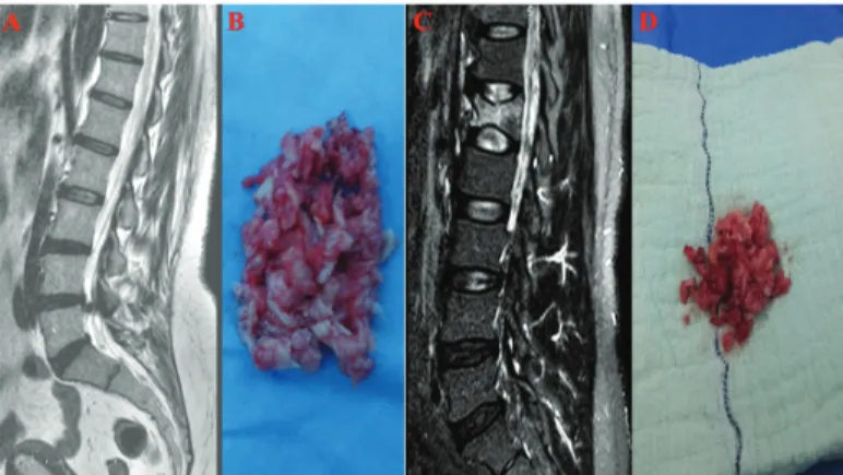
The cases in the experimental group were those determined to have degenerated disc hernias, which compresses nerve roots and the spinal cord (Figure 1A). Ratings of disc degeneration were determined by using modified Pfirrmann classification by means of T2 weighted MRI (23).
It was aimed to include 15 cases in the study. However, in cases where the patient had malignancy (n=1), where a biological agent was applied (n=1), had addictions to alcohol and smoking (n=2), took beta-blocker drugs (n=2), took cytochrome CYP 3A4 grouped drugs such as erythromycin (n=1), or took fluoxetine (n=2) were excluded from the study. Tissues were obtained from six cases (average age 42.17±13.78 years) by means of microdiscectomy. The tissues were placed in Falcon tubes for the purpose of collection of human primary NP/AF cell cultures (Figure 1B). The control group included 15 samples at the start of the research. These cases were volunteers who had applied in
the emergency department following a spinal trauma. After cases with vertebral fracture and spinal cord compression were brought into the study, primary cultures were prepared (Figure 1C).
Following preoperative lumbar MRI evaluations, six cases of spinal instability or traumatic intervertebral disc hernia and were treated with posterior transpedicular stabilization, laminectomy or discectomy were included in the study. The control group comprised cases with non-degenerated and healthy tissues (Figure 1D). We paid attention to select similar cases in both the control and experimental groups in terms of gender, age, and body mass index (average age 44.74±12.41 years).
Figure 1. a-d. Sagittal (a) T2-weighted MR images show L4-5
sequestrated intervertebral disc herniation, (b) degenerated intervertebral disc tissue resected after lumbar microdiscectomy from this case, sagittal (c) T2-weighted MR images show L1 compression fracture after spinal trauma, (d) intact intervertebral disc tissue resected after surgery from this case.
Resection of Tissues and Preparation of Primary Cell Cultures
Disc and granulation tissues were transferred to the laboratory in culture medium (Dulbecco’s Modified Eagle’s Medium [DMEM], Cat#41965062, Gibco, Dublin, Ireland) supplemented with 1% penicillin-streptomycin (Cat#15140122, Gibco, Dublin, Ireland), 15% fetal bovine serum ([FBS], Cat#10082147, Gibco, Dublin, Ireland), and 1% L-glutamine (Cat# 25030081, Gibco, Dublin, Ireland). Tissue samples were irrigated with 0.9% isotonic sodium chloride solution in the laminar flow cabinet and clarified from the red blood cells. Next tissues were dissected into 0.4 cm3 pieces, washed in Hank’s balanced salt solution (HBSS-1X, Cat#14025, Gibco, Dublin, Ireland), and transferred to Falcon tubes. Collagenase type II enzyme in an amount of 0.375 mg (Cat#17101015, Gibco, Dublin, Ireland) dissolved in a complete medium were added, and the samples were incubated with 5% CO2 at 37 0C overnight (1,10,13,15).
Next, the samples were centrifuged at 1200 rpm for 10 m. The obtained cell pellets were resuspended by adding cell culture medium and then transferred to T75 flasks and incubated to obtain primary cell cultures.
Confluent primary cultures were passaged and stained
with trypan blue. The cells were then counted and planted into 96 well plates at 1.4×10⁴ cell/well for MTT analysis, 24 well plates at 3.4×10⁴ cell/well for AO/PI analysis, and 100 mm petri dish as 4.4×10⁶ cell/dish for RNA isolation. At the end of a 24-hour incubation period, confluent cell cultures, which stick to the bottom, were selected for drug treatment.
Preparation of Drugs to be added to Culture Environment
The main stock solution was freshly prepared as 30 mg/ ml of nimodipine in DMEM medium into a flow cabin. Afterward, the main stock solutions were subject to color coding, which enabled researchers to be blind to analyses. The final concentration of nimodipine was applied to human primary NP/AF cell cultures at 100 µMolar. The drug dose administered to the cultures was calculated before carrying out analyses. The doses of nimodipine applied to the cultures began at 1 µMolar and proceeded at 50, 100, and 1000 µMolar. It has been previously reported that cell proliferation totally stopped in cultures where nimodipine at concentrations of greater than 100 µMolar were applied. Therefore, the necessary doses of nimodipine at concentrations allowing proliferation were applied to the cell classes found in the cultures. The groups were separated into two main groups, non-degenerated culture (iAF/NP) and degenerated culture (dAF/NP). These were designated Group 1 and Group 2, respectively, and the drug was not applied to the cell classes found in these groups.
Two subgroup cell class samples were then formed, and Groups 1 and 2 did not have the drug applied. Group 3 contained iAF/NP cell samples to which nimodipine had been applied. Group 4 contained dAF/NP cell samples to which nimodipine was applied (Table 1).
Table 1. Experimental design : AFCs/NPCs Nimodipine
Group ID Sample* Nimodipine
application Duration of applica-tion/Analyzes**
Group 1 iAFC/NPC - 0h 24h 48h
Group 2 dAFC/NPC - 0h 24h 48h
Group 3 iAFC/NPC + 24h 48h
Group 4 dAFC/NPC + 24h 48h
MTT-ELISA viability and toxicity proliferation analyses and AO/PI staining
Cells that adhered to the surfaces of flasks and showed chondrocytic activity were evaluated under an inverted microscope. Cell viability was analyzed using a commercial MTT kit (Vybrant MTT Cell Proliferation Assay, Cat#V13154, Thermo Fisher Scientific, Waltham, MA, USA) in line with the manufacturer’s instruction. MTT analyses were performed before and after the drug was added in all experimental groups, and results were obtained using an enzyme-linked immunosorbent assay ([ELISA]/540 nm) microplate reader (Mindray MR 96 A, PRC, Georgia Tech, Atlanta, GA, USA). Letter labels designated for the individual samples were transcribed by the project coordinator.
Vitality of the control groups (Group 1, control group for iAF/NP—Intact AFC/NP cell tissue, and Group 2, control group for dAF/NP—degenerated AF/NP cell tissue) were assumed to be 100% before the transfer of nimodipine (0 h) into the culture medium. At 24 h post administration of nimodipine, viability absorbance of the cells was recorded as nm. Subsequent proliferation analyses were recorded hourly between 24 h and 48 h.
To determine cell viability and confirm MTT results, nucleic acid binding dyes AO and PI were used. AO stain all nucleated cells whether alive or dead, generating green fluorescence. PI penetrates only dead cells with poor membrane integrity and stains nucleated cells to generate red fluorescence. When stained with both AO and PI, all live nucleated cells fluoresce green, and all dead nucleated cells fluoresce red (24).
To prepare the AO/PI stain, 4 mg AO (dissolved in 2 ml 99% ETOH), 10 g sodium-ethylenediaminetetraacetic acid, 4 mg PI, and 50 ml FBS were mixed well and sterile distilled water was added to reach a 200-ml final volume (24).
RT-PCR analyses
Total ribonucleic acid (RNA) was extracted from cultured iAF/NP and dAF/NP using a PureLink RNA Mini Kit (Ambion, Inc., Cat#12183018A, Foster City, CA, USA) and 2 Mercaptoethanol (Cat#31350010, Thermo Fisher Scientific, Waltham, MA, USA). To obtain complementary DNA (cDNA), 50 ng RNA were reversely transcribed with a High-Capacity cDNA Reverse Transcription Kit (Cat#4368814, Thermo Fisher Scientific, Waltham, MA, USA) using a thermal cycler (ProFlex, Thermo Fisher Scientific, Waltham, MA, USA). PCR reaction was performed using 10× reverse transcription buffer (10 μl), 25× deoxyribonucleotide triphosphate (4 μl), 10× random primers (10 μl), 50 U/μl reverse transcriptase (5 μl), nuclease-free water (5 μl), and RNA (20 μl) reaction mix. Reaction protocol was as follows: Hold for 10 m at 25 0C and hold for 120 m at 37 0C.
All genes were amplified using TaqMan Gene Expression Assays for CHAD (Hs00154382_m1, Cat#4448892), HIF 1α (Hs00153153_m1, Cat#4453320), COL2A1 (Hs00264051_ m1, Cat#4453320), and an internal control gene of housekeeping genes–actin beta (ACTB) (Hs99999903_ m1, Cat#4453320).
The RT-PCR reaction mix was prepared with 1 μl TaqMan Gene Expression Assay, 10 μl TaqMan Gene Expression Master Mix (Cat#4369016), 4 μl cDNA template, and UltraPure DNase/RNase-Free Distilled Water (Cat#10977035) for each gene in MicroAmp Fast Optical 96-Well Reaction Plates (Cat#4346906). Reaction protocol was as follows: 2 m hold at 50 0C, 10 m hold at 95 0C, 15 s at 95 0C, and 1 m at 60 0C for 40 cycles. The protocol was performed with the Applied Biosystems 7300/7500 Real-Time PCR System (24).
Statistical Analysis
Data on proliferation of nimodipine was gathered using Minitab 16 Statistical Software (Minitab Inc., State College,
PA). The data were evaluated with 95% confidence interval. Descriptive statistics were given as mean ± standard deviation.
Initially, it was intended to evaluate the data by repeated ANOVA test because it collects data from time points and can be used for comparing more than 3 group averages. However, repeated ANOVA could not be used because proliferations were compressed after 24 h, and gene expression decreased. Instead, one-way variance analysis (one-way ANOVA) was applied, and the F-value obtained from this analysis was used. Afterward, the post-hoc Tukey HSD method was used to determine the difference between groups. Alpha significance value was considered to be <0.05.
RESULTS
Morphological Analysis of Chondrocyte Cultures: Inverted Microscopy and AO/PI Staining
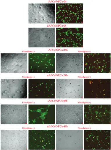
Cell cultures composed of intact and degenerated tissues were evaluated by inverted microscopy for cell proliferation follow-up and morphological analysis at 24 h and 48 h. The micrographs are given in Figure 2. AO/PI staining was applied to evaluate the effects of nimodipine application on cell vitality in the same cultures and to support MTT analyses. Micrographs that were obtained after AO/PI staining are given in Figure 2.
Figure 2. Inverted light/fluorescent microscopy and acridine
In cultures that were obtained from intact tissue at 0 h, cell proliferation and extracellular matrix development were higher than cells comprising degenerated AFC/NP cell tissues, as expected (Figure 2C). Cells in both cultures preserved their vitality and specific morphology (Figure 2B and 2D).
It was observed that cell density increased on a time-dependent basis and preserved vitality. Additionally, it was seen that proliferation was suppressed and necrosis increased in nimodipine-applied cultures.
Cultures from degenerated AF/NP cell tissue were most affected by nimodipine application (Figure 2K and L). However, it was observed that this negative effect had an about-turn at 48 h post application (Figure 2T and U).
Statistical Evaluation of Proliferation from MTT Data
The data obtained after MTT analyses were evaluated at a 95% confidence interval. By means of ANOVA (p=0.000), it was shown that cell proliferation differed between the intact AF/NP cell and degenerated AF/NP cell groups as well as the dosed group and the non-dosed group at 0 h, 24 h, and 48 h (Table 2).
Cultures prepared with intact AF/NP cells and degenerated AF/NP cells were evaluated in terms of nimodipine application and application time and then grouped accordingly. It was found that cell proliferation increased in intact AF/NP cell cultures and non-nimodipine groups at 24 h. In addition, it was reported that cell numbers were similar at 48 h to those at 24 h. It was statistically confirmed that nimodipine compressed proliferation at 24 h in the intact AF/NP cell experimental group; however,
the proliferation continued and reached the same cell numbers at 48h as it was at 0 h.n the degenerated AF/ NP cell groups, it was determined that cell proliferation increased in a time-dependent manner in the non-nimodipine cultures. However, it was found that when nimodipine was applied, proliferation was repressed at 24 h and then restarted at 48 h.
Table 2. The assessment of the data obtained from nimopidine administered to the degenerative and intact tissues by means of the post-hoc Turkey Pairwise Comparison test after the repeated measures one-way ANOVA
Application (mean ± SD)Time (hour) p
0h 24h 48h Control degenerate tissue 0.270 ± 0.01 0.312 ± 0.02 0.360 ± 0.07 0.00 Degenerate tissue 0.198 ± 0.01 0.224 ± 0.01 0.185 ± 0.01 Control intact tissue 0.270 ± 0.02 0.312 ± 0.02 0.317 ± 0.05 0.00 Intact tissue 0.230 ± 0.01 0.246 ± 0.01 0.216 ± 0.01 IRT-PCR Evaluation
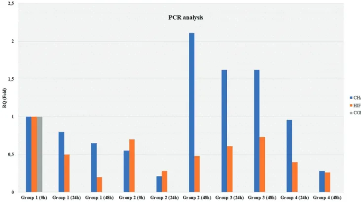
RNA isolation followed by RT-PCR analyses were performed at 0 h, 24 h, and 48 h in Groups 1 and 2, the non-nimodipine groups, whereas they were conducted at 24 h and 48 h in Groups 3 and 4, the nimodipine groups. In Group 1, intact AFC/NP cell tissue (iAF/NP) culture at 0 h was considered to be the reference, and the gene expression level was considered to be RQ=1 (100%). The gene expression levels of other experimental groups were calculated as RQ namely fold (Figure 3).
COL2A1 gene expression was not found in any of the groups. CHAD and HIF 1α expression decreased on a time-dependent basis in Group 2, which comprised NP/AF cell tissues (dAF/NP) that did not have nimodipine application. However, while CHAD and HIF 1α expression were found to decrease at 24 h (RQ=0.21 and 0.28, respectively), they began to increase at 48 h, which was different from intact tissue.
It was observed that CHAD expression was 2.11 times greater. CHAD expression in intact tissues that did not have nimodipine applied was stable at 24 h and 48 h and was 1.62 times higher than the control group. Additionally, although the HIF 1α level was lower than the control group, there was a slight increase at 48 h. In degenerated NP/AF cell cultures where nimodipine was used, CHAD and HIF 1α expressions decreased over time, different from the non-nimodipine group.
DISCUSSION
One of the major characteristics of drugs is their selectiveness [25]. However, it is not possible for a drug to show only the desired effect. From the point of view of clinical practice, effects of drugs are divided into two categories, desired effects and adverse effects. Most adverse effects are harmful regardless of the drug’s indications of drugs, and these effects are called toxic (4,8,26-31).
Drug toxicity is a serious complication in treatments (4,8,26-31). these toxicities may result in dosage increases, delays, termination of treatments, or death. Increases in preventive treatment and developments in supportive care contribute to improvements and favorable results in treatments because the prime purpose is to provide the desired benefits with less toxicity (4,8,26-31).
The current study primarily aimed to determine if nimodipine, among calcium channel blockers, which is indicated in prophylaxis and treatment of ischemic neurological disorders caused by cerebral vasospasm that develops after aneurysmal subarachnoidal bleeding, was toxic in NP/AF cultures.
The secondary aim was to analyze the effects of nimodipine on CHAD, which is related to the development between spinal cord and dorsal colon development; COL2A1, which is responsible for extracellular matrix development; and HIF 1α, a continuous expression of the NP-specific marker (24).
After pharmaceutical preparations are used, changes at the level of electrolytes such as Na+, K+, Ca2+, and Cl are epitoms for toxic biochemical effects. These are generally bound to pharmacodynamic drug effects (32). Structural toxic effects of drugs are toxicities that appear in tissues and cells in morphological disorders. The toxicity of medicines and other chemical agents or their metabolites causes an irreversible degradation of subcellular structures or macromolecules, such as DNA, RNA, and enzymes, which are functional in the life of cells (33).
Various fundamental structural changes occur in cells under the influence of direct toxicity of drugs. Effects such as deformation in volume correction, deterioration of metabolism, fragmentation of lysosomes, lipid peroxidation, and triglyceride accumulation in cells can be observed (34).
Median lethal dose (LD50) is a quantitative indicator used in evaluating acute toxicity of drugs in laboratory animals. This dose kills 50% of laboratory animals when applied in a group. Drugs are examined in terms of subacute (subchronic) and chronic toxicities after they are used for in recurrent doses for an extended period of time. For subacute tests, a drug is given orally to laboratory animals regularly for a period of one-sixth of its average life span (90 days for rats and mice). In chronic toxicity analyses, a drug is given for a period which is almost equal to the animal’s average life span (2 years for rats and mice). In this study, we performed our analyses putting nimodipine in each well at a final concentration of 100 µMolar, and analyses were made at 0 h, 24 h, and 48 h. It is known that the rating of toxic effects is associated with reactions that are based on idiosyncrasy and genetic differences in the people who took the drug. A drug that is toxic to one person may not be toxic to another. In other words, reactions may occur with use of a drug that may differ from person to person, and some people may have severe reactions (4,8, 24-31). For instance, in some studies, cigarette and alcohol users were more sensitive than non-smokers to the bronchoconstrictor effects of beta-blockers (35,36).
Therefore, this study did not include tissues from patients who smoke or drink alcohol. Also excluded were those who took medications to inhibit cytochrome CYP3A4 (37), which may interact with nimodipine and anyone who drank grapefruit juice within the last 48 hours. Additionally, other causes for exclusion from this study were patients who were using macrolide-grouped antibiotics such as erythromycin; anti-HIV protease inhibitors such as ritonavir; anti-antimycotics containing azole ring such as ketoconazole; antidepressants such as nefazodone and fluoxetine; and cimetidine and valproic acid active ingredient drugs.
Nimodipine is a pharmacologic agent that is a calcium antagonist in the 1,4-dihidropiridin group (38). In animal experiments, it was found that nimodipine had a more profound effect on cerebral arteries than any other places in the body, which results from its exceeding of the blood-brain barrier because it is lipophilic (39,40).
After nimodipine is taken orally, it is completely absorbed. Peak plasma concentrations for elderly people are (Cmaks) 7.3–43.2 ng/ml which is typically reached in 0.6 h to 1.6 h. After a single dose of 30 mg or 60 mg nimodipine, the average peak for young volunteers was 16±8 ng/ml and 31±12 ng/ml, respectively. Peak plasma concentration and the area under the curve increase proportionally until the maximum dose that is tested (90 mg). Nimodipine
is tied to plasma proteins at a level of 97%–99%, even in animal experiments, and the drug has been reported to be transferred to the placenta (41). Despite the limited number of studies carried out in human beings, it is probable that the same distributions would be shown. We have been unable to find a study in the literature indicating this drug has any effects with characteristics of cerebral antivasoconstrictive, antiischemic, neuropharmacological, or psychopharmacologic activity on NP/AF cells. In our in vitro study, we observed that cell proliferation was suppressed, and necrosis rate increased. Cultures from degenerated NP/AF cell tissue that were subject to nimodipine application for 24 hours were especially affected. However, it was observed that this negative effect did an about-turn at 48 hours. This showed that some of the cultured cells were able to tolerate the drug. However, it should be taken into consideration that increasing necrosis might be the case for tissues that are continuously exposed to nimodipine. Another milestone of our research was to examine the effects of nimodipine application on COL2A1, CHAD, and HIF 1α expressions. COL2A1 gene expression wasn’t found in any experimental groups. It was reported that in iAF/NP cell cultures where nimodipine was not applied, CHAD and HIF 1α expression decrease over time.
CHAD and HIF 1α expression were found to decrease at 24 h (RQ=0.21 and 0.28, respectively) in dAF/NP cell cultures. These began to increase at 48 h, which supports microscopy data.
It was found that CHAD expression in intact tissues where nimodipine was applied were stable at 24 and 48 h, and they were 1.62 times higher than the control group. Also, although HIF 1α levels were lower than the control group, there was a slight increase at 48 h.
In degenerated NP/AF cell cultures where nimodipine was used, CHAD and HIF 1α expressions decreased on a time-dependent basis, which was different from the non-nimodipine groups. All RT-PCR data were evaluated at large, and there was no correlation between nimodipine application and gene expression in terms of increase or decrease.
This experimental setup also demonstrated that the cell gene expressions that were repressed at 24 h had increased at 48 h post application of nimodipine. It is thought that this increase could have stemmed from the in vitro experimental setup. It should be noted, however, that a drug dose applied clinically to a living organism must be continuous during the treatment process. Additionally, compensatory mechanisms play a role in minimizing toxic effects that might be stemming from a pharmacological agent as the first pass effect taking place in the liver and gastrointestinal tract. Furthermore, electrochemical and hormonal response mechanisms come into play against a drug in tissues. On the other hand, it has been observed that toxic effects encountered in an in vitro experimental setup (in a laboratory) were significantly less than those
encountered clinically for the following reasons: a) absence of the compensatory mechanism, and b) single-dose application of the pharmacological agent at the beginning of the experiment but absence of continual dosing. However, if the doses were applied continuously, as is done clinically, the gene expressions would change, and proliferation might stop.
Gene expression is affected by individual differences, so more cases would serve to have more clear-cut results. However, we would like to underline that nimodipine had sufficient impact to change gene expression.
As a result, primary human cell cultures that we used in our study are important with regard to the comparison of effects of nimodipine on vitality of NP/AF cells.
CONCLUSION
The data obtained from in vitro experiments would not directly affect clinical practice. Using developments from pharmaceutical technology in clinical practices in the future will reduce the frequency and severity of complications because patients will receive treatment using personalized doses and schedules. Because frequently prescribed drugs may suppress proliferation and differentiation of other cells and/or tissues, a clinician should always consider this risk.
Competing interests: The authors declare that they have no competing interest.
Financial Disclosure: There are no financial supports
Ethical approval: Namik Kemal University School of Medicine Local Ethic Committee; 2017/41/04/01
REFERENCES
1. Zhu JJ, Xu YQ, He JH, Yu HP, Huang CJ, Gao JM, et al. Human cardiotoxic drugs delivered by soaking and microinjection induce cardiovascular toxicity in zebrafish. J Appl Toxicol 2014;34(2):139-48.
2. Moreira CD, Carvalho SM, Mansur HS, Pereira MM. Thermogelling chitosan-collagen-bioactive glass nanoparticle hybrids as potential injectable systems for tissue engineering. Mater Sci Eng C Mater Biol Appl 2016; 58:1207-16.
3. Yan Z, Yin L, Wang Z, Ye J, Zhang Z, Li R, et al. A novel organ culture model of mouse intervertebral disc tissues. Cells Tissues Organs 2016;201(1):38-50.
4. Gumustas F, Yilmaz I, Sirin DY, Gumustas SA, Batmaz AG, Isyar M, et al. Chondrocyte proliferation, viability and differentiation is declined following administration of methylphenidate utilized for the treatment of attention$deficit/hyperactivity disorder. Hum Exp Toxicol 2017;36(9):981-92.
5. Oh CD, Im HJ, Suh J, Chee A, An H, Chen D. Rho-Associated Kinase Inhibitor Immortalizes Rat Nucleus Pulposus and Annulus Fibrosus Cells: Establishment of Intervertebral Disc Cell Lines With Novel Approaches. Spine (Phila Pa 1976) 2016;41(5):255-61.
6. van den Akker GG, Surtel DA, Cremers A, Rodrigues-Pinto R, Richardson SM,Hoyland JA, et al. Novel immortal human cell lines reveal subpopulations in the nucleus pulposus. Arthritis Res Ther 2014;16(3):R135.
7. Kraus P, Lufkin T. Bovine annulus fibrosus cell lines isolated from intervertebral discs. Genom Data 2016;10:83-4.
8. Gumustas SA, Yilmaz I, Isyar M, Sirin DY, Batmaz AG, Ugras AA, et al. Assessing the negative impact of phenyl alkanoic acid
derivative, a frequently prescribed drug for the suppression of pain and inflammation, on the differentiation and proliferation of chondrocytes. J Orthop Surg Res 2016;11(1):70.
9. Navarro-Gonzalez MF, Grayson TH, Meaney KR, Cribbs LL, Hill CE. Non-L-type voltage-dependent calcium channels control vascular tone of the rat basilar artery. Clin Exp Pharmacol Physiol 2009;36(1):55-66.
10. St-Onge M, Dubé PA, Gosselin S, Guimont C, Godwin J, Archambault PM, et al. Treatment for calcium channel blocker poisoning: a systematic review. Clin Toxicol (Phila) 2014;52(9):926-44.
11. Behroozi-Lak T, Zarei L, Moloody-Tapeh M, Farhad N, Mohammadi R. Protective effects of intraperitoneal administration of nimodipine on ischemia-reperfusion injury in ovaries: Histological and biochemical assessments in a rat model. J Pediatr Surg 2017;52(4):602-8.
12. Stein G, Srivastava MK, Merker HJ, Neubert D. Effects of calcium channel blockers on the development of early rat post implantation embryos in culture. Arch Toxicol 1990;64(8):623-38.
13. Wang GH, Liu Y, Wu XB, Lu Y, Liu J, Qin YR, et al. Neuroprotective effects of human umbilical cord-derived mesenchymal stromal cells combined with nimodipine against radiation-induced brain injury through inhibition of apoptosis. Cytotherapy 2016;18(1):53-64.
14. Ben Fredj N, Grange J, Sadoul R, Richard S, Goldberg Y, Boyer V. Depolarization-induced translocation of the RNA-binding protein Sam68 to the dendrites of hippocampal neurons. J Cell Sci 2004;117(Pt 7):1079-90.
15. Albanna W, Weiss M, Conzen C, Clusmann H, Schneider T, Reinsch M, et al. Systemic and Cerebral Concentration of Nimodipine During Established and Experimental Vasospasm Treatment. World Neurosurg 2017;102:459-65.
16. Livonia MI. Nimodipine capsule package insert. Major Pharmaceutical. (2015). http://medlibrary.org/lib/rx/meds/ nimodipine-1/page/4/2018/01/01. accessed date 18.03.2018 17. Porchet F, Chiolero R, de Tribolet N. Hypotensive effect of
nimodipine during treatment for aneurysmal subarachnoid haemorrhage, Acta Neurochir (Wien) 1995;137(1-2): 62-9. 18. Subramani K, Ghrew M. Severe myocardial depression
following intravenous nimodipine for aneurysmal subarachnoid haemorrhage. Intensive Care Med 2004;30(7):1498-9.
19. Ehlicke F, Freimark D, Heil B, Dorresteijn A, Czermak P. Intervertebral disc regeneration: influence of growth factors on differentiation of human mesenchymal stem cells (hMSC). Int J Artif Organs 2010;33(4):244-52.
20. Rosenzweig DH, Tremblay Gravel J, Bisson D, Ouellet JA, Weber MH, Haglund L. Comparative analysis in continuous expansion of bovine and human primary nucleus pulposus cells for tissue repair applications. Eur Cell Mater 2017;33:240-51.
21. Zhou X, Tao Y, Liang C, Zhang Y, Li H, Chen Q. BMP3 alone and together with TGF-β promote the differentiation of human mesenchymal stem cells into a nucleus pulposus-like phenotype. Int J Mol Sci 2015;16(9):20344-59.
22. Feng G, Jin X, Hu J, Ma H, Gupte MJ, Liu H, et al. Effects of hypoxias and scaffold architecture on rabbit mesenchymal stem cell differentiation towards a nucleus pulposus-like phenotype. Biomaterials 2011;32(32):8182-9.
23. Hanley EN Jr DR, McCulloch JA. Surgical Indication and Techniques International Society for the Study of the Lumbar Spine: The Lumbar SpineThe Lumbar Spine 2nd edition. Philadelphia PA: WB Saunders. 1996; p. 492-524.
24. Karaarslan N, Yilmaz I, Ozbek H, Sirin Yasar D, Kaplan N, Akyuva Y, et al. Are specific gene expressions of extracellular matrix and nucleus pulposus affected by primary cell cultures prepared
from intact or degenerative intervertebral disc tissues? Turk Neurosurg 2018;22.
25. Kruse AC, Weiss DR, Rossi M, Hu J, Hu K, Eitel K, et al. Muscarinic receptors as model targets and antitargets for structure$based ligand discovery. Mol Pharmacol 2013;84(4):528-40.
26. Finsterer J, Scorza FA. Effects of antiepileptic drugs on mitochondrial functions, morphology, kinetics, biogenesis, and survival. Epilepsy Res 2017;136:5-11.
27. Sirin DY, Karaarslan N. Evaluation of the effects of pregabalin on chondrocyte proliferation and CHAD, HIF-1α, and COL2A1 gene expression. Arch Med Sci 2018.
28. Oznam K, Sirin DY, Yilmaz I, Kaya YE, Isyar M, Gumustas SA, et al. Iopromide-and gadopentetic acid-derived preparates used in MR arthrography may be harmful to chondrocytes. J Orthop Surg Res 2017;2(1):98.
29. Karaarslan N, Yilmaz I, Isyar M, Ozbek H, Akyuva Y, Sirin DY, et al. Do Nitric Oxide Synthases Enzyme Inhibitors Affect Chondrocyte Activity? Merit Res J Med Med Sci 2017;5(12):635-45.
30. Guzelant AY, Isyar M, Yilmaz I, Sirin DY, Cakmak S, Mahirogullari M. Are chondrocytes damaged when rheumatologic inflammation is suppressed? Drug Chem Toxicol 2017;40(1):13-23.
31. Gumustas SA, Yilmaz I, Isyar M, Sirin DY, Batmaz AG, Gonultas A, et al. Is it possible to expedite ressearches for effects of pharmacological agents on primary cell culture which is obtained from high grade fibular osteosarcoma? J Clin Exp Oncol 2016;5(6):1-8.
32. Inrig JK, Molina C, D’Silva K, Kim C, Van Buren P, Allen JD, et al. Effect of low versus high dialysate sodium concentration on blood pressure and endothelial-derived vasoregulators during hemodialysis: a randomized crossover study. Am J Kidney Dis 2015;65(3):464-73.
33. Goodwin WH. The use of forensic DNA analysis in humanitarian forensic action: The development of a set of international standards. Forensic Sci Int 2017;278:221-7.
34. Begriche K, Massart J, Robin MA, Borgne-Sanchez A, Fromently B. Drug-induced toxicity on mitochondria and lipid metabolism: mechanistic diversity and deleterious consequences for the liver. J Hepatol 2011;54(4):773-94.
35. Cohn A, Ehlke S. Association of Current and Lifetime DSM-IV Drug use Disorder Diagnoses to 6-Month Changes in Smoking Behavior in Risky Drinking Smokers: A Pilot Study. Subst Use Misuse 2017;52(13):1784-94.
36. Lasner TM, Weil RJ, Riina HA, King JT Jr, Zager EL, Raps EC, et al. Cigarette smoking-induced increase in the risk of symptomatic vasospasm after aneurysmal subarachnoid hemorrhage. J Neurosurg 1997;87(3):381-4.
37. Zhou YT, Yu LS, Zeng S, Huang YW, Xu HM, Zhou Q. Pharmacokinetic drug-drug interactions between 1,4-dihydropyridine calcium channel blockers and statins: factors determining interaction strength and relevant clinical risk management. Ther Clin Risk Manag 2014;10:17-26. 38. Shen H, Liang P, Qiu S, Zhang B, Wang Y, Lv P. The role of Na(+),
K(+)-ATPase in the hypoxic vasoconstriction in isolated rat basilar artery. Vascul Pharmacol 2016;81:53-60.
39. Wang F, Yin YH, Jia F, Jiang JY. Antagonism of R-type calcium channels significantly improves cerebral blood flow after subarachnoid hemorrhage in rats. J Neurotrauma 2010;27(9):1723-32.
40. Monzani D, Genovese E, Pini LA, Di Berardino F, Alicandri Ciufelli M, Galeazzi GM, et al. Nimodipine in otolaryngology: from past evidence to clinical perspectives. Acta Otorhinolaryngol Ital 2015;35(3):135-45.
41. Product information of nimodipine. http://www.bayerresources. com.au/resources/uploads/pi/file9406.pdf Accessed date 31.03.2018


