Contents lists available atScienceDirect
Journal of Drug Delivery Science and Technology
journal homepage:www.elsevier.com/locate/jddstImproving therapeutic e
fficacy of voriconazole against fungal keratitis:
Thermo-sensitive in situ gels as ophthalmic drug carriers
Neslihan Üstünda
ğ Okur
a,∗, Vildan Yozgatl
ı
b, Mehmet Evren Okur
c, Aysegül Yolta
ş
d,
Panoraia I. Siafaka
b,eaUniversity of Health Sciences, Faculty of Pharmacy, Department of Pharmaceutical Technology, Uskudar, 34668, Istanbul, Turkey bIstanbul Medipol University, School of Pharmacy, Department of Pharmaceutical Technology, Beykoz, 34810, Istanbul, Turkey cUniversity of Health Sciences, Faculty of Pharmacy, Department of Pharmacology, Uskudar, 34668, Istanbul, Turkey
dEge University, Faculty of Science, Department of Biology, Fundamental and Industrial Microbiology Division, Bornova, 35100, Izmir, Turkey eAristotle University of Thessaloniki, Department of Chemistry, 54124, Thessaloniki, Greece
A R T I C L E I N F O
Keywords: Voriconazole
Thermo-sensitive in situ gel Ocular drug delivery Microbiological study Ex vivo
Rabbit
Ocular irritation test
A B S T R A C T
The aim of this research was to evaluate the potential use of in situ gel formulations for voriconazole ocular delivery as fungal keratitis treatment. The in situ gelling system was applied to increase residence time and the bioavailability of voriconazole in the ocular mucosa. Temperature-triggered in situ gel formulations were pared by cold method. Poloxamer 188, poloxamer 407 and carboxymethylcellulose were used for the pre-paration of thermosensitive in situ ocular gel. Voriconazole concentration in formulations was 0.1% (w/w). The prepared gels were evaluated for clarity, sol-gel transition temperature, gelling capacity, pH, viscosity, FT-IR and drug content. The gelation temperatures of all the formulations were within the range of 29–34 °C. All for-mulations exhibited fairly uniform drug content. Furthermore, sterility, antifungal activity, stability, in vitro drug release, ex vivo permeation, and penetration and in vivo study of these formulations were also examined. Drug release results indicated that all formulations showed sustained release properties. Irritation studies showed that no ocular damage or clinically abnormal signs were observed in the cornea, conjunctiva or iris upon adminis-tration of the formulation. In conclusion, voriconazole loaded in situ gels could be offered as a promising strategy as ocular carriers for the treatment of fungal keratitis.
1. Introduction
The anatomy, physiology, and biochemistry of the eye render this organ exquisitely impervious to foreign substances such as active in-gredients. Consequently, compared with conventional routes of ad-ministration (oral, intravenous etc), ocular drug delivery has to over-come significant problems [1–3]. Conventional eye drops could generate eye irritation due to the shape and particle size while eyes different barriers such as lachrymal fluid-eye and blood-ocular barriers might induce lachrymation, nasolachrimal drainage system, enzymatic metabolism [4,5]. The extent of ocular drug absorption is seriously limited by physiological constraints. Ocular absorption is mainly re-stricted by the relatively impermeable corneal barrier. It has been well referred that cornea is comprised from three membranes: the epithe-lium, the endotheepithe-lium, and inner stroma which act as main absorptive barriers. The epithelium facing the tears with lipophilic cellular layers plays the role of a barrier to ion transport. The tight junctions of the
corneal epithelium act as the barrier for small molecules preventing the diffusion of macromolecules via the paracellular route. In further, 90% of the cornea consists of the stroma beneath the epithelium which is a highly hydrophilic layer. The corneal endothelium is responsible for maintaining normal corneal hydration [6–8].
Fungal infections, generally occurring in both animals and plants, are often observed in immunocompromised eyes after corneal trauma, surgery, or topical corticosteroid treatment [9]. Fungal keratitis is the major cause of ocular morbidity and blindness. Although fungal infec-tions are distributed worldwide, they are more frequently found in tropical and subtropical regions [10,11]. In previous reports, the most common organism isolated from culture-proven fungal endophthalmitis was the yeast species Candida albicans, followed by molds such as As-pergillus species. A limited number of Colletotrichum species have been reported to induce infection in humans. More specifically, Colleto-trichum dermatium and ColletoColleto-trichum truncatum were responsible for keratitis with few endophthalmitis cases [12]. In fungal keratitis, early
https://doi.org/10.1016/j.jddst.2018.12.005
Received 21 September 2018; Received in revised form 26 November 2018; Accepted 4 December 2018 ∗Corresponding author.
E-mail address:[email protected](N. Üstündağ Okur).
Available online 05 December 2018
1773-2247/ © 2018 Elsevier B.V. All rights reserved.
diagnosis and antifungal therapy are essential to prevent further com-plications such as hypopyon formation, endophthalmitis, or loss of vi-sion. Several drugs have been applied against fungal infections with oral, intravenous or topical eye solution of voriconazole to win the race for clinicians [13]. The majority of the reports noted that 1% w/v voriconazole eye drops is efficacious for the management of ophthalmic fungal keratitis, though thefinal voriconazole concentration achieved in the eye was not high enough to treat all types of fungal keratitis [11,14].
Voriconazole (VCZ) (UK-109,496) is a potent new triazole deriva-tive with a broad spectrum of antifungal activity against many oppor-tunistic fungal pathogens including Candida, Cryptococcus, and Aspergillus species [15–18]. The in vitro susceptibility opposed to var-ious fungal isolates associated with keratitis and endophthalmitis has been found nearly 100%. Moreover, various studies indicated a superior efficacy of VCZ against several ocular mycoses following topical ad-ministration [11]. It should also be mentioned that the clinical results which have been achieved following VCZ treatment in cases of fungal keratitis and endophthalmitis caused by Aspergillus spp., Scedosporium apiospermum, Paecilomyces lilacinus, Candida spp., and Fusarium spp. are outstanding [19,20].
Clinicians, so as to treat eye infections, usually choose topical ad-ministration [1,21] since the 90% of the marketed ophthalmic for-mulations are in the form of eye drops. Among others, features like patient compatibility, tolerability, the easier production method, and economical cost make eye drops acceptability wider. However, the meager bioavailability (< 5%) of conventional eye drops (suspensions, solutions, etc.) is one of the major concern for pharmaceutical tech-nologists and companies [22]. This low bioavailability is mainly due to rapid drainage of the molecules by tears and corneal epithelium membranes impermeability. Additionally, the temporary residence in the conjunctival fornix and non-productive absorption in the nasal cavity are also pledged for the low drug dose in intraocular tissues. Several research groups suggest that innovative systems such as sus-pensions, ointments, inserts, hydrogels, polymeric micelles, and lipid-based nanocarriers could be effective on meeting the challenges re-sulted from ocular delivery [1,23–25].
Generally, ocular ointments were studied for their excellent drug bioavailability due to the improved contact time, minimum tear re-moval as well as their resistance on nasolacrimal drainage. On the contrary, ointments could provoke blurred vision, limiting their use during the night or against infections on the outside and edges of the eyelids [26]. Controlled drug delivery systems using erodible non-toxic polymers seems to be the solution for the ocular problems since the primal ophthalmic solutions, suspensions, and ointments dosage forms are insufficient in ophthalmic diseases management [2].
In situ gelling systems, involving various polymers have been re-searched because they present prolonged contact time onto the ocular surface and improved drug corneal permeability [27–31]. In situ acti-vated gel-forming systems can be described as viscous liquids which by changing physiological conditions (pH, temperature, ionic strength, UV) can shift to a gel phase [32]. In situ gels, in contrast to already gelled formulations, are advantageous formulations considering that can be accurately administered in reproducible quantities, promoting precorneal retention. For the production of in situ gels several both natural and synthetic polymers have been applied [33]. Poloxamers are triblock copolymers between polyethylene polypropylene oxide-polyethylene oxide (PEO-PPO-PEO) which have the ability to form optical clear gels in aqueous media. Their high water content which leads to the swellable hydrogels have a positive effect on drugs ab-sorption. Their reversed thermal gelation behavior (solution in 3–4 °C shifting to gel under physiological conditions) in addition to their mucomimetic properties classified poloxamers as optimal candidates for ophthalmic delivery gels systems [8,34,35]. Poloxamer 407 (P407) and Poloxamer 188 (P188) are used for several pharmaceutical pro-ducts due to their non-toxicity, safety [36] and suitability as
controlled-release agents [37]. In order to achieve better mucoadhesive properties and controlled diffusion, hydrophilic polymers, such as water-soluble derivatives of cellulose, polyacrylic acid and chitosan are incorporated in gels [38,39]. Carboxymethylcellulose (CMC) is water-soluble deri-vative of cellulose, sensitive in ionic strength and pH. It is widely known for its ionization and mucoadhesiveness because its -CH2COOH groups are substituted on the glucose units of the cellulose chain through an ether linkage [40].
The aim of this work was to develop new ocular in situ gel for-mulations containing VCZ for the treatment of fungal keratitis and to evaluate them as effective topical ocular delivery. For this purpose, physicochemical characterization, rheological studies, stability, in vitro release, microbiological studies, ex vivo and in vivo studies of ocular in situ gels were assessed.
2. Materials and methods 2.1. Materials
VCZ was purchased from Sigma-Aldrich, Germany. Poloxamer 407 and poloxamer 188 were kindly gifted from BASF, Turkey. Carboxymethyl cellulose (CMC) was purchased from ZAG, Turkey. Benzalkonium chloride (BZC) was supplied from Sigma-Aldrich, Germany. High-pressure liquid chromatography (HPLC) grade acet-onitrile (Sigma, Germany) was used for HPLC experiments. All the other solvents and chemicals were of analytical or HPLC grade. Dialysis membrane (Spectra/por 4, diameter 16 mm, the molecular weight of 12–14 kDa) was purchased from Spectrum. Distilled water was used throughout the study.
For microbiological studies,fluid thioglycollate medium was pur-chased from Merck Millipore Corporation, Darmstadt Germany, soya-bean casein digest medium (CM0129) purchased from Oxoid, Thermo Fisher Scientific Inc. was used. Roswell Park Memorial Institute (RPMI 1640) medium was purchased from Sigma-Aldrich, Germany. Dimethyl sulfoxide purchased from Merck Millipore, Germany. 96 well microtiter plates were purchased from LP, Italiana. The McFarland standard was acquired from Biomerieux. While Mueller-Hinton agar and D-glucose were purchased from BD Difco, methylene blue dye was obtained from SSI Diagnostica, Hillerød, Denmark.
2.2. Preparation of in situ gel formulations
The polymeric solutions were prepared by dispersing the required quantity of P407 and P188 in water using a magnetic stirrer until the poloxamers completely dissolve. Aqueous solutions were stirred for about two hours by using magnetic stirrer [33].
In situ gels were selected according to pH values and gelling tem-peratures of formulations. For the preparation of ocular in situ gel, CMC, VCZ, BZC as well as sodium chloride were incorporated in aqueous solutions containing P407, P188, and distilled water. BZC (0.02% w/w) was added as a preservative to the solutions. Sufficient amount of so-dium chloride (0.9% w/w) was added to the mixture to maintain the isotonicity. Voriconazole concentration was 0.1% (w/w).
2.3. Characterization of in situ gels
2.3.1. Determination of sol-gel temperature (Tsol-gel)
20 g of cold sample solution was put into a beaker and placed in a temperature-controlled stirrer. A thermometer (JG-220 Digital Thermometer (−50 + 260 °C), ± °1C accuracy) was immersed in the sample solution for constant monitoring. The solution was heated at the rate of 2 °C/min with the continuous with stirring at 200 rpm. The temperature at which the magnetic bar stopped moving due to gelation was reported as the gelation temperature. The maximum limit for ge-lation was checked up to 60 °C.
2.3.2. Gelling capacity
The gelling capacity of the prepared formulation was determined by placing a drop of the formulation in a beaker at 32 ± 0.5 °C and it was visually observed for gelling time [2,41].
2.3.3. Determination of pH
pH is one of the most important parameters involved in ophthalmic formulations. The pH of the gel was measured using calibrated pH meter (Mettler Toledo, Switzerland). Determination of pH was carried out in triplicate and the average of these determinations was taken as the pH of the prepared gels.
2.3.4. Drug content
0.125 mL of the developed formulations (IS-1-8-VCZ) were dis-solved in 25 mL mobile phase followed by HPLC estimation of the ali-quot to determine drug concentration [2].
2.3.4.1. HLPC analysis. The HPLC system consisted of a gradient pump and a UV detector supplied by Agilent 1100. C18 column (5μm, 150 × 4.6 mm) was used. The samples were analyzed at 256 nm with 1 mL/min flow rate at 25 °C. The mobile phase was a mixture of acetonitrile: ultrapure water (50:50). The retention time of VCZ was 4.098 min (Üstündağ Okur et al., 2016b). The method was validated for linearity, limit of detection (LOD) and limit of quantitation (LOQ), precision, accuracy and specificity, selectivity and stability. The linearity between peak area and concentration was analyzed using calibration curve obtained from standard solutions of VCZ (1–30 μg/ mL). The accuracy of an analytical method is the closeness of test results obtained by the method to the true value and is defined recovery. The prepared standard solutions were injectedfive times at different levels as a test sample. Eight μg/mL solution was injected ten times in order to evaluate method precision, standard deviation (SD) and coefficient of variation. The Limit of Detection (LOD) and Limit of Quantitation (LOQ) tests for the procedure are performed on samples containing very low concentrations of analytes.The used method for VCZ analysis was found to be linear. The standard deviation of the slope and intercept were low. It is well known that if the standard deviation is less than the acceptance criteria (2%), the analysis system for the determination of assay is to verify.
2.3.5. Spreadability of VCZ loaded in situ gels
To determine spreadability of VCZ loaded in situ gels, 0.1 g of VCZ loaded in situ gels were transferred to the center of a glass plate (10 cm × 10 cm), which this glass plate temperature 32 ± 0.5 °C and this glass plate was compressed under another glass plate of the same size. Thus, the gel was spread out in between the plates. After one minute, the weight was removed and the diameter of the spread area (cm) was measured. The measurement was performed in triplicate [42]. 2.3.6. Stability of VCZ loaded in situ gels
In order to check physical stability, VCZ in situ gels were stored at 4 ± 1 °C in the refrigerator for 3 months. After 3 months storage visual appearance, clarity, pH, gelation time of in situ gels and VCZ content were investigated. The experiments were repeated three times. 2.3.7. FT-IR spectroscopy of in situ gels
In situ gels were characterized via FT-IR spectroscopy using FTIR––2000 spectrometer (Perkin Elmer, Germany). KBr disks (thick-ness of 500μm) technique was applied to check the possible interac-tions between drug and excipients. The spectra were recorded from 4000 to 400 cm−1 (Resolution of 2 cm−1 was applied, 32 co–added scans). Baseline corrected and converted to the absorbance mode spectra are shown herein.
2.3.8. Rheological studies
The viscosity measurements were carried out using Brookfield
viscometer LVDV-E model with a cylindrical spindle. The in situ gel formulations were placed in the sample tube. The samples were ana-lyzed with 50 rpm at 4 ± 0.5 °C by a circulating bath connected to the viscometer adaptor prior to each measurement [2].
2.4. In vitro drug release studies
In vitro release study of in situ gel solution was carried out in si-mulated tearfluid at 50 rpm. Simulated tear fluid (composition: sodium chloride 0.68 g, sodium bicarbonate 0.22 g, calcium chloride dihydrate 0.008 g, potassium chloride 0.14 g, and distilled deionized water to 100 mL) was used as the medium for in vitro release study [1,43]. The temperature was maintained at 32 ± 0.5 °C to mimic eye temperature. 5 g of formulations were separated from release media using dialysis membrane (Spectra/por, MW of 12–14 kDa) and capped with closures. 1 mL of sample was withdrawn at a predetermined time interval of 30 min to 12 h and the same volume of fresh medium was replaced. The samples were analyzed with HPLC for the drug content.
2.5. Microbiological studies of in situ gel 2.5.1. Sterility studies
Pure in situ gels as well as VCZ in situ gels were prepared at Laminar airflow Cabinet (Haier HR40-IIA2). To check the sterility of the pre-pared ocular formulations sterility control testing were performed. Sterility testing of the in situ gel formulations with or without vor-iconazole was carried out under aseptic conditions according to the international pharmacopoeia. For anaerobic bacteria, fluid thiogly-collate medium was used. For fungi and aerobic bacteria, soya-bean casein digest medium was used. 1 mL of formulation solution was added to each medium and incubated in BINDER GmbH incubator at 35 °C for bacteria and 25 °C for fungi for 14 days. No growth of the micro-organisms occurred.
To check the suitability of the used mediums for the sterility testing promotion test were performed. For growth promotion test of aerobes, anaerobes, and fungi, fluid thioglycollate media (using a separate portion of media for each microorganism) were inoculated with 100 CFU of Staphylococcus aureus ATCC 6538, Clostridium sporogenes ATCC 19404 and Candida albicans ATCC 10231. Media were incubated at 35 °C for 48 h. Both microorganisms showed visible growth. 2.5.2. Determination of MIC of VCZ
The broth microdilution test was done in accordance with CLSI guidelines forfilamentous fungi (CLSI document M38-A2) and yeasts (CLSI document M27-A3). VCZ was dissolved in dimethyl sulfoxide, final dilutions of formulations were made in RPMI 1640 medium buf-fered to pH 7.0 with 0.165 M MOPS buffer [3-(N-morpholino) propa-nesulfonic acid] andfinal concentrations were 250–0.125 μg/mL. Using the spectrophotometric method of inoculum preparation, a suspension with a 0.5 McFarland standard was utilized for yeasts and 0.4–5 × 10⁴ spores/mL for moulds, and RPMI 1640 medium buffered with MOPS was used. C. albicans ATCC 10231, C. tropicalis RSKK 2412, A. fumigatus ATCC 204305, A. niger ATCC 10864, A. terreus ATCC 20542 and A. flavus ATCC 204304 which could be possible causes of fungal keratitis were used to evaluate antifungal activity of developed formulations [44,45]. A 0.1 mL inoculum was added to each well of the microdilu-tion trays. The MICs were determined after 48 h of incubamicrodilu-tion. The plates were shaken before the comparison of growth in wells. With an aid of a reading mirror, growth in each well was compared with the voriconazole free growth control well. The MIC endpoints were eval-uated for the lowest drug concentration that showed a prominent re-duction (50%) of the growth in the control well.
2.5.3. Disk diffusion testing
Disk diffusion testing was performed according to CLSI standard M44-A2 for yeasts and M51-A forfilamentous fungi. Mueller-Hinton
agar supplemented with 2% w/v glucose and 0.5μg/mL methylene blue dye was used. Blank disks that were 12.1 mm in diameter were im-pregnated with 20μL of formulations at the final concentration of 1 μg/ disk and allowed to dry at room temperature in aseptic conditions. C. albicans ATCC 10231 and C. tropicalis RSKK 2412, A. fumigatus ATCC 204305 A. niger ATCC 10864, A. terreus ATCC 20542 and A.flavus ATCC 204304 were used to evaluate antifungal activity of developed for-mulations. The mold inocula were prepared at optical densities ranging from 80 to 82% and a suspension with a 0.5 McFarland standard was utilized for yeasts. The plates were incubated at 35 °C and inhibition zone (in millimeters) diameters were read by using a digital ruler at 24 and 48h.
2.6. Ex vivo studies (permeation and penetration studies)
Vertical diffusion cells with an effective area of 0.63 cm2were used for the corneal permeation and penetration experiments The excised goat corneas were clamped between the donor and the receptor chamber of cells. An exact amount of 0.5 mL in situ gels and 9 mL si-mulated tear fluid were filled into the donor and receptor chamber, respectively. The cells temperature was maintained at 32 ± 2 °C and magnetic stirring was also applied. The samples were collected at 0.5, 1, 2, 3, 4, 5, 6, 7, 8, 10, 12 and 24 h. Sink conditions were maintained in the receptor compartment during ex-vivo permeation studies. To ensure the sink condition 1 mL Tween 80 was used.
After 24 h of contact time, each cornea was washed for penetration study. Each cornea was placed in 10 mL of methanol for 24 h in a mixer to determine the amount of penetration VCZ. Penetrated drug and permeated drug were assayed by means of HPLC. The experiment was performed in triplicate.
2.7. In vivo studies 2.7.1. Animals
New Zealand albino rabbits ranging in weight from 2.5 to 3.5 kg and with no signs of ocular inflammatory were used for in vivo studies. The in vivo experimental protocol was approved by the Ethical-Scientific Committee of Bezmialem Vakıf University (approval number: 2018/ 118). All animal experiments comply with ARRIVE guidelines and were carried out in accordance with the UK animals and associated guide-lines such as EU directive 2010/63/EU. Rabbits were housed in a room maintained at 22 ± 1 °C and humidity in air-conditioned chambers with an alternating 12 h light-dark cycle was used. Animals had free access to a pellet diet and water ad libitum. During the in vivo studies, the rabbits were placed in boxes, and their heads and eyes movements were not restricted.
2.7.2. Determination of the VCZ in tear
Six rabbits were used in the study and, during the experiments, the rabbits were allowed to move their heads freely, and their eye move-ments were not restricted. Schirmer Tear Test Strip (ERC®) was used for the take tears. Fifty microliters of sterilized VCZ loaded in situ gel for-mulation (IS-1-VCZ) was administered in the lower conjunctival sac of the right eye only one time. The same amount of suspension of VCZ was administered to the lower conjunctival sac of the left eye only one time. After the formulations were applied to the eye, tears were collected at 0.5, 1, 2, 3, 4, 6, 8, and 24 h using tear strip. During the samples were taken, the tear strip placed into the conjunctival sac of the eye and marked tear strip was expected to reach a certain level. Strips were immediately placed in eppendorf tubes with 2 mL methanol and stored at + 4 °C until HPLC analyses. The HPLC was used to determine drug amount in tears [35,46].
2.7.3. Ocular irritation test
The possible ocular irritancy and/or damaging effects of VCZ loaded in situ gels were evaluated according to the modified Draize test. The in
situ gel formulation (0.01 mL) was instilled directly onto the cornea of the right eye every 30 min for 6 h (12 treatments). Left eye served as control and was treated with 0.9% w/v sodium chloride. The conges-tion, swelling, discharge, and redness of each rabbit eyes were graded on a scale from 0 to 3, 0 to 4, 0 to 3, and 0 to 3, respectively. Irritation and corneal opacity were graded on a scale from 0 to 4 [47]. 2.7.4. Statistical data analysis
Statistical data analysis was performed using the student's t-test with P < 0.05 as the minimal level of significance.
3. Results and discussion
3.1. Preparation of in situ gel formulations
Temperature sensitive in situ gels were successfully prepared by the cold method using poloxamer 407, poloxamer 188 and CMC. Cold method is one of the preferred methods because it provides clear so-lution for in situ gel and it does not form polymer lumps as it has been reported when hot process is applied [48]. The prepared formulations were coded as IS-1-8. Amount of 0.1% VCZ (w/w) was loaded to in situ gel formulations and 0.3% CMC was added in poloxamer solutions (in case of IS-5-8) with continuous stirring until complete dissolution while as a preservative to the solutions, BZC 0.02% (w/w) was added. Suffi-cient amount of sodium chloride was added to the solution to maintain isotonicity. Thefinal concentration of VCZ in formulations was 0.1% (w/w). The dispersion was kept in a refrigerator for 48 h to get a clear solution. The components of ocular in situ gels absence or presence of CMC are shown inTable 1.
3.2. Characterization of in situ gel formulations
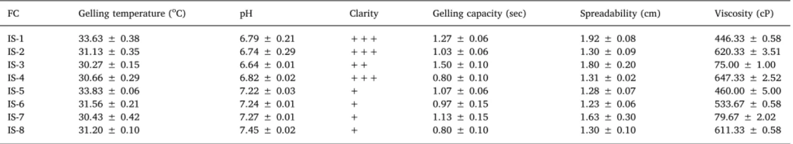
Physicochemical characterization of in situ gel formulations is an important issue to be considered in the formulation stage, especially when the formulations intended for ocular administration. The physi-cochemical parameters of in situ gels without VCZ are reported in Table 2and physicochemical parameters of in situ gels with VCZ are seen inTable 3. Clarity is one of the most crucial factors of ophthalmic preparations. All developed formulations were assessed for clarity by visual observation against a black and white background, demon-strating that the clarity of all the formulations was sufficient. Gelation temperature of prepared in situ gel formulations without VCZ was changed as about 30.27 ± 0.15 °C to 33.83 ± 0.06 °C and gelation temperature of prepared in situ gel formulations with VCZ was changed as about 29.73 ± 0.21 °C to 34.13 ± 0.32 °C. It can be safely con-cluded that mucoadhesive polymers (like CMC) played a reverse role on gelation temperature [49,50]. This indicates that the formulations can be converting to gel when they installed the eye surface. Between 32 and 34 ± 2 °C, the solutions are converted into gels with high visc-osity. The gelling capacity data of prepared formulations with and without VCZ are presented in Tables 2 and 3, respectively. It was Table 1
Formulation codes (FC) and components.
Composition of the prepared formulations
FC P 407 (g) P 188 (g) CMC (g) VCZ (g) BZC (% w/w) Physiological Saline (q.s) (g) IS-1-VCZ 15 25 – 0.1 0.02 100 IS-2-VCZ 18 22 – 0.1 0.02 100 IS-3-VCZ 20 5 – 0.1 0.02 100 IS-4-VCZ 20 20 – 0.1 0.02 100 IS-5-VCZ 15 25 0.3 0.1 0.02 100 IS-6-VCZ 18 22 0.3 0.1 0.02 100 IS-7-VCZ 20 5 0.3 0.1 0.02 100 IS-8-VCZ 20 20 0.3 0.1 0.02 100
established that the formulations had immediate gelation existing for two seconds.
The pH of an ophthalmic formulation is significant for patient compliance considering that if pH value is beyond 4–8 which condoned by eye, the patient could feel discomfort. In further, the eye could be irritated, and drug bioavailability can be further decreased because of increased tearing [26]. The pH of the prepared formulations without VCZ ranged between 6.64 ± 0.01 and 7.45 ± 0.02 and pH of the prepared formulations with VCZ ranged between 6.63 and 7.24 which is appropriate for ocular delivery since they were iso-hydric. This fact reveals the non-irritancy of the formulation in the ocular mucosa. When a formulation is administered to the eye, it stimulates theflow of tears. Tearfluid is capable of quickly diluting and buffering small volumes of added substances, thus the eye can tolerate a fairly wide pH range. Ophthalmic solutions may range from 6.5 to 8.5 [51,52]. Among others, the formulation should present an optimum viscosity, which would allow its instillation into the eye as a liquid, which afterward would undergo rapid sol-gel transition due to temperature change. When the in situ solutions were installed the 32–34 ± 2 °C surface temperature, the solutions were converted to gel form after only a few seconds. Additionally, so as to facilitate sustained release of the drug to the ocular tissue, the gel formed in situ should preserve its integrity without dissolving or eroding for a prolonged period of time.Tables 2 and 3 shows the gelling capacity of all formulations. The gelling ca-pacity increases by increasing the concentration of the gelling agent. Similarly to our study, Patil et al. prepared norfloxacin loaded in situ gels for the treatment of conjunctivitis which evaluated for their gelling Table 2
Physicochemical properties of neat in situ gels.
FC Gelling temperature (oC) pH Clarity Gelling capacity (sec) Spreadability (cm) Viscosity (cP)
IS-1 33.63 ± 0.38 6.79 ± 0.21 +++ 1.27 ± 0.06 1.92 ± 0.08 446.33 ± 0.58 IS-2 31.13 ± 0.35 6.74 ± 0.29 +++ 1.03 ± 0.06 1.30 ± 0.09 620.33 ± 3.51 IS-3 30.27 ± 0.15 6.64 ± 0.01 ++ 1.50 ± 0.10 1.80 ± 0.20 75.00 ± 1.00 IS-4 30.66 ± 0.29 6.82 ± 0.02 +++ 0.80 ± 0.10 1.31 ± 0.02 647.33 ± 2.52 IS-5 33.83 ± 0.06 7.22 ± 0.03 + 1.07 ± 0.06 1.28 ± 0.07 460.00 ± 5.00 IS-6 31.56 ± 0.21 7.24 ± 0.01 + 0.97 ± 0.15 1.23 ± 0.06 533.67 ± 0.58 IS-7 30.43 ± 0.42 7.27 ± 0.01 + 1.13 ± 0.15 1.63 ± 0.30 79.67 ± 2.02 IS-8 31.20 ± 0.10 7.45 ± 0.02 + 0.80 ± 0.10 1.30 ± 0.10 611.33 ± 0.58 Table 3
Physicochemical properties of VCZ loaded in situ gels.
FC Gelling temperature (oC) pH Clarity Gelling capacity (sec) Spreadability (cm) Viscosity (cP) Drug content (%)
IS-1-VCZ 34.13 ± 0.32 6.80 ± 0.03 +++ 1.77 ± 0.20 1.6 ± 0.08 533.33 ± 5.77 92.55 ± 0.19 IS-2-VCZ 31.83 ± 0.25 6.85 ± 0.02 +++ 1.27 ± 0.25 1.48 ± 0.07 668.33 ± 7.64 92.64 ± 0.22 IS-3-VCZ 30.00 ± 0.10 6.63 ± 0.01 ++ 1.90 ± 0.10 1.81 ± 0.06 71.67 ± 7.64 106.87 ± 0.19 IS-4-VCZ 30.77 ± 0.80 6.81 ± 0.01 +++ 1.03 ± 0.06 1.40 ± 0.13 613.33 ± 7.64 93.69 ± 0.53 IS-5-VCZ 33.73 ± 0.20 7.22 ± 0.01 + 1.17 ± 0.29 1.21 ± 0.09 371.67 ± 7.63 90.96 ± 0.07 IS-6-VCZ 31.20 ± 0.26 7.24 ± 0.01 + 0.80 ± 0.10 1.23 ± 0.06 401.67 ± 2.89 92.88 ± 0.11 IS-7-VCZ 30.06 ± 0.15 7.13 ± 0.01 + 1.37 ± 0.23 1.28 ± 0.15 78.33 ± 1.44 97.78 ± 0.08 IS-8-VCZ 29.73 ± 0.20 7.24 ± 0.01 + 0.73 ± 0.20 1.32 ± 0.07 568.33 ± 7.64 93.30 ± 0.22 Table 4
Stability of VCZ loaded in situ gels for drug content and pH at 4 ± 1 °C.
FC Drug content (% w/w) Drug content (% w/w) (after 3 months) pH pH (after 3 months) IS-1-VCZ 92.55 ± 0.19 93.97 ± 0.38 6.80 ± 0.03 6.90 ± 0.02 IS-2-VCZ 92.64 ± 0.22 95.56 ± 0.34 6.85 ± 0.02 6.83 ± 0.04 IS-3-VCZ 100.88 ± 0.19 61.55 ± 0.09 6.63 ± 0.01 6.72 ± 0.01 IS-4-VCZ 93.69 ± 0.53 94.08 ± 0.41 6.81 ± 0.01 6.84 ± 0.01 IS-5-VCZ 90.96 ± 0.07 92.36 ± 0.16 7.22 ± 0.01 7.04 ± 0.02 IS-6-VCZ 92.87 ± 0.11 91.25 ± 0.23 7.24 ± 0.01 7.07 ± 0.02 IS-7-VCZ 97.78 ± 0.08 81.84 ± 0.35 7.13 ± 0.01 7.30 ± 0.08 IS-8-VCZ 93.30 ± 0.22 90.59 ± 0.42 7.24 ± 0.01 7.27 ± 0.01
Fig. 1. a) FT-IR spectroscopy of unloaded in situ gels (inset: P407, P188, and CMC neat polymers) and b) loaded with VCZ in situ gels (inset: pure VCZ).
capacity. They found analogous results with our study as gelling ca-pacity depends on polymer conglutination of the formulations [53]. Moreover, spreading diameter of the developed formulations demon-strated that is similar for all formulations. However, IS-1 and IS-3 spreadability is higher than the other formulations. This could be ex-plained by their increased viscosity since Chaudhary et al. found that by increasing the concentration of the polymer, viscosity of the solution was also improved but the spreadability of the formulation was reduced [54].
3.2.1. Stability
The stability studies were carried out at 4 ± 1 °C, for a period of 3 months using the refrigerator. The samples were analyzed periodically on every month, and it was found that there are no changes in visual appearance, clarity, and gelation time. After 3 months, pH values of IS-1-VCZ–IS-8-VCZ formulations were established as 6.73 ± 0.01–7.30 ± 0.08, respectively.
Drug content was analyzed with HPLC using a validated method. The analytical method has good linearity, accuracy, precision, se-lectivity, and stability. The determination coefficient R2 for the regression line is 0.999 with the slope of 13.764x and yintercept of -0.656 for standard solution of VCZ. The percentage of recovery was almost 100% and the standard deviation was less than the acceptance criteria. Moreover, the LOD was 0.022μg/mL, LOQ was 0.065 μg/mL
and the assay exhibited a linear range of 1–30 μg/mL. The drug en-trapment was high in all tests ranging from 90 to 100% whereas 61–95% of initial drug content of formulations was detected after the studied period of 3 months (Table 4). No indication of aggregation/ precipitation or pH change was observed over a period of 90 days. This fact revealed that the formulations were stable after such period.
3.2.2. FT-IR spectroscopy of in situ gels
FT-IR spectroscopy belongs to the most useful techniques of phar-maceutical formulations characterization given that it can provide in-formation concerning interactions between carriers and active mole-cules as well as compatibility of drug and excipients [55–58]. In this work, various polymers were used for the preparation of in situ gels. P407 and P188 are copolymers of poly(propylene oxide) and poly (ethylene oxide). IR spectra of poloxamer 188 and 407 are character-ized by principal absorption peaks at 3400 (hydroxyl groups), 2889 and 2891 cm−1(C-H stretch aliphatic), 1348.15 cm−1and 1339 cm−1 (in-plane O-H bend), as well as at 1108 cm−1(C-O stretch), respectively. IR spectrum of CMC exhibits a broad absorption band at 3432 cm−1, due to the–OH group stretching. The observed peak at 2909 cm−1is as-signed to C–H stretching vibration while the strong absorption band at 1603 cm−1indicates the presence of COO−group. The bands at 1423 and 1325 cm−1 are attributed to –CH2 scissoring and –OH bending vibration, respectively whereas at 1061 cm−1 is depicted the CH–O–CH2stretching (Fig. 1-a) [59–61].
In the case of unloaded in situ gels, the presence of water [62] and BZC is depicted by the main peaks at 2096 and 1646 (alkenes) cm−1, respectively (Fig. 1-a). The other peaks attributed to poloxamers are similarly observed with small changes, identifying their compatibility. The addition of CMC in IS-5-8 formulations did not affect the spectra since it was in low content. The broadening of OH groups band could be due to the formulation method. ObservingFig. 1b, it can be said that VCZ spectrum exhibits broad peaks at 3200 and 3121 cm−1 corre-sponds to the hydroxyl groups and aromatic rings, respectively. Aro-matic C–C stretch peaks are seen at 1615 and 1586 cm−1while the presence of (Fluoro) groups is indicated by sharp peaks from 690 to 515 [17]. In most studies found in literature, similar in situ gels do not ex-hibit new peaks after the addition of drug in gels [58]. According to this fact the drugs and excipients are compatible (Fig. 1-b). Similarly, in our research, the absence of newly recorded peaks proves the compatibility of the gels [18]. However, it can be concluded that there are some in-teractions between the polymers and VCZ due to the shift in lower wavenumbers of some peaks [1].
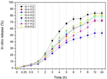
Fig. 2. In vitro release studies of VCZ from the prepared in situ gels (n:3, ± STD).
Table 5
Inhibition zone diameters (mm) of blank and VRZ loaded in situ gels against several types of microorganisms (Candida albicans, Candida tropicalis, Aspergillusflavus, Aspergillus fumigates, Aspergillus terreus, Aspergillus niger).
Formulations Microorganisms
C.tropicalis C.albicans A.fumigatus A.flavus A.niger A.terreus
IS-1 19 0 0 0 0 0 IS-2 20 0 0 0 0 0 IS-3 0 0 0 0 0 0 IS-4 19 0 0 0 0 0 IS-5 15 0 0 0 0 0 IS-6 19 0 0 0 0 0 IS-7 20 0 0 0 0 0 IS-8 17 0 0 0 0 0 IS-1-VCZ 38 26 59 54 52 57 IS-2-VCZ 41 22 58 55 54 55 IS-3-VCZ 42 25 56 55 52 56 IS-4-VCZ 43 23 54 57 50 60 IS-5-VCZ 41 19 60 54 56 55 IS-6-VCZ 43 24 61 59 55 59 IS-7-VCZ 45 26 59 54 53 57 IS-8-VCZ 42 23 54 56 57 58
3.2.3. In vitro drug release studies
The prepared VCZ loaded in situ gel formulations IS-1-VCZ– IS-8-VCZ were evaluated for their in vitro release, using simulated tearfluid of pH 7.4 as release medium. In vitro drug release results of VCZ loaded in situ gels are shown inFig. 2. VCZ API belongs to class II drugs on biopharmaceutics classification system while its low solubility in gastric fluids has already been reported in our previous studies [17,18]. This Fig. 3. Zone inhibition diameters of VCZ loaded and unloaded formulations.
Table 6
Ex vivo permeation and penetration studies of IS-1-VCZ and IS-5-VCZ.
Formulations % Permeation from the cornea (after 24 h)
% Penetration to the cornea
IS-1-VCZ 42.92 ± 6.81 4.99 ± 1.06 IS-5-VCZ 38.34 ± 2.55 7.57 ± 0.26
problem is a challenge for pharmaceutical technologists, nonetheless, in this study, VCZ release was effectively improved. Probably, the possible interactions found in FTIR spectroscopy could have lead to drug effi-cient dissolution or the drug was dispersed in small molecules. More specifically, all formulations showed sustained drug release for a period of 12 h with the absence of burst phenomenon (Fig. 2). Within 1st hour of the experiment only 5–10% of the drug has been released providing that gels can control the release of VCZ. After 60 min a progressive release rate can be seen. In general, it seems that viscosity plays an important role for in vitro release of in situ gels by lowering the release [63]. However, in this work viscosity is not the leading cause of slowing release rate.
At the end of the 12 h, in vitro VCZ release from IS-1-VCZ, IS-8-VCZ, IS-4-VCZ, and IS-6-VCZ formulations was found as 83.5%, 80.5%, 78.6%, 77.4%, respectively (p > 0.05). IS-5-VCZ formulation demon-strated slower release than the other formulations which can be at-tributed to the higher concentration of both P407 and CMC. In addition, all the formulations containing CMC present lower release rates. Mandal et al. prepared moxifloxacin hydrochloride loaded in situ gel using sodium alginate and hydroxypropyl methyl cellulose as polymers. They found that when sodium alginate and hydroxypropyl methyl cellulose concentration was increased, the release rate was decreased [2]. IS-1-VCZ presents the higher release at 83.5% after 24 h and for this reason, it is selected as the optimal carrier for in vivo studies.
3.4. Microbiological studies
The prepared in situ gels containing VCZ were tested in terms of their antifungal ability. Several fungal cultures were used as it can be seen (Table 5,Fig. 3). In order to check the sterility of the prepared formulations sterility control testing influid thioglycollate medium for anaerobic and soya-bean casein digest medium for fungi and aerobic bacteria were used and after incubation period for 14 days, no growth of any microorganism was seen. To check the suitability of the used mediums for the sterility testing, promotion test was performed.
For disk diffusion testing, yeasts such as C. albicans, C. tropicalis and filamentous fungi such as A. fumigatus, A. terreus, A. niger and A. flavus
were used as they are the common organisms causing ocular fungal infections [44,45]. Above mentioned microorganisms were inoculated to media and after incubation period both microorganisms showed visible growth. The MIC endpoints were evaluated for the lowest drug concentration that showed a prominent reduction (90% and 50%) of the growth in the control well. MIC90values of VCZ against C. albicans, C. tropicalis, A. fumigatus, A. niger, A. terreus, and A.flavus were 2, 1, 0.5, 0.5, 0.5 and 0.5μg/mL respectively and MIC50values were 0.25μg/mL for all of the microorganisms.
Antifungal results of the VCZ loaded and unloaded in situ gels are given inTable 5. Clear zones of inhibition were obtained. In situ gels were found to be more effective on filamentous fungi than yeasts. While most of the test organisms were not affected by the blank gels, a minor inhibition zone was seen in C. tropicalis when blank gels except IS-3 were applied to it. This fact can reveal that some of the gel components could affect the growth of C. tropicalis. Since the interpretive break-points of VCZ for susceptible species is≥ 17 mm and for resistant is≤ 13 mm, it can be said that all of the organisms are susceptible to the in situ gels loaded VCZ formulations [64].
The antifungal study shows that VCZ retained its antifungal efficacy against the selected microorganisms, when formulated as a gel-forming ophthalmic system. It can be concluded that according to microbiology data, IS-1-VCZ and IS-5-VCZ are the best candidates to be applied on the eye. More specifically, physicochemical properties as gelling tempera-ture, pH, clarity, gelling capacity, spreadability, viscosity and drug content parameters of formulations IS-1-VCZ and IS-5-VCZ are optimal and therefore formulations IS-1 and IS-5 have chosen for ex vivo studies.
3.4. Ex vivo studies (permeation and penetration studies)
The prepared in situ gelling formulations of VCZ, IS-1-VCZ, and IS-5-VCZ, were evaluated for their ex vivo permeation and penetration stu-dies, using simulated tearfluid of pH 7.4 as the medium. Ex vivo per-meation and penetration results of VCZ loaded in situ gels, IS-1-VCZ and IS-5-VCZ are shownTable 6. As it was stated at previous section, IS-1-VCZ and IS-5-IS-1-VCZ formulations were chosen as the optimal candidates to use for ex vivo studies considering their characterization parameters Fig. 4. VCZ concentrations of VCZ loaded in situ gel (IS-1-VCZ) and VCZ solution in the tearfluid A) at 1, 30, 60, 120, 180 and 240 min, B) at 120, 180 and 240 minutes
and in vitro release. Goat corneas were used for permeation and pene-tration studies.
Table 6shows VCZ % diffusion and cumulative VCZ % remaining in the cornea after 24 h. It was ruled out that afte 24 h the amount of VCZ permeated through the cornea from IS-1-VCZ and IS-5-VCZ was de-tected as 42.92 ± 6.81%, 38.34 ± 2.546%, respectively. So, IS-1-VCZ formulation depicts improved permeation from cornea compared to IS-5-VCZ formulation due to the absence of CMC. Similarly, Paavola et al. studied lidocaine loaded poloxamer gel using poloxamer 407, CMC, HPMC and dextran. It was found that cellulose additives prolong the release [65]. In further, when the penetration to the corneal tissue was examined, it was seen that the amount of VCZ penetrated, in case of IS-1-VCZ and IS-5-VCZ were 4.99 ± 1.06% and 7.57 ± 0.26%, respec-tively. Finally, it is indicated that IS-5-VCZ formulation provides more penetration to the cornea, due to the presence of CMC which presents mucoadhesive properties. In a similar way, Sassi et al. prepared topical halofuginone loaded gel formulations using different concentrations of CMC and they found that by increasing the concentration of CMC effect increases the penetration of the drug [66].
3.5. In vivo studies
3.5.1. Determination of the VCZ in tear
IS-1-VCZ was selected in order to be studied for in vivo studies. This fact was the result of the obtained characterization data. Although, IS-5-VCZ presents desirable physicochemical characteristics as well as per-meation and penetration ability, its low release rate (50%) after 24 h, can limit its application for further studies. Therefore, IS-1-VCZ was chosen for in vivo studies due to its improved release rate, as well as permeation and penetration studies.
Fig. 4depicts VCZ concentration in the tearfluid as in vivo studies demonstrated. After instillation of 50μL of IS-1-VCZ and VCZ solution to the eye, the samples were taken from tear using Schirmer tear strip. In the initial samples (atfirst minute), the amount of VCZ established as 41.370 ± 10.05μg/mL and 34.465 ± 5.269 μg/mL for IS-1-VCZ and VCZ solution, respectively. After detecting thefirst drug concentration, tear samples have been taken at 0.5, 1, 2, 3, 4, 6, 8 and 24 h. At 30 min, the cumulative concentration of VCZ was detected as 22.344 ± 6.19μg/mL and 12.488 ± 3.88 μg/mL from IS-1-VCZ and VCZ solution, respectively. At the end of the 4th hour, VCZ was de-tected in case of in situ gel treated eyes, whereas VCZ could not be detected in the VCZ solution treated eyes. In further, after 4 h, the higher amount of VCZ is found in the gel applied eye than VCZ solution applied eye. Finally, at the end of the 6th hour, no VCZ has been de-tected to both in situ gel applied eyes and in the VCZ solution applied eyes. To conclude, the maximum concentration of VCZ obtained from in situ gel (IS-1-VCZ) instead of neat VCZ solution. This is a quite rational result given that the gel is stable after hours.
3.5.2. Ocular irritation test
It has been reported that in situ gels, in most cases, do not show irritability or undesirable effects on eye [67,68]. Ocular contact is one of the most probable routes of human exposure. Therefore, the de-termination of the eye irritating potential is a rational basis for risk assessment in man. The common method assessing the ocular irritation potential of substances is the in vivo Draize Rabbit Eye Test [69]. Consequently, the above mentioned test was chosen to study the ideal formulation herein (IS-1-VCZ). New Zealand white rabbits are most commonly used in this test, as they have large eyes with a well-de-scribed anatomy and physiology, are easily handled, readily available and are relatively inexpensive [70]. In this research, no ocular damage or clinically abnormal signs were observed in the cornea, conjunctiva or iris upon administration of IS-1-VCZ and VCZ solution. Only grades 0 and occasionally 1 were recorded, according to modified Draize test. No difference between control and treated eyes for each group of rabbits were observed (Fig. 5). The eye irritation scores for all groups were less than 1, indicating the excellent ocular tolerance of IS-1-VCZ. No irri-tation was seen after application of VCZ loaded in situ gel to healthy rabbit eyes. The hydrogel properties of in situ gels can be criticized as suitable for application to the eye.
4. Conclusion
In the present study, the potential of VCZ loaded in situ gels as drug carriers for ocular delivery was evaluated. It was ruled out that in situ gels of VCZ can be successfully prepared with the cold method. The clarity, pH, gelation time and drug content of all formulations were found to be satisfactory. The developed in situ gels demonstrated anti-fungal activity against C. albicans, C. tropicalis, A. fumigatus, and A. flavus. In addition, the formulations were found to be stable for 3 months. FT-IR spectroscopy studies did not reveal undesirable effects and provide useful information such as compatibilty of drug and ex-cipients. Further, the formulations showed sustained drug release for a period of 8 h, which is satisfying in order to treat ocular diseases. According to their physicochemical properties, formulations IS-1-VCZ and IS-5-VCZ were criticized as the most suitable for eye applications. Fig. 5. Results of irritation test at 0, 1, 3 and 6 h after the administration of
Ex vivo permeation and penetration studies, revealed that both of the carriers diffuse the drug. However, IS-5-VCZ exhibited low release rate and for this reason, IS-1-VCZ was applied for in vivo studies. Determination of the VCZ in tear study showed that maximum con-centration of VCZ obtained from in situ gel (IS-1-VCZ) which was higher in comparison to neat VCZ solution. Finally, from in vivo Draize Rabbit Eye Test no ocular damage or clinically abnormal signs were observed in the cornea, conjunctiva or iris upon administration of IS-1-VCZ and VCZ solution. In conclusion, this research can open up a window on ophthalmicfield since in situ gels loaded with VCZ, found to be a better alternative to conventional eye drops in the treatment of fungal kera-titis.
Declaration of interest
The authors declare no conflict of interest. Acknowledgements
The authors would like to thank the BASF for providing the polox-amers.
References
[1] P.I. Siafaka, A. Titopoulou, E.N. Koukaras, M. Kostoglou, E. Koutris, E. Karavas, D.N. Bikiaris, Chitosan derivatives as effective nanocarriers for ocular release of timolol drug, Int. J. Pharm. 495 (2015) 249–264,https://doi.org/10.1016/j. ijpharm.2015.08.100.
[2] S. Mandal, G. Prabhushankar, M. Thimmasetty, M. Geetha, Formulation and eva-luation of an in situ gel-forming ophthalmic formulation of moxifloxacin hydro-chloride, Int. J. Pharm. Investig. 2 (2012) 78,https://doi.org/10.4103/2230-973X. 100042.
[3] D.S. Aldrich, C.M. Bach, W. Brown, W. Chambers, J. Fleitman, D. Hunt, M.R.C. Marques, Y. Mille, A.K. Mitra, S.M. Platzer, T. Tice, G.W. Tin, Ophthalmic preparations, stimuli to revis, Process - US Pharmacopeial Conv. 39 (2013) 1–21. [4] S. Rewar, B.K. Bansal, C.J. Singh, Review on : intraocular drug delivery system, Int.
J. Res. Dev. Pharm. Life Sci. 3 (2014) 1236–1243.
[5] K. Puranik, A. Tagalpallewar, Voriconazole in situ gel for ocular drug delivery, SOJ Pharm. Pharm. Sci. 2 (2015) 1–10.
[6] R. Gaudana, H.K. Ananthula, A. Parenky, A.K. Mitra, Ocular drug delivery, AAPS J. 12 (2010) 348–360,https://doi.org/10.1208/s12248-010-9183-3.
[7] F.A. Maulvi, T.G. Soni, D.O. Shah, A review on therapeutic contact lenses for ocular drug delivery, Drug Deliv. 23 (2016) 3017–3026,https://doi.org/10.3109/ 10717544.2016.1138342.
[8] C.L. Bourlais, L. Acar, H. Zia, P.A. Sado, T. Needham, R. Leverge, Ophthalmic drug delivery systems–recent advances, Prog. Retin. Eye Res. 17 (1998) 33–58http:// www.ncbi.nlm.nih.gov/pubmed/9537794.
[9] A. Ogawa, Y. Matsumoto, T. Yaguchi, S. Shimmura, K. Tsubota, Successful treat-ment of Beauveria bassiana fungal keratitis with topical voriconazole, J. Infect. Chemother. 22 (2016) 257–260,https://doi.org/10.1016/j.jiac.2015.10.008. [10] D. Moemen, T. Bedir, E.A. Awad, A. Ellayeh, Fungal keratitis: rapid diagnosis using
methylene blue stain, Egypt, J. Basic Appl. Sci. 2 (2015) 289–294,https://doi.org/ 10.1016/j.ejbas.2015.08.001.
[11] N. Üstündağ Okur, E.Ş. Çağlar, V. Yozgatli, Development and validation of an Hplc method for voriconazole active substance in bulk and its pharmaceutical formula-tion, Marmara Pharm. J. 20 (2016) 79,https://doi.org/10.12991/mpj. 20162076793.
[12] V. Squissato, Y.H. Yucel, S.E. Richardson, A. Alkhotani, D.T. Wong, N. Nijhawan, C.C. Chan, Colletotrichum truncatum species complex: treatment considerations and review of the literature for an unusual pathogen causing fungal keratitis and endophthalmitis, Med. Mycol. Case Rep. 9 (2015) 1–6,https://doi.org/10.1016/j. mmcr.2015.06.001.
[13] D. Al-Badriyeh, C.F. Neoh, K. Stewart, D.C.M. Kong, Clinical utility of voriconazole eye drops in ophthalmic fungal keratitis, Clin. Ophthalmol. 4 (2010) 391–405,
https://doi.org/10.2147/OPTH.S6374.
[14] D. Al-Badriyeh, L. Lok, T. Roydhouse, J. Li, M. Daniell, R.O. Fullinfaw, G.E. Davies, K. Stewart, D.C.M. Kong, 2% voriconazole eye drops for the management of oph-thalmic fungal keratitis, Int. J. Infect. Dis. 12 (2008) e285,https://doi.org/10. 1016/j.ijid.2008.05.764.
[15] M. Murphy, E.M. Bernard, T. Ishimaru, D. Armstrong, Activity of voriconazole (UK-109,496) against clinical isolates of Aspergillus species and its effectiveness in an experimental model of invasive pulmonary aspergillosis, Antimicrob. Agents Chemother. 41 (1997) 696–698http://www.ncbi.nlm.nih.gov/pubmed/9056016. [16] M. Sandherr, G. Maschmeyer, Pharmacology and metabolism of voriconazole and Posaconazole in the treatment of invasive aspergillosis: review of the literature, Eur. J. Med. Res. 16 (2011) 139–144http://www.ncbi.nlm.nih.gov/pubmed/21486727. [17] P. Siafaka, N. Üstündağ Okur, M. Mone, S. Giannakopoulou, S. Er, E. Pavlidou,
E. Karavas, D. Bikiaris, Two different approaches for oral administration of
voriconazole loaded formulations: electrospunfibers versus β-cyclodextrin com-plexes, Int. J. Mol. Sci. 17 (2016) 282,https://doi.org/10.3390/ijms17030282. [18] N. Üstündağ Okur, M. Filippousi, M.E. Okur, Ş. Ayla, E.Ş. Çağlar, A. Yoltaş,
P.I. Siafaka, A novel approach for skin infections: controlled release topical mats of poly(lactic acid)/poly(ethylene succinate) blends containing Voriconazole, J. Drug Deliv. Sci. Technol. 46 (2018) 74–86,https://doi.org/10.1016/j.jddst.2018.05.005. [19] M.A. Thiel, A.S. Zinkernagel, J. Burhenne, C. Kaufmann, W.E. Haefeli, Voriconazole concentration in human aqueous humor and plasma during topical or combined topical and systemic administration for fungal keratitis, Antimicrob. Agents Chemother. 51 (2007) 239–244,https://doi.org/10.1128/AAC.00762-06. [20] T.J. Walsh, T. Driscoll, P.A. Milligan, N.D. Wood, H. Schlamm, A.H. Groll, H. Jafri,
A.C. Arrieta, N.J. Klein, I. Lutsar, Pharmacokinetics, safety, and tolerability of voriconazole in immunocompromised children, Antimicrob. Agents Chemother. 54 (2010) 4116–4123,https://doi.org/10.1128/AAC.00896-10.
[21] H.M. El-Mofty, M.A.S.E. Abdelhakim, M.F. El-Miligi, M.A. El-Nabarawi, I.A.H. Khalil, A new combination formula for treatment of fungal keratitis: an ex-perimental study, J. Ophthalmol. 2014 (2014) 1–7,https://doi.org/10.1155/2014/ 173298.
[22] R. Kumar, V.R. Sinha, Preparation and optimization of voriconazole microemulsion for ocular delivery, Colloids Surfaces B Biointerfaces 117 (2014) 82–88,https://doi. org/10.1016/j.colsurfb.2014.02.007.
[23] N. Üstündağ-Okur, E.H. Gökçe, D.İ. Bozbıyık, S. Eğrilmez, G. Ertan, Ö. Özer, Novel nanostructured lipid Carrier-based inserts for controlled ocular drug delivery: evaluation of corneal bioavailability and treatment efficacy in bacterial keratitis, Expet Opin. Drug Deliv. 12 (2015) 1791–1807,https://doi.org/10.1517/ 17425247.2015.1059419.
[24] N. Üstündaǧ-Okur, E.H. Gökçe, D.I. Bozbiyik, S. Eǧrilmez, Ö. Özer, G. Ertan, Preparation and in vitro-in vivo evaluation of ofloxacin loaded ophthalmic nano structured lipid carriers modified with chitosan oligosaccharide lactate for the treatment of bacterial keratitis, Eur. J. Pharmaceut. Sci. 63 (2014) 204–215,
https://doi.org/10.1016/j.ejps.2014.07.013.
[25] S. Yu, Q.-M. Wang, X. Wang, D. Liu, W. Zhang, T. Ye, X. Yang, W. Pan, Liposome incorporated ion sensitive in situ gels for opthalmic delivery of timolol maleate, Int. J. Pharm. 480 (2015) 128–136,https://doi.org/10.1016/j.ijpharm.2015.01.032. [26] P. Baranowski, B. Karolewicz, M. Gajda, J. Pluta, ophthalmic drug dosage forms: characterisation and research methods, Sci. World J. (2014) 1–14,https://doi.org/ 10.1155/2014/861904(2014).
[27] O. Séchoy, G. Tissié, C. Sébastian, F. Maurin, J.Y. Driot, C. Trinquand, A new long acting ophthalmic formulation of carteolol containing alginic acid, Int. J. Pharm. 207 (2000) 109–116http://www.ncbi.nlm.nih.gov/pubmed/11036236. [28] Y. Liu, J. Liu, X. Zhang, R. Zhang, Y. Huang, C. Wu, In situ gelling gelrite/alginate
formulations as vehicles for ophthalmic drug delivery, AAPS PharmSciTech 11 (2010) 610–620,https://doi.org/10.1208/s12249-010-9413-0.
[29] N. Patel, V. Thakkar, V. Metalia, L. Baldaniya, T. Gandhi, M. Gohel, Formulation and development of ophthalmic in situ gel for the treatment ocular inflammation and infection using application of quality by design concept, Drug Dev. Ind. Pharm. 42 (2016) 1406–1423,https://doi.org/10.3109/03639045.2015.1137306. [30] K. Patel, B. Sundara Raj, Y. Chen, X. Lou, Cytotoxicity of folic acid conjugated
hollow silica nanoparticles toward Caco2 and 3T3 cells, with and without en-capsulated DOX, Colloids Surfaces B Biointerfaces 140 (2016) 213–222,https://doi. org/10.1016/j.colsurfb.2015.12.046.
[31] S. Ganguly, A.K. Dash, A novel in situ gel for sustained drug delivery and targeting, Int. J. Pharm. 276 (2004) 83–92,https://doi.org/10.1016/j.ijpharm.2004.02.014. [32] M.P. Venkatesh, P.K. Liladhar, T.M.P. Kumar, H.G. Shivakumar, In situ gels based
drug delivery systems, Curr. Drug Ther. 6 (2011) 213–222,https://doi.org/10. 2174/157488511796392004.
[33] N. Üstündaǧ-Okur, A. Yoltas, V. Yozgatli, Development and characterization of voriconazole loaded in situ gel formulations for ophthalmic application, Turk J Pharm Sci 13 (2016) 311–317,https://doi.org/10.4274/tjps.2016.05.
[34] G. Dumortier, J.L. Grossiord, F. Agnely, J.C. Chaumeil, A review of poloxamer 407 pharmaceutical and pharmacological characteristics, Pharm. Res. 23 (2006) 2709–2728,https://doi.org/10.1007/s11095-006-9104-4.
[35] H. Qi, W. Chen, C. Huang, L. Li, C. Chen, W. Li, C. Wu, Development of a poloxamer analogs/carbopol-based in situ gelling and mucoadhesive ophthalmic delivery system for puerarin, Int. J. Pharm. 337 (2007) 178–187,https://doi.org/10.1016/j. ijpharm.2006.12.038.
[36] S. Singh-Joy, V. McLain, Safety assessment of poloxamers 101, 105, 108, 122, 123, 124, 181, 182, 183, 184, 185, 188, 212, 215, 217, 231, 234, 235, 237, 238, 282, 284, 288, 331, 333, 334, 335, 338, 401, 402, 403, and 407, poloxamer 105 benzoate, and poloxamer 182 dibenzoate as use, Int. J. Toxicol. 27 (2008) 93–128,
https://doi.org/10.1080/10915810802244595.
[37] D.A. Chiappetta, A. Sosnik, Poly(ethylene oxide)–poly(propylene oxide) block co-polymer micelles as drug delivery agents: improved hydrosolubility, stability and bioavailability of drugs, Eur. J. Pharm. Biopharm. 66 (2007) 303–317,https://doi. org/10.1016/j.ejpb.2007.03.022.
[38] R.M. Farid, M.A. Etman, A.H. Nada, A.E.A.R. Ebian, Formulation and in vitro evaluation of salbutamol sulphate in situ gelling nasal inserts, AAPS PharmSciTech 14 (2013) 712–718,https://doi.org/10.1208/s12249-013-9956-y.
[39] G.G. Pereira, F.A. Dimer, S.S. Guterres, C.P. Kechinski, J.E. Granada, N.S.M. Cardozo, Formulation and characterization of poloxamer 407®: thermo-reversible gel containing polymeric microparticles and hyaluronic acid, Quim. Nova 36 (2013) 1121–1125,https://doi.org/10.1590/S0100-40422013000800008. [40] A. Fini, V. Bergamante, G.C. Ceschel, Mucoadhesive gels designed for the controlled
release of chlorhexidine in the oral cavity, Pharmaceutics 3 (2011) 665–679,
https://doi.org/10.3390/pharmaceutics3040665.
system of Midazolam hydrochloride using 32 full factorial design, J. Drug Deliv. Sci. Technol. 30 (2015) 154–162,https://doi.org/10.1016/j.jddst.2015.10.010. [42] N. Üstündağ Okur, E.Ş. Çağlar, M.D. Arpa, H.Y. Karasulu, Preparation and
eva-luation of novel microemulsion-based hydrogels for dermal delivery of benzocaine, Pharmaceut. Dev. Technol. (2016) 1–11,https://doi.org/10.3109/10837450.2015. 1131716.
[43] M. Paulsson, H. Hägerström, K. Edsman, Rheological studies of the gelation of deacetylated gellan gum (Gelrite) in physiological conditions, Eur. J. Pharmaceut. Sci. 9 (1999) 99–105http://www.ncbi.nlm.nih.gov/pubmed/10494003. [44] Z. Ansari, D. Miller, A. Galor, Current thoughts in fungal keratitis: diagnosis and
treatment, Curr. Fungal Infect. Rep. 7 (2013) 209–218,https://doi.org/10.1007/ s12281-013-0150-110.1007/s12281-013-0150-1.
[45] P.K. Shukla, M. Kumar, G.B.S. Keshava, Mycotic keratitis: an overview of diagnosis and therapy, Mycoses 51 (2008) 183–199http://www.ncbi.nlm.nih.gov/pubmed/ 18399899.
[46] R.M.D. Byrro, G. de Oliveira Fulgêncio, A. da Silva Cunha, I.C. César, P.R. Chellini, G.A. Pianetti, Determination of ofloxacin in tear by HPLC-ESI-MS/MS method: comparison of ophthalmic drug release between a new mucoadhesive chitosanfilms and a conventional eye drop formulation in rabbit model, J. Pharmaceut. Biomed. Anal. 70 (2012) 544–548,https://doi.org/10.1016/j.jpba.2012.05.003. [47] Y.S. Chhonker, Y.D. Prasad, H. Chandasana, A. Vishvkarma, K. Mitra, P.K. Shukla,
R.S. Bhatta, Amphotericin-B entrapped lecithin/chitosan nanoparticles for pro-longed ocular application, Int. J. Biol. Macromol. 72 (2015) 1451–1458,https:// doi.org/10.1016/j.ijbiomac.2014.10.014.
[48] P. Borole, Y. Chaudhari, S. Dharashivkar, S. Kumavat, K. Shenghani, P. Shah, Preparation and evaluation of in situ gel of levofloxacin hemihydrate for treatment of peridontal disease, Int. J. Pharm. Res. Bio-Sci. 2 (2013) 185–196.
[49] S. Jagdale, N. Shewale, B.S. Kuchekar, Optimization of thermoreversible in situ nasal gel of timolol maleate, Scientifica (Cairo) (2016) 1–11,https://doi.org/10. 1155/2016/6401267(2016).
[50] A.P. Gadad, P.D. Wadklar, P. Dandghi, A. Patil, Thermosensitive in Situ Gel for Ocular Delivery of Lomefloxacin, (2016), p. 50,https://doi.org/10.5530/ijper.50. 2.24.
[51] R. Gonnering, H.F. Edelhauser, D.L. Van Horn, W. Durant, The pH tolerance of rabbit and human corneal endothelium, Invest. Ophthalmol. Vis. Sci. 18 (1979) 373–390http://www.ncbi.nlm.nih.gov/pubmed/34576.
[52] N. Üstündag-Okur, E.H. Gökçe, S. Eğrilmez, Ö. Özer, G. Ertan, Novel ofloxacin-loaded microemulsion formulations for ocular delivery, J. Ocul. Pharmacol. Therapeut. 30 (2014) 319–332,https://doi.org/10.1089/jop.2013.0114. [53] S. Patil, A. Kadam, S. Bandgar, S. Patil, Formulation and evaluation of an in situ gel
for ocular drug delivery of anticonjunctival drug, Cellul. Chem. Technol. 49 (2015) 35–40.
[54] B. Chaudhary, S. Verma, Preparation and evaluation of novel in situ gels containing acyclovir for the treatment of oral Herpes simplex virus infections, Sci. World J. (2014) 1–7,https://doi.org/10.1155/2014/280928(2014).
[55] P.J.P. Sundari, R. Maddela, T. State, Formulation and evaluation of thermosenstive ocular insitu gels for sustained release of balofloxacin 4 (2015), pp. 835–845. [56] U. Hani, H. Shivakumar, Development of Miconazole Nitrate Thermosensitive
Bioadhesive Vaignal Gel for Vaginal Candidiasis, Ajadd.Co.Uk. (n.d.).http://ajadd. co.uk/PA-500116-[16].pdf.
[57] D. Pandurangan, P. Bodagala, V. Palanirajan, S. Govindaraj, Formulation and evaluation of voriconazole ophthalmic solid lipid nanoparticles in situ gel, Int. J. Pharm. Investig. 6 (2016) 56–62,https://doi.org/10.4103/2230-973X.176488. [58] S.B. Makwana, V.A. Patel, S.J. Parmar, Development and characterization of in-situ
gel for ophthalmic formulation containing ciprofloxacin hydrochloride, Results Pharma Sci. 6 (2016) 1–6,https://doi.org/10.1016/j.rinphs.2015.06.001. [59] D.R. Biswal, R.P. Singh, Characterisation of carboxymethyl cellulose and
poly-acrylamide graft copolymer, Carbohydr. Polym. 57 (2004) 379–387,https://doi. org/10.1016/j.carbpol.2004.04.020.
[60] N. Haleem, M. Arshad, M. Shahid, M.A. Tahir, Synthesis of carboxymethyl cellulose from waste of cotton ginning industry, Carbohydr. Polymer 113 (2014) 249–255,
https://doi.org/10.1016/j.carbpol.2014.07.023.
[61] R. Girija Aswathy, B. Sivakumar, D. Brahatheeswaran, S. Raveendran, T. Ukai, T. Fukuda, Y. Yoshida, T. Maekawa, D. Nair Sakthikumar, Multifunctional bio-compatiblefluorescent carboxymethyl cellulose nanoparticles, J. Biomaterials Nanobiotechnol. 03 (2012) 254–261,https://doi.org/10.4236/jbnb.2012.322031. [62] P. Innocenzi, L. Malfatti, D. Carboni, M. Takahashi, Sol-to-Gel transition in fast
evaporating systems observed by in situ time-resolved infrared spectroscopy, ChemPhysChem 16 (2015) 1933–1939,https://doi.org/10.1002/cphc.201500126. [63] R. Rajalakshmi, C. Padmaja, N. Radhika, P. Kumuda, P. Kumar, B. Ujjwala,
V. Vinesha, Formulation and assessment of gemifloxacin mesylate ocular in situ gelling system, Int. Res. J. Pharm. 4 (2013) 33–38, https://doi.org/10.7897/2230-8407.041009.
[64] J.H. Rex, B.D. Alexander, D. Andes, B. Arthington-Skaggs, S.D. Brown, V. Chaturvedi, M.A. Ghannoum, A. Espinel-Ingroff, C.C. Knapp, L. Ostrosky-Zeichner, M.A. Pfaller, D.J. Sheehan, T.J. Walsh, Reference method for broth di-lution antifungal susceptibility testing of yeasts: approved standard - third edition, Clin. Lab. Stand. Inst. (2008) 1–25.
[65] A. Paavola, J. Yliruusi, P. Rosenberg, Controlled release and dura mater perme-ability of lidocaine and ibuprofen from injectable poloxamer-based gels, J. Contr. Release 52 (1998) 169–178,https://doi.org/10.1016/S0168-3659(97)00206-X. [66] A. Sassi, B. Kacem, A. Hassairi, M. Jaidane, Formulation and evaluation of topical
halofuginone gel using a novel ex vivo model, 17 (2018) 755–760.
[67] N. Khan, M. Aqil, S.S. Imam, A. Ali, Development and evaluation of a novel in situ gel of sparfloxacin for sustained ocular drug delivery: in vitro and ex vivo char-acterization, Pharmaceut. Dev. Technol. 20 (2015) 662–669,https://doi.org/10. 3109/10837450.2014.910807.
[68] J. Song, H. Bi, X. Xie, J. Guo, X. Wang, D. Liu, Preparation and evaluation of si-nomenine hydrochloride in situ gel for uveitis treatment, Int. Immunopharm. 17 (2013) 99–107,https://doi.org/10.1016/j.intimp.2013.05.020.
[69] C.S. Reshma, S. Sruthi, S. Syama, V. Gayathri, P.V. Mohanan, Assessing the systemic toxicity in rabbits after sub acute exposure to ocular irritant chemicals, Toxicol. Res. 31 (2015) 49–59,https://doi.org/10.5487/TR.2015.31.1.049.
[70] S.L. Wilson, M. Ahearne, A. Hopkinson, An overview of current techniques for ocular toxicity testing, Toxicology 327 (2015) 32–46,https://doi.org/10.1016/J. TOX.2014.11.003.