The Protective Effect of The Interleukin 1 Receptor
Antagonist on Chronic Thromboembolic Pulmonary
Hypertension Model
Wpływ ochronny antagonisty receptora interleukiny
1 w modelu przewlekłego zakrzepowo-zatorowego
nadciśnienia płucnego
Serdar Kalemci
1, , , ,, Arife Zeybek
2,, Serap Cilaker Mıcılı
3,, Aydın Sarıhan
4, ,,
Meryem Çalışır
5,, Abdullah Şimşek
6,, Fatih Akın
7, , ,, Alperen İhtiyar
8, ,,
Osman Yılmaz
5,1 Gebze Medical Park Hospital, Department of Chest Illnesses, Gebze, İzmit, Turkey
2 Muğla Sıtkı Koçman University, Faculty of Medicine, Department of Chest Surgery, Muğla, Turkey 3 Dokuz Eylül University, School of Medicine, Department of Histology, Izmir, Turkey
4 Manisa State Hospital, Emergency Medicine, Manisa, Turkey
5 Dokuz Eylül University, School of Medicine, Clinical Animals Multidisciplinary Laboratory, Izmir, Turkey 6 Bursa Chest Disease Hospital, Department of Chest Diseases, Bursa, Turkey
7 Mugla Sitki Kocman University, Faculty of Medicine, Departmant of Cardiology, Mugla, Turkey 8 Izmir Katip Celebi University, Department of Clinical Biochemistry İzmir, Turkey
Summary
Chronic thromboembolic pulmonary hypertension (CTEPH) is one of the main reasons of severe pulmonary hypertension and has significantly higher morbidity and mortality rates. The pathogenesis of the disease is characterized by the incomplete resolution of acute embolisms. The elevated inflammatory conditions after the acute embolism are one of the critical factors. Therefore, we aimed to investigate whether or not anakinra is an option for treating CTEPH in an animal model.
We studied twenty-one rats in this study They were randomly divided into three groups con-taining seven animals: the control group: saline-treated control; the embolism group: CTEPH + normal saline; the anakinra group: CTEPH + anakinra.
We have observed that the layers of the segmental arteries and the alveolar were normal in the control group. In the cardiac tissue it was observed that muscular tissues and connective tissue were normal in the right ventricle. In embolism group, we detected a widening of the alveolar septum, a surrounding the alveolar infiltrates and a thickening of the segmental arter-ies in the muscular layer and a hypertrophy in the right ventricle tissues. We have determined that the lung and cardiac tissue specimens in the anakinra group are similar to control group. We have showed that anakinra was useful option for the CTEPH model in rats. Anakinra may be considered as protective effect and the regression of the increased inflammation in CTEPH. The effectiveness of anakinra will continue to be subject to the further experimental and clinical studies.
Received: 30.08.2018 Accepted: 27.03.2019 Published: 31.12.2019 Original Article
e-ISSN 1732-2693
Aim:
Material/Methods:
Conclusions:
Results:
Authors’ Contribution: Study Design Data Collection Statistical Analysis Data Interpretation Manuscript Preparation Literature Search Funds Collectionment [12]. Previously, in patients with pulmonary arterial hypertension, it has been reported that serum levels of interleukin (IL) 1beta were increased and correlated with a worse outcome, and anakinra (IL-1 beta receptor antag-onist) has been shown beneficial in the treatment of pul-monary hypertension in a patient with Adult-Onset Still’s Disease (AOSD) [8, 27]; however, the efficacy of the IL-1 beta receptor antagonist (anakinra) on CTEPH remains unknown [7]. Our aim is to evaluate the effectiveness of anakinra on an experimental rat model of CTEPH. MATERIAL AND METHODS
Experimental Design
We studied twenty-one rats in this study. They were two--month-old males weighing 200–300 gr. The rats were obtained from the Dokuz Eylul University Clinical Experi-mental Animals Multi-Disciplinary Laboratory. The animals were given standard rat chow food and water ad libitum. All animals were caged at 18°C to 20°C in 65% to 70% relative humidity and on a 12h light-dark cycle. All experimental procedures complied with the requirements of the Animal Care and Ethics Committee of the Dokuz Eylul University (03/15/2017, protocol no: 05/2017) before the experiments. All participants were certified for the care and use of lab-oratory animals. Following a week adaptation period, the animals were weighed and alloc ated into three groups; con-trol, embolism and anakinra groups (total 21, n = 7/group). No deaths were encountered during the study.
The rat model of CTEPH was modified from those previ-ously described [23]. The animals were anesthetized with an intraperitoneal application of ketamine/xylazine (40 mg/kg+5 mg/kg). To generate an embolism, 2 ml of blood was collected from the tail veins of each animals. After keeping the blood samples in a petri dish for 1 day in 370C, 15 thromboses/petri dishes were generated (dimensions of thromboses: 1x3 mm). Fifteen thromboses were placed into a serum physiologic solution.
In the control group, the rats were injected with an intraperitoneal serum physiologic solution through INTRODUCTION
Chronic thromboembolic pulmonary hypertension (CTEPH) is a life-threatening result of acute pulmon-ary embolism. Although the incidence of CTEPH after pulmonary embolism is very low, there is a high risk of morbidity or mortality [11, 25]. Without treatment or complete resolution of acute pulmonary embolism, the pulmonary arteries become clogged with progress-ive organized fibrotic materials that cause pulmonary artery hypertension and progressive right heart fail-ure and death [3]. Although the mechanisms related to the incomplete resolution of acute embolisms and pro-gression of CTEPH are not yet fully understood, several responsible mechanisms are considered.
CTEPH is correlated with the high prevalence of inflamma-tory conditions and inflammainflamma-tory diseases [6]. After the embolism appears, the physiologic response to a throm-bus is organized by leukocyte migration into the embolic area through fibrinolysis and angiogenesis [18]. It has been shown that inflammatory cells (e.g. CD45+) increased in the area of an incomplete embolism, and collagen-secret-ing cells were found in the vascular walls of patients with CTEPH [33]. In addition, it has been reported that tumor necrosis factor-alfa, monocyte chemoattractant protein (MCP)-1 and C-reactive protein levels platelet EC adhesion molecule 1 (PECAM-1) are increased in that area [18, 25]. A significant positive correlation between MCP-1 and pul-monary vascular resistance is certain [25]. Zabini et al. [34] reported high levels of inflammatory factors in the pulmo-nary thromboendarterectomy materials and serums. Bon-derman et al. [4] showed the relationship between delayed thrombosis resolution and staphylococcus infections. Pulmonary thromboendarterectomy is a curative sur-gical choice for the treatment of CTEPH [11]. However, there is limited access to surgical centers, experienced staff and equipment, even if the patient can undergo the operation [24]. Though many medical treatment alter-natives have been utilized for inoperable patients, it has been proven that only the soluble guanylate cyclase stimu lator, known as riociguat, is beneficial in the
treat-Aydin Sarihan MD, Manisa State Hospital, Emergency Medicine, Manisa, Turkey; e-mail: aydinsarihan@ yahoo.com
Author’s address:
GICID
Word count:
DOI:
Tables:
Figures:
References:
01.3001.0013.7878 10.5604/01.3001.0013.7878 3925 2 2 34pulmonary hypertension • pulmonary endarterectomy • interleukin-1 receptor antagonist • chronic pulmonary thromboembolism • pulmonary arteriopathy
formalin for 48 h and subsequently embedded into the paraffin. The paraffin blocks were placed into a rotary microtome (RM 2255, Leica, Germany). Sections of 5 µm thickness were obtained. After deparaffinization and rehydration, all sections were stained with hematoxy-lin-eosin (H-E) and Masson’s trichrome stain.
Masson’s Trichrome Stain: Lung and cardiac tissues were deparaffinized and rehydrated with 100% alco-hol, 95% alcohol 70% alcoalco-hol, respectively. The tissues were rinsed under running tap water for 5–10 min-utes to remove the yellowish color. The tissues were stained with Wiegert’s iron hematoxylin working solu-tion for 10 minutes. After washing, the tissues were stained with Biebrich scarlet-acid fuchsin solution for 10–15 minutes. The sections were washed in distilled water again and differentiated in phosphomolybdic-phosphotungstic acid solution for 10-15 minutes. The tissues were transferred into the anil ine blue solution for 5–10 minutes. After rinsing, they were differen-tiated in 1% acetic acid solution for 2–5minutes. The tissues were dehydrated using the increasing alcohol series. They were mounted with the help of a mounting medium (Nova Ultra Special Stain Kits).
All sections from cardiac and lung tissues were evalu-ated by the same pathologist, blinded from groups.
Statistical Analysis
All analyses were performed using SPSS 16.0 for Win-dows (v16.0, SPSS Inc., Chicago, Illinois). The data was expressed as categorical classification based on the func-tional classification of World Health Organization (WHO--FC), unless otherwise stated. Fisher’s Exact test and McNemar’s test were used to classify the groups before and after the experiments separately. P <0.05 was con-sidered as statistically significant.
the tail veins on day 0 and 14 of the experiment. In the embolism group, 15 thromboses in 2 ml physiologic saline solutions were injected through the tail veins on day 0 and 14 of the experiment. Additionally, physiolo-gic saline solution was applied intraperitoneally all days, expect on day 0 and 14. In anakinra group, 15 throm-boses in 2 ml physiologic saline solutions were injected through the tail veins on day 0 and 14 of the experiment, as was the case in the embolism group. Additionally, anakinra (10 mg/kg/day) [16] was applied intraperito-neally on all days, expect on day 0 and 14. In the control group, an intraperitoneal serum physiologic solution was injected to rats through the tail veins at the ini-tiation of the experiment and on day 14 of the exper-iment. In the embolism group, 15 thromboses in 2 ml physiologic saline solutions were injected through the tail veins at the beginning of the study and on day 14 of the experiment. Additionally, physiologic saline solution was applied intraperitoneally on all days, expect at the initiation of the experiment and on day 14. In the ana-kinra group, 15 thromboses in 2 ml physiologic saline solutions were injected through the tail veins at initia-tion of the experiment and on day 14 of the experiment like in the embolism group. Additionally, anakinra (10 mg/kg/day) [16] was applied intraperitoneally all days expect the beginning of the study and on day 14. The injection of thrombus was well-tolerated in this study. Cyanosis and shortness of breath developed in rats and disappeared after a short time. Additionally, the flow rate was measured (0.5 ml/min). The experiment ended day 28. The rats were sacrificed after four weeks, and the lung and cardiac tissue samples were harvested.
Histological Analysis
After the tissues were harvested from the animals, the lung and cardiac tissues were fixed in a 10% buffered Table 1. The evaluation of symptoms of all animals is made according to WHO-FC
Groups
Control Embolism Anakinra p
Before Experiments Class 1 7(100) 7(100) 7(100) N/A Class 2 0(0) 0(0) 0(0) Class 3 0(0) 0(0) 0(0) Class 4 0(0) 0(0) 0(0) After Experiments Class 1 7(100) 2(28.57) 6(85.71) 0.017 Class 2 0(0) 0(0) 0(0) Class 3 0(0) 4(57.14) 1(14.29) Class 4 0(0) 1(100) 0(0)
pathologist, blinded from groups. Changes in histological findings between groups were listed in Table 2.
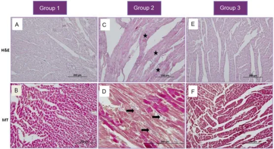
In sections of the embolism group, thickening of the seg-mental arteries muscular layer, atrophy of endothelial structure, hypertrophy in the right ventricular tissue, and an increase in the interstitial tissue samples were reported in all animals (p <0.01). The light microscopic examina-tions of the control lung secexamina-tions revealed that the alve olar structures and segmental arterial layers were intact and not disrupted (Fig. 1A). Additionally, the muscular and con-nective tissue structures were intact and not disrupted in the evaluation of the right ventricular sections of the heart (Fig. 2A, B). The specimens of both lung and cardiac tissue in the anakinra group yielded similar findings as those in the control group (Fig. 1C, 2E, 2F).
However, the light microscopic examinations of the embolism group sections revealed that the alveo-lar septum was widened in the lung sections due to the increase in the interstitial tissue and widespread RESULTS
We evaluated all the animals according to WHO-FC for pulmonary hypertension symptoms both at the initi-ation of the experiments and at day 28. In all groups, none of the animals showed any symptoms related to WHO-FC at the beginning of the experiments.
At the beginning of the study, all the animals were accepted as Class-I. In the control group, none of the animals had pulmonary hypertension symptoms at the end of day 28. Additionally, all the control animals were accepted class-I at the end of the experiment. In the embolism group, 4 rats of 7 were classified as class-3, one of them classified as class-4 and the others were classi-fied as class-1. In the anakinra group, only 1 rat of 7 was classified as class-3 while the others were classified as class-1 at the end of experiments (Table 1).
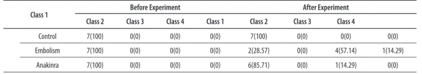
After harvesting the tissues and preparing the sections, a gross light microscopy evaluation was made by one same Table 2. Comparison of percentile of all animals is made according to WHO-FC
Class 1 Before Experiment After Experiment
Class 2 Class 3 Class 4 Class 1 Class 2 Class 3 Class 4
Control 7(100) 0(0) 0(0) 0(0) 7(100) 0(0) 0(0) 0(0) Embolism 7(100) 0(0) 0(0) 0(0) 2(28.57) 0(0) 4(57.14) 1(14.29)
Anakinra 7(100) 0(0) 0(0) 0(0) 6(85.71) 0(0) 1(14.29) 0(0) McNemar test is used.
Fig. 1. The H&E staining of the lung is shown as following; a: Alveolus; the black arrow: the thickened alveolar septum; the star: mononuclear leukocytes infiltration; the yellow
Zabini et al. [34] and Mercier et al. [22] have demon-strated the critical role of many inflammatory cytokines in the formation of obstructive structures in the pulmo-nary arteries of CTEPH and reported the high levels of growth factors (fibroblast growth factor 2) and inflamma-tory cytokines (IL 1 beta, IL 6, monocyte chemoattractant protein 1), and cell adhesion molecules in CTEPH [22, 34]. They also showed the effects of these factors on the devel-opment of CTEPH [22, 34].
IL-1 is a pro-inflammatory cytokine that has wide range of effects on metabolism and such mechanisms as pain sensitivity and tissue damage. The stress exposure of cell triggers a deployment of inflammasomes, which are molecular platforms that lead to caspase-1 activa-tion. These signals are essential for the activation of IL 1-beta [26]. It has been shown that chronic inflam-mation leads to cystic fibrosis and lung fibrosis in the an imal models and the indication of treatment with IL-1 receptor antagonists was revealed [7, 16]. There is con-siderable clinical evidence and studies suggesting that the blockade of IL-1 decreases the severity of diseases and reverses inflammation related loss of organ function in autoinflammatory syndromes [10]. Thus, blockade of IL-1 can be an opportunity for the treatment of those auto-inflammatory syndromes. Anakinra is an approved recombinant form of IL-1 receptor antagonist and a new treatment option for a wide range of diseases associ-ated with inflammatory conditions, such as rheumatoid arthritis [1], familial Mediterranean fever [2], and dia-betes [20]; however, the usage of anakinra is not limited with those diseases. The effectiveness of anakinra has been described in the case of pulmonary arterial hyper-tension [8] as well as in a series of AOSD patients [26]. Elevated IL-1 beta serum levels in PAH correlate with worse outcomes [27]. However, to our knowledge, there poly morph nuclear leukocytes infiltration; the
mus-cular layer of the segmental arteries were thickened, endothelial cell structure were atrophic (Figure 1B); the right ventricular tissue was hypertrophic; inflam-matory cells were infiltrated and the connective tissue fibrils were increased (Fig. 2C, 2D).
DISCUSSION
In this study, we have assessed the effect of anakinra on CTEPH model in rats. The animals were evaluated at the beginning and the end of experiment accord-ing to WHO-FC for pulmonary hypertension. After the experiments, the lung and cardiac tissues of the rats were collected and analyzed histologically among groups for the purpose of evaluating the effects of anakinra on the tissue level. The findings in tissue evaluation and symptoms of embolism in embolism group were significantly different than those in the control and anakinra groups. Additionally, we have shown that histological parameters were decreased in the ana kinra group compared to those of the embo-lism group. There are only a few studies performed on animal models (dog and porcine) for CTEPH. In these models, prepared thromboses are directly applied through pulmonary arteries in animals in order to incite an embolism, so that the CTEPH model can pro-gress in animals [9, 15]. Li et al. [21] has used an animal model in which 15 thromboses (each measuring 1x3 mm) were injected through the jugular veins of rats to mimic embolism conditions. They also reported that the rats showed cyanosis and shortness of breath after the application [21]. We developed an animal model with CTEPH by inducing of 15 thromboses. Our results confirmed the presence of the model with showing WHO-FC class 3 to 4 individuals in embolism group.
Fig. 2. The Masson’s trichrome staining of cardiac tissue is shown as following: the star: polymorph nuclear leukocytes infiltration; the black arrow: fiber increase in
an oral cyclic guanosine monophosphate stimulator, riociguat was applied to patients with non-operable CTEPH or persistent/recurrent pulmonary hyperten-sion after a pulmonary endarterectomy. Riociguat was observed to be useful via enhanced 39 meters the 6-min-ute walking test. A CTEPH team, which included at least one experienced surgeon, has recommended riociguat for the treatment of symptomatic patients with inoper-able CTEPH or with persistent or recurrent CTEPH after surgical treatment [13].
The pathogenesis of CTEPH has not yet been fully under-stood. Despite this fact, its relation to venous throm-boembolism raises the following question: Is CTEPH a disorder of misguided vascular remodeling after pulmo-nary thromboembolism? In some cases, genetic factors can play a significant role leading to a larger throm-bus (non-O- blood groups, rare thrombophilia, dysfi-brinogenemia) in combination with elev ated factor VIII and platelet activation [16]. Recently, these factors should take into consideration about disease phatogen-esis. These conditions suppress the two key pathways: thrombosis angiogenesis and innate immune cell func-tion. It is not clear whether similar mechanisms contrib-ute to secondary pulmonary ar teriopathy. Alleviating the major vessel obstruction in CTEPH can provide an understanding of the mech anisms of regression of sec-ondary pulmonary ar teriopathy and can lead to new treatment objectives.
CONCLUSION
We have showed that anakinra was a useful option for the CTEPH model in rats. Anakinra may be considered to have a protective effect and can contribute to the regression of the increased inflammation in CTEPH. The effectiveness of anakinra will continue to be subject to the further experimental and clinical studies.
are not any studies about the use of anakinra in Group 4 pulmonary artery hypertension or CTEPH. In our study, we aimed to investigate whether or not anakinra is an option for the treatment of CTEPH in an animal model. CTEPH is an intimal disease of organized thrombus and vascular remodeling, with progressive intimal thick-ening (collagenous, inflammatory, atherosclerotic, hemosiderosis and calcific) resulting in right ventricu-lar pressure overload and remodeling with ultimately decompensation, right ventricle failure and death [19]. Normally, acute pulmonary embolisms resolve within 6 months. However, in 0.1-3.8 % of cases, the transi-tion of thrombosis to a fibrous tissue leads to pulmo-nary hypertension [3]. Other risk factors for chronic pulmonary thromboembolism are elevated Factor VIII, antiphospholipid antibody, lupus anticoagulants, sple-nectomy and cancer [5, 6, 32]. In addition, ventricular-atrial (VA) shunts, which are used in the treatment of hydrocephaly, may result in CTEPH. In some cases there are no risk factors for venous thromboembolism, and there may be a relationship between vascular remode-ling and staphylococcal infections [4]. In addition to the up-regulation of transforming growth factors and IL-1 beta, the staphylococcal infections can lead to a delay in thrombosis resolution [28].
For a period of 16 weeks, the dual endothelin antagonist, bosentan was evaluated in 157 patients with inoperable CTEPH or persistent/recurrent pulmonary hypertension after a pulmonary endarterectomy [17]. This study indic-ated that the decrease in pulmonary vascular resistance and increase in the 6-minute walking capacity could not reach the ultimate goal [17]. However, in the MERIT-1 study, patients with inoperable CTEPH were treated with another endothelin receptor antagonist (macitentan), and macitentan significantly improved pulmonary vas-cular resistance [14]. In addition, for a 16-week period, REFERENCES
[1] Abramson S.B., Amin A.: Blocking the effects of IL-1 in rheumatoid arthritis protects bone and cartilage. Rheumatology, 2002; 41: 972–980 [2] Bilginer Y., Ayaz N.A., Ozen S.: Anti-IL-1 treatment for secondary amyloidosis in an adolescent with FMF and Behçet’s disease. Clin. Rheumatol., 2010; 29: 209–210
[3] Blauwet L.A., Edwards W.D., Tazelaar H.D., McGregor C.G.: Surgi-cal pathology of pulmonary thromboendarterectomy: a study of 54 cases from 1990 to 2001. Hum Pathol., 2003; 34: 1290–1298 [4] Bonderman D., Jakowitsch J., Redwan B., Bergmeister H., Renner M.K., Panzenböck H., Adlbrecht C., Georgopoulos A., Klepetko W., Kneussl M., Lang I.M.: Role for staphylococci in misguided throm-bus resolution of chronic thromboembolic pulmonary hypertension. Arterioscler. Thromb. Vasc. Biol., 2008; 28: 678–684
[5] Bonderman D., Turecek P.L., Jakowitsch J., Weltermann A., Adl-brecht C., Schneider B., Kneussl M., Rubin L.J., Kyrle P.A., Klepetko W., Maurer G., Lang I.M.: High prevalence of elevated clotting factor VIII in chronic thromboembolic pulmonary hypertension. Thromb. Haemost., 2003; 90: 372–376
[6] Bonderman D., Wilkens H., Wakounig S., Schäfers H.J., Jansa P., Lindner J., Simkova I., Martischnig A.M., Dudczak J., Sadushi R.,
Sko-ro-Sajer N., Klepetko W., Lang I.M.: Risk factors for chronic throm-boembolic pulmonary hypertension. Eur. Respir. J., 2009; 33: 325-331 [7] Burgy O., Bellaye P.S., Causse S., Beltramo G., Wettstein G., Bouta-nquoi P.M., Goirand F., Garrido C., Bonniaud P.: Pleural inhibition of the caspase-1/IL-1β pathway diminishes profibrotic lung toxicity of bleomycin. Respir Res., 2016; 17: 162
[8] Campos M., Schiopu E.: Pulmonary arterial hypertension in adult--onset Still’s disease: Rapid response to anakinra. Case Rep. Rheu-matol., 2012; 2012: 537613
[9] Deng C., Wu D., Zhai Z., Lin Q., Zhong Z., Yang Y., Chen Q., Lian N., Gao S., Yang M., Liu K., Wang C.: Close concordance between pulmonary angiography and pathology in a canine model with chronic pulmonary thromboembolism and pathological mechanisms after lung ischemia reperfusion injury. J. Thromb. Thrombolysis, 2016; 41: 581–591 [10] Dinarello C.A., Simon A., van der Meer J.W.: Treating inflamma tion by blocking interleukin-1 in a broad spectrum of diseases. Nat. Rev. Drug Discov., 2012; 11: 633–652
[11] Fedullo P., Kerr K.M., Kim N.H., Auger W.R.: Chronic thrombo embolic pulmonary hypertension. Am. J. Respir. Crit. Care Med., 2011; 1830: 1605–1613
[23] Li C.Y., Deng W., Liao X.Q., Deng J., Zhang Y.K., Wang D.X.: The effects and mechanism of ginsenoside Rg1 on myocardial remo-deling in an animal model of chronic thromboembolic pulmonary hypertension. Eur. J. Med. Res., 2013; 18: 16
[24] Mercier O., Arthur Ataam J., Langer N.B., Dorfmüller P., Lamrani L., Lecerf F., Decante B., Dartevelle P., Eddahibi S., Fadel E.: Abnormal pulmonary endothelial cells may underlie the enigmatic pathoge-nesis of chronic thromboembolic pulmonary hypertension. J. Heart Lung Transplant., 2017; 36: 305–314
[25] Moser K.M., Cantor J.P., Olman M., Villespin I., Graif J.L., Konopka R., Marsh J.J., Pedersen C.: Chronic pulmonary thromboembolism in dogs treated with tranexamic acid. Circulation, 1991; 83: 1371–1379 [26] Pepke-Zaba J., Delcroix M., Lang I., Mayer E., Jansa P., Ambroz D., Treacy C., D’Armini A.M., Morsolini M., Snijder R., Bresser P., Torbicki A., Kristensen B., Lewczuk J., Simkova I., et al.: Chronic thromboembolic pulmonary hypertension (CTEPH): results from an international prospective registry. Circulation, 2011; 124: 1973–1981 [27] Quarck R., Nawrot T., Meyns B., Delcroix M.: C-reactive protein: a new predictor of adverse outcome in pulmonary arterial hyper-tension. J. Am. Coll. Cardiol., 2009; 53: 1211–1218
[28] Scannapieco F.A., Ng P., Hovey K., Hausmann E., Hutson A., Wac-tawski-Wende J.: Salivary biomarkers associated with alveolar bone loss. Ann. N. Y. Acad. Sci., 2007; 1098: 496–497
[29] Simonneau G., Gatzoulis M.A., Adatia I., Celermajer D., Denton C., Ghofrani A., Gomez Sanchez M.A., Krishna Kumar R., Landzberg M., Machado R.F., Olschewski H., Robbins I.M., Souza R.: Updated clinical classification of pulmonary hypertension. J. Am. Coll. Car-diol., 2013; 62: D34–D41
[30] Soon E., Holmes A.M., Treacy C.M., Doughty N.J., Southgate L., Machado R.D., Trembath R.C., Jennings S., Barker L., Nicklin P., Walker C., Budd D.C., Pepke-Zaba J., Morrell N.W.: Elevated levels of inflammatory cytokines predict survival in idiopathic and familial pulmonary arterial hypertension. Circulation, 2010; 122: 920–927 [31] Svensson S., Forsberg M., Hulander M., Vazirisani F., Palmquist A., Lausmaa J., Thomsen P., Trobos M.: Role of nanostructured gold surfaces on monocyte activation and Staphylococcus epidermidis bio film formation. Int. J. Nanomedicine, 2014; 9: 775–794 [32] Wolf M., Boyer-Neumann C., Parent F., Eschwege V., Jaillet H., Meyer D., Simonneau G.: Thrombotic risk factors in pulmonary hy-pertension. Eur. Respir. J,. 2000; 15: 395–399
[33] Yao W., Firth A.L., Sacks R.S., Ogawa A., Auger W.R., Fedullo P.F., Madani M.M., Lin G.Y., Sakakibara N., Thistlethwaite P.A., Jamieson S.W., Rubin L.J., Yuan J.X.: Identification of putative endothelial pro-genitor cells (CD34+CD133+Flk-1+) in endarterectomized tissue of pa-tients with chronic thromboembolic pulmonary hypertension. Am. J. Physiol. Lung Cell Mol. Physiol., 2009; 296: L870–L878
[34] Zabini D., Nagaraj C., Stacher E., Lang I.M., Nierlich P., Klepetko W., Heinemann A., Olschewski H., Bálint Z., Olschewski A.: Angiosta-tic factors in the pulmonary endarterectomy material from chronic thromboembolic pulmonary hypertension patients cause endothe-lial dysfunction. PLoS One, 2012; 7: e43793
The authors have no potential conflicts of interest to declare.
[12] Fitzgerald A.A., Leclercq S.A., Yan A., Homik J.E., Dinarello C.A.: Rapid responses to anakinra in patients with refractory adult-onset Still’s disease. Arthritis Rheum., 2005; 52: 1794–1803
[13] Galiè N., Humbert M., Vachiery J.L., Gibbs S., Lang I., Torbicki A., Simonneau G., Peacock A., Vonk Noordegraaf A., Beghetti M., Gho-frani A., Gomez Sanchez M.A., Hansmann G., Klepetko W., Lancellotti P., et al.: 2015 ESC/ERS Guidelines for the diagnosis and treatment of pulmonary hypertension: The Joint Task Force for the Diagnosis and Treatment of Pulmonary Hypertension of the European Socie-ty of Cardiology (ESC) and the European Respiratory SocieSocie-ty (ERS): Endorsed by: Association for European Paediatric and Congenital Cardiology (AEPC), International Society for Heart and Lung Trans-plantation (ISHLT). Eur. Heart J., 2016; 37: 67–119
[14] Ghofrani H.A., D’Armini A.M., Grimminger F., Hoeper M.M., Jansa P., Kim N.H., Mayer E., Simonneau G., Wilkins M.R., Fritsch A., Neuser D., Weimann G., Wang C., CHEST-1 Study Group: Riociguat for the treatment of chronic thromboembolic pulmonary hypertension. N. Engl. J. Med., 2013; 369: 319–329
[15] Ghofrani H.A., Simonneau G., D’Armini A.M., Fedullo P., Howard L.S., Jaïs X., Jenkins D.P., Jing Z.C., Madani M.M., Martin N., Mayer E., Papadakis K., Richard D., Kim N.H., MERIT Study Investigators: Ma-citentan for the treatment of inoperable chronic thromboembolic pulmonary hypertension (MERIT-1): results from the multicentre, phase 2, randomised, double-blind, placebo-controlled study. Lancet Respir Med., 2017; 5: 785–794
[16] Guihaire J., Haddad F., Boulate D., Capderou A., Decante B., Flécher E., Eddahibi S., Dorfmüller P., Hervé P., Humbert M., Ver-hoye J.P., Dartevelle P., Mercier O., Fadel E.: Right ventricular plasti-city in a porcine model of chronic pressure overload. J. Heart Lung Transplant., 2014; 33: 194–202
[17] Homoncik M., Gessl A., Ferlitsch A., Jilma B., Vierhapper H.: Al-tered platelet plug formation in hyperthyroidism and hypothyro-idism. J. Clin. Endocrinol. Metab., 2007; 92: 3006–3012
[18] Iannitti R.G., Napolioni V., Oikonomou V., De Luca A., Galosi C., Pariano M., Massi-Benedetti C., Borghi M., Puccetti M., Lucidi V., Co lombo C., Fiscarelli E., Lass-Flörl C., Majo F., Cariani L., et al.: IL-1 recep tor antagonist ameliorates inflammasome-dependent in-flammation in murine and human cystic fibrosis. Nat. Commun., 2016; 7: 10791
[19] Jaïs X., D’Armini A.M., Jansa P., Torbicki A., Delcroix M., Ghofrani H.A., Hoeper M.M., Lang I.M., Mayer E., Pepke-Zaba J., Perchenet L., Morganti A., Simonneau G., Rubin L.J., Bosentan Effects in iNopE-rable Forms of chronIc Thromboembolic pulmonary hypertension Study Group: Bosentan for treatment of inoperable chronic throm-boembolic pulmonary hypertension: BENEFiT (Bosentan Effects in iNopErable Forms of chronIc Thromboembolic pulmonary hyper-tension), a randomized, placebo-controlled trial. J. Am. Coll. Cardiol,. 2008; 52: 2127–2134
[20] Kellermair J., Redwan B., Alias S., Jabkowski J., Panzenboeck A., Kellermair L., Winter M.P., Weltermann A., Lang I.M.: Platelet endothelial cell adhesion molecule 1 deficiency misguides venous thrombus resolution. Blood, 2013; 122: 3376–3384
[21] Lang I.M., Dorfmüller P., Vonk Noordegraaf A.: The pathobiolo-gy of chronic thromboembolic pulmonary hypertension. Ann. Am. Thorac. Soc., 2016; 13: S215–S221
[22] Larsen C.M., Faulenbach M., Vaag A., Ehses J.A., Donath M.Y., Man-drup-Poulsen T.: Sustained effects of interleukin-1 receptor antago-nist treatment in type 2 diabetes. Diabetes Care, 2009; 32: 1663–1668