Full Terms & Conditions of access and use can be found at
https://www.tandfonline.com/action/journalInformation?journalCode=ibih20
Biotechnic & Histochemistry
ISSN: 1052-0295 (Print) 1473-7760 (Online) Journal homepage: https://www.tandfonline.com/loi/ibih20
Effect of a honey and
arginine-glutamine-hydroxymethylbutyrate mixture on the healing
of colon anastomosis in rats immunosuppressed
with tacrolimus
S. Akbulut, Z. Dogan, A. Baskiran, H. Elbe & Y. Turkoz
To cite this article: S. Akbulut, Z. Dogan, A. Baskiran, H. Elbe & Y. Turkoz (2019) Effect of a honey and arginine-glutamine-hydroxymethylbutyrate mixture on the healing of colon anastomosis in rats immunosuppressed with tacrolimus, Biotechnic & Histochemistry, 94:7, 514-521, DOI: 10.1080/10520295.2019.1601257
To link to this article: https://doi.org/10.1080/10520295.2019.1601257
Published online: 15 Apr 2019.
Submit your article to this journal
Article views: 120
View related articles
Effect of a honey and arginine-glutamine-hydroxymethylbutyrate mixture on
the healing of colon anastomosis in rats immunosuppressed with tacrolimus
S. Akbulut a, Z. Doganb, A. Baskirana, H. Elbec, and Y. Turkozd
aDepartment of Surgery and Liver Transplant Institute, Inonu University Faculty of Medicine, Malatya, Turkey;bDepartment of Anatomy,
Adiyaman University Faculty of Medicine, Adiyaman, Turkey;cDepartment of Histology and Embryology, Sıtkı Kocman University Faculty of
Medicine, Mugla, Turkey;dDepartment of Medical Biochemistry, Inonu University Faculty of Medicine, Malatya, Turkey
ABSTRACT
We compared the effect of honey and a mixture of arginine-glutamine-hydroxymethylbutyrate (AGHMB) on healing of a descending colon anastomosis in rats that were immunosuppressed with tacrolimus (Tac). Sprague-Dawley rats were divided into four groups: untreated control, Tac, Tac + honey and Tac + AGHMB. Colon resection and anastomosis were performed on day 14 and re-laparotomy was performed on the day 21 of the study. Anastomotic bursting pressure, macroscopic adhesion score, weekly body weight changes, histopathological features and immunohistochemical staining of TGF-β1 were determined for all groups. We found no significant difference in anastomotic bursting pressure among the experimental groups. We found significant weekly increases in body weight for the Tac + honey group. We found no significant difference in the weekly body weight measurements for the Tac + AGHMB group. We found significant increases in TGF-β1 expression in the Tac + honey group compared to the control and Tac groups. No significant differences in inflammatory cell infiltration, fibroblast proliferation or collagen deposition were found between the Tac + honey and Tac + AGHMB groups; however, a significant difference in neovascularization between these groups was found. Neovascularization in the Tac + honey group was significantly greater than for the Tac + AGHMB group. We found that both honey and the AGHMB mixture were beneficial for anastomotic wound healing in rats that were immunosuppressed using Tac.
KEYWORDS Anastomosis; arginine; bursting pressure; colon; glutamine; honey; hydroxymethylbutyrate; tacrolimus
The major causes of morbidity and mortality following colorectal surgery are complications of anastomosis such as fistula formation and leakage (Raptis et al. 2012). These complications are due to anemia, blood transfusion, chemoradiotherapy, diabetes, hypoalbuminemia, inflammatory bowel disease, malignant disorders, malnutrition, obesity, poor surgical technique, prolonged surgery, smoking and use of immunosuppressive medication (Raptis et al. 2012, 2018; Ekinci et al. 2018). Increased frequency of organ transplantation and incidence of autoimmune disorders have increased the use of immunosuppressive agents including corticosteroids and tacrolimus (Tac, FK506, fujimycin). Tac is a potent immunosuppressive agent that was isolated in 1985 from the fungus, Streptomyces tsukubaensis (Raptis et al. 2012, 2018; Zheng et al. 2017). Although Tac delayed wound healing in some clinical and experimental studies, it has been suggested that Tac actually promotes healing by inducing release of growth factors such as
transforming growth factor beta (TGF-β) and vascular endothelial growth factor (VEGF), which stimulate collagen synthesis and angiogenesis, respectively (Saber 2010; Raptis et al.2012).
Dietary supplementation with amino acids has been investigated for accelerating wound healing including arginine, glutamine, β-hydroxyl-β-methyl butyrate (HMB) (Raptis et al. 2012; Yaman et al. 2013; Bozkırlı
et al.2015). A mixture of arginine, glutamine and HMB (AGHMB) has been reported to promote wound healing and prevent apoptosis.
Honey also has been studied for its effect on wound healing (Gollu et al. 2008; Ergul and Ergul2010; Saber
2010; Gencay et al. 2008). Honey has been used since antiquity for treating wounds and surgical incisions (Gollu et al. 2008; Gencay et al. 2008). The literature contains many reports of local or oral application of honey for accelerating wound healing. Although honey has been reported to promote wound healing in experimental gastrointestinal anastomosis models, we have found no report of the effects of honey in an
CONTACTS. Akbulut [email protected] Department of Surgery and Liver Transplant Institute, Inonu University Faculty of Medicine, Malatya 44280, Turkey
2019, VOL. 94, NO. 7, 514–521
https://doi.org/10.1080/10520295.2019.1601257
immunosuppressed rat model (Gollu et al. 2008; Gencay et al. 2008). Consequently, we compared the effects of both honey and AGHMB on healing of a descending colon anastomosis in rats that had been immunosuppressed by Tac.
Materials and methods
Experimental protocol
All procedures were in compliance with the National Health Institute’s Animal Research Guidelines. Our investigation was approved by the Inonu University Animals Research Ethics Committee (2011/A-110). We used 48 10–11-week-old, 230–310 g male Sprague-Dawley rats obtained from Inonu University Laboratory Animals Production and Research Center. The rats were housed at 21 ± 2 °C, 60 ± 5% humidity with a 12 h light:12 h dark cycle and free access to rat food and water. The animals were divided randomly into four groups of 12. The control group was not subjected to any experimental treatment. The Tac group was treated daily with 0.5 mg/kg/day Tac (Prograf; Astellas Phama Inc., Tokyo, Japan) by gavage from day 1 of the study. The rats in the groups other than the control group were administered a single dose of 0.5 mg/kg/day Tac by gavage to induce immunosuppression using the method of Uysal et al. (2017). The Tac + honey group was given 10 g/kg/day liquid honey (Balparmak, Altiparmak Pazarlama Koll Sti, Istanbul, Turkey) by gavage in addition to standard rat feed from day 1 of Tac treatment. The Tac + AGHMB group was given a mixture containing 308 g/kg glutamine, 308 g/kg arginine and 54 g/kg β-hydroxyl-β-methyl butyrate (Abound; Abbot, Lakeview, CA) at a dose of 0.24 g/350 g by gavage in addition to standard rat feed from the day 1 of Tac treatment.
All rats were weighed at weeks 1, 2 and 3 of the study. On days 14 and 21, the rats were anesthetized for surgical procedures by intraperitoneal (i.p.) administration of 40 mg/kg ketamine hydrochloride (Ketalar; Eczacibasi Warner-Lambert Laboratories, Istanbul, Turkey) and 5 mg/kg xylazine hydrochloride (Rompun, Bayer Laboratories, Istanbul, Turkey). On day 14, all rats were anesthetized and placed in a supine position. After shaving the anterior abdominal wall and cleaning with povidone iodine, a 4 cm abdominal incision was made to expose the abdominal cavity. After resecting a small segment of the descending colon, a colo-colonic anastomosis was created using 6/0 polyglactin 910 (Vicryl; Ethicon, Edinburgh, UK). Abdominal fascia and skin were sutured with 4/0 polyglactin 910 suturing material
(Vicryl; Ethicon). On day 21 all rats were re-anesthetized as described above. After another laparotomy, intra-abdominal adhesions were checked macroscopically as described below. After resecting a large colonic segment that included the anastomosis line, all rats were sacrificed using an overdose of ketamine/xylazine.
Anastomotic bursting pressure
Measurement was based on the methods of Ergul and Ergul (2010), Gollu et al. (2008) and Raptis et al. (2012). The distal end of the resected colon segment was bound firmly with a suture material. The tube from a sphygmomanometer was inserted into the lumen from the proximal end of the resected colon segment. The colon segment was submerged in a water-filled container and the blood pressure cuff inflated. The pressure reading (mm Hg) at which air bubbles first emerged through the anastomosis line was recorded as the burst pressure.
Macroscopic scoring of adhesion
Adhesions were scored using the adhesion severity criteria described by Evans et al. (2008). The scoring system was: 0, no adhesion; 1, spontaneously separating adhesion; 2, separation of adhesion with traction; 3, separation of adhesion with sharp dissection. Scoring was conducted by an experienced surgeon who did not participate otherwise in the study.
Histopathology
The colon tissues were fixed in 10% neutral buffered formalin for 48 h. Tissues were washed in running water, then dehydrated through 50, 75, 96 and 100% ethanol. After dehydration, specimens were cleared in xylene and embedded in paraffin wax. Sections were cut at 5 μm, mounted on slides and stained with both hematoxylin and eosin (H & E) and periodic acid-Schiff (PAS)-Alcian blue (Bancroft and Gamble 2008). Stained sections were examined using light microscopy and graded using a scale that was modified from Ehrlich and Hunt’s scoring system (Ehrlich et al. 1973). We evaluated collagen deposition, fibroblast proliferation, inflammatory cell infiltration and neovascularization (Ergul and Ergul
2010; Raptis et al. 2012; Ekinci et al. 2018). To do this, each parameter was assessed individually using the following scale: 0, no evidence; 1, occasional evidence; 2, moderate evidence; 3, abundant evidence; 4, confluent cells or fibers. All sections were examined BIOTECHNIC & HISTOCHEMISTRY 515
using a Nikon Eclipse 80i light microscope and Nikon Image Analysis system (Nikon, Tokyo, Japan)
Immunohistochemistry
For immunohistochemical analysis, sections were mounted on polylysine coated slides. After rehydrating, samples were transferred to citrate buffer, pH 7.6, and heated in a microwave oven for 20 min for antigen retrieval. After cooling for 20 min to room temperature, the sections were washed with phosphate-buffered saline (PBS). Sections were placed in 0.3% (v/v) H2O2for 7 min,
then washed with PBS. Sections were incubated with anti-rabbit anti-TGF-β1 antibody (sc-146; Santa Cruz Biotechnology, Dallas, TX) for 60 min according to the manufacturer’s instructions. Sections then were rinsed in PBS and incubated with biotinylated goat antipolyvalent IgG for 10 min and streptavidin peroxidase for 10 min at room temperature according to the manufacturer’s instructions. After 20-fold dilution of the AEC chromogenic substrate with AEC diluent solution, staining was completed with the commercial ready-to-use liquid AEC (3-amino-9-ethylcarbazole) chromogenic substrate solution (sc-24979; Santa Cruz Biotechnology) by incubating 15 min at room temperature. Staining was completed using ready-to-use AEC HRP Red chromogen + substrate (sc-24979; Santa Cruz Biotechnology) for 15 min at 25 °C and slides were counterstained with Mayer’s hematoxylin for 1 min, rinsed in tap water, and dehydrated. Staining for anti-TGF-β1 was identified by brown coloration. The relative intensity of TGF-β1 immunostaining was scored as: 0, absent; 1, slight; 2, moderate; 3, strong. All sections were examined using a Nikon Eclipse 80i light microscope and Nikon Image Analysis system (Nikon).
Statistical analysis
Statistical analyses were carried out using the IBM SPSS Statistics v25.0 software package. Body weight and anastomotic burst pressure were recorded as means ± SD, median and minimum−maximum values. The numerical information describing histopathologic and immunohistochemical data were recorded as means ± SD. Adhesion scores were reported as numbers and percent. The Pearson chi-squared test was used to compare qualitative data. The Shapiro-Wilk test was used to test the normality of quantitative data. The Kruskal Wallis test was used to compare non-normally distributed quantitative data. To demonstrate which groups created statistical significance, Kruskal Wallis 1-way ANOVA (k samples) all pairwise multiple comparisons test, or the Mann Whitney-U test, was
used. The Friedman test was used for repeated intra-group measurements. The Wilcoxon-signed rank test was used to determine which measurements reached statistical significance between repeated measurements. Values for p ≤ 0.05 were considered statistically significant.
Results
Adhesion scores
Adhesion scores among the study groups did not differ significantly.
Anastomotic bursting pressure
The median anastomotic bursting pressures of the control, Tac + AGHMB, Tac + honey and Tac groups were 220.5 (189−241) mm Hg, 180 (110−220) mm Hg, 142.5 (84−220) mm Hg and 110 (48−210) mm Hg, respectively (p = 0.001). Statistical significance was as follows: control group vs. Tac group (p < 0.001) and control group vs. Tac + honey group (p = 0.005). None of the rats developed anastomosis dehiscence.
Body weight changes
We found significant differences in body weight among the groups during the first (p = 0.002), second (p = 0.006) and third weeks (p = 0.003). The first week body weight for the Tac group was significantly greater than for the control (p = 0.01) and Tac + AGHMB (p = 0.003) groups. No significant difference in first week body weight was found between the Tac and Tac+ honey groups. The second week body weights for the Tac group were significantly greater than for the Tac + AGHMB group (p = 0.006). The second week body weight for the Tac + honey group was significantly greater than for the Tac + AGHMB group (p = 0.035). We found no significant difference in second week body weights between the Tac and Tac + honey groups. Third week body weight of the Tac + honey group was significantly greater than for the Tac + AGHMB group (p = 0.01). We found no significant difference in third week body weights among the control, Tac and Tac + honey groups.
Intra-group body weight changes also were evaluated. Significant increases were found between the weekly body weight measures for the control group (1st week vs. 2nd week, p = 0.012; 1st week vs. 3rd week, p = 0.012). We found significant decreases between the weekly body weight measures for the Tac group (1st week vs. 3rd week, p = 0.002; 2nd week vs. 3rd week, p = 0.002). We also found significant
increases between the weekly body weights for the Tac + honey group (1st week vs. 2nd week,p = 0.004; 1st week vs. 3rd week, p = 0.004; 2nd week vs. 3rd week, p = 0.015). We found no significant difference between the weekly body weight changes for the Tac + AGHMB group (p > 0.05 for 1st week vs. 2nd week, 1st week vs. 3rd week and 2nd week vs. 3rd week). Details for body weights for all groups are presented in Table 1.
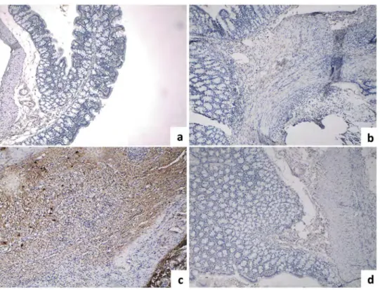
Histopathology
The control group exhibited normal histological structure (Figure 1a). In the Tac group, the submucosa exhibited numerous dilated blood vessels, the glands were dilated at the site of anastomosis and inflammatory cell infiltration in the lamina propria and submucosa were evident (Figure 1b–d). Histopathological differences between the control and Tac groups were statistically significant (p < 0.05). In the Tac + honey and Tac + AGHMB groups, the colon tissue appeared nearly normal (Figure 1e,f). The amount of inflammatory cell infiltration in the Tac (p < 0.05), Tac + honey (p < 0.005) and Tac + AGHMB (p < 0.005) groups was significantly greater than the control group. Collagen deposition in the Tac (p < 0.01), Tac + honey (p < 0.001) and Tac + AGHMB (p < 0.001) groups was significantly greater than the control group. We found no significant difference in the amount of inflammatory cell infiltration or collagen deposition among groups. Neovascularization in the Tac (p < 0.05), Tac + honey (p < 0.001) and Tac + AGHMB (p < 0.001) groups was significantly greater than the control group. Neovascularization in the Tac + honey (p < 0.01) and Tac + AGHMB (p < 0.01) groups was significantly greater
than for the Tac group. Neovascularization in the Tac + honey group was significantly greater than for the Tac + AGHMB group (p < 0.05). Fibroblast proliferation in the Tac (p < 0.01), Tac + honey (p < 0.001) and Tac + AGHMB (p < 0.005) groups was significantly greater than for the control group. Fibroblast proliferation in Tac + honey (p < 0.05) and Tac + AGHMB (p < 0.05) groups was significantly greater than for the Tac group. We found no significant difference in infiltration, fibroblast proliferation and collagen deposition between the Tac + honey and Tac + AGHMB groups. Neovascularization in Tac + honey group was significantly greater than for the Tac + AGHMB (p < 0.05). The histopathological evaluation score for each group is presented inTable 2.
Immunohistochemistry
TGF-β1 immunostaining in the control group showed weakly stained normal cells. In the Tac group, TGF-β1 immunostaining was evident in connective tissue. In the same areas in Tac + honey group, the intensity of TGF-β1 immunostaining increased compared to the Tac group (p < 0.05) (Figure 2). Slightly increased intensity of TGF-β1 immunostaining in the Tac + AGHMB group was not statistically significant compared to the Tac group. The TGF-β1 immunostaining scores are presented in Table 3.
Discussion
Our investigation addresses a common clinical problem. More than 200 liver transplantation procedures are performed annually at our center, and
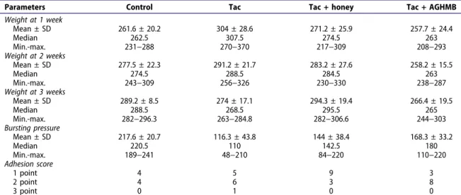
Table 1.Comparison of experimental groups in terms of body weight, anastomotic bursting pressure and adhesion score.
Parameters Control Tac Tac + honey Tac + AGHMB
Weight at 1 week Mean ± SD 261.6 ± 20.2 304 ± 28.6 271.2 ± 25.9 257.7 ± 24.4 Median 262.5 307.5 274.5 263 Min.-max. 231−288 270−370 217−309 208−293 Weight at 2 weeks Mean ± SD 277.5 ± 22.3 291.2 ± 21.7 283.2 ± 27.6 258.2 ± 15.5 Median 274.5 288.5 284.5 263 Min.-max. 243−309 256−326 230−330 238−287 Weight at 3 weeks Mean ± SD 289.2 ± 8.5 274 ± 17.1 294.3 ± 19.4 266.4 ± 19.5 Median 288.5 268.5 295.5 265 Min.-max. 282−296.3 263−284.8 282−306.6 244−303 Bursting pressure Mean ± SD 217.6 ± 20.7 116.3 ± 43.8 144 ± 38.4 168.3 ± 33.2 Median 220.5 110 142.5 180 Min.-max. 189−241 48−210 84−220 110−220 Adhesion score 1 point 4 5 9 3 2 point 4 6 3 8 3 point 0 1 0 0
Weights are in grams. Bursting pressure is in mm Hg. n = 12 for each group. Weight at 1 week: Tac vs. Tac + AGHMB (p = 0.003), Tac vs. control (p = 0.01). Weight at 2 weeks: Tac + AGHMB vs. Tac + honey (p = 0.035), Tac + AGHMB vs. Tac (p = 0.006). Weight at 3 weeks: Tac + AGHMB vs. Tac + honey (p = 0.01). Bursting pressure: control vs. Tac (p < 0.01); control vs. Tac + honey (p = 0.005).
Tac is the main immunosuppressive agent. Gastrointestinal complications including hollow organ perforation, anastomotic dehiscence and anastomotic leakage may develop during the early or late postoperative periods in these patients. Most of these problems require gastrointestinal anastomosis or
anastomosis repair. It has been suggested that to achieve optimal anastomotic healing in transplant patients with colon anastomosis, the dose of immunosuppressive agent should be reduced or stopped at certain stages (Uysal et al. 2017); however, this increases the risk of rejection of the liver graft.
Figure 1.Histological appearance of colon tissues. a) Control group showing normal histological appearance. H & E staining. 10 x objective lens. b) TAC group showing increased fibroblasts and collagen fibers in the submucosal layer. H & E staining. 10 x objective lens. c,d) Tac group showing inflammatory cell infiltration in the lamina propria and submucosa. PAS-Alcian blue staining. 40 x objective lens. e) TAC + honey group showing fibroblast proliferation and synthesis of new collagen in the submucosal layer. H & E staining. 10 x objective lens. f) TAC + AGHMB group showing histological findings similar to TAC + honey group. H & E staining. 10 x objective lens.
Table 2.Histopathological evaluation scores of experimental groups.
Score Control Tac Tac + honey Tac + AGHMB
Inflammatory cell infiltration 0.00 ± 0.00 0.50 ± 0.32a 0.87 ± 0.22b 0.62 ± 0.26b
Fibroblast proliferation 0.00 ± 0.00 0.75 ± 0.25c 1.25 ± 0.25d, e 1.12 ± 0.29b, d
Neovascularization 0.00 ± 0.00 0.50 ± 0.18a 1.62 ± 0.18e, f 1.12 ± 0.12e− g Collagen deposition 0.00 ± 0.00 0.75 ± 0.30c 1.50 ± 0.18e 1.50 ± 0.18e Data are means ± SD.ap < 0.05 vs. control,bp < 0.005 vs. control,cp < 0.01 vs. control,dp < 0.05 vs. Tacep < 0.001 vs. control,
f
Colorectal anastomosis leaks are the leading cause of postoperative morbidity and mortality. Rates of anastomosis leaks between 3 and 6% generally are regarded as acceptable; the actual rates reported in the literature range from 0.5 to 30% (Raptis et al. 2012,
2018; Yaman et al. 2013). Several factors influence leaks; nutritional status is a major factor that affects wound healing (Bozkırlı et al.2015).
Arginine is an amino acid that is required for patients who undergo major surgical procedures or trauma (Yaman et al. 2013; Bozkırlı et al. 2015). Dietary intake of arginine creates a positive nitrogen balance, increases the tissue hydroxyproline level, promotes collagen accumulation at the bowel anastomosis line, exerts an antioxidant effect and promotes anastomotic healing (Yaman et al. 2013). Arginine also participates in the inflammatory phase of anastomotic healing.
Glutamine is an amino acid that serves as an energy source for gastrointestinal epithelial and immune system
cells (Yaman et al.2013; Bozkırlı et al.2015). Despite its abundance, its level is reduced by trauma, infection and major surgical procedures (Yaman et al. 2013; Bozkırlı
et al. 2015). Glutamine also participates in nucleotide synthesis in fibroblasts and macrophages, and it also is a strong immunomodulatory agent and therefore hastens wound healing (Yaman et al.2013; Bozkırlı et al.2015).
HMB is formed during the metabolism of leucine; it participates in regulating protein synthesis and maintaining nitrogen balance (Yaman et al. 2013; Bozkırlı et al. 2015). The AGHMB mixture of arginine, glutamine and HMB is widely used clinically for tumor cachexia, exercise-induced muscular injury and trauma (Seker et al. 2013; Yaman et al. 2013; Bozkırlı et al. 2015; Kusabbi et al. 2015). We have found only three reports, however, concerning the effects of AGHMB on wound healing using an experimental anastomosis model; none of these used an immunosuppressed rat model (Seker et al. 2013; Yaman et al.2013; Kusabbi et al.2015).
Figure 2.Immunohistochemical staining of TGF-β1. a) Control group showing weakly stained normal cells. TGF-β1 staining. 10 x objective lens. b) Tac group showing TGF-β1 immunostaining in connective tissue. TGF-β1 staining. 10 x objective lens. c) Tac + honey group showing intensity of TGF-β1 immunostaining greater than the TAC group. TGF-β1 staining. 10 x objective lens. d) TAC + AGHMB group showing less intensity of TGF-β1 immunostaining than for the Tac + honey group. TGF-β1 staining. 10 x objective lens.
Table 3.TGF-β1 immunostaining score of all experimental groups.
Score Control TAC TAC + honey TAC + AGHMB
TGF-β1 immunostaining 0.25 ± 0.16 0.62 ± 0.18 1.37 ± 0.18a,b 0.87 ± 0.22 Data are means± SD.ap < 0.005 vs. control,bp < 0.05 vs. Tac
Honey is a nutrient that has been widely studied using experimental wound healing models. Honey is made by bees from plant nectars and therefore exhibits seasonal and geographical variation. Honey exhibits a wide range of clinically useful effects (Basbug et al. 2011; Gollu et al. 2008; Gencay et al.
2008), although it is not entirely clear how it accelerates wound healing (Hadagali and Chua 2014). Nevertheless, the value of systemic or topical application of honey for healing gastrointestinal anastomosis has been reported for several experimental models (Gollu et al. 2008; Ergul and Ergul2010; Saber2010).
Problem-free wound healing is important for gastrointestinal anastomoses (Raptis et al.2012). During the inflammatory phase of healing, neutrophils and macrophages migrate into the anastomosis line; optimal numbers are reached by about 48 h (Raptis et al.2012). During the proliferative phase, neovascularization, fibroblast proliferation and migration, collagen synthesis, and crosslinking between collagen fibers occur (Raptis et al. 2012). Fibroblasts contribute to collagen synthesis and construction of extracellular matrix (Uysal et al. 2017). Fibroblasts are vital for forming a strong and durable anastomosis.
VGEF is important for regulation of neovas-cularization, while TGF-β is important factor for fibrogenic activity (Raptis et al. 2012). TGF-β1 is the
main cytokine that stimulates VGEF synthesis. Changes in wound healing phases can be demonstrated by histopathological and immunohistochemical analysis. We found that the greatest TGF-β1 expression at the anastomosis line was in Tac + honey and Tac + AGHMB groups (Table 3).
Tac exerts immunosuppressive effects by inhibiting IL-2 gene expression and disrupting apoptosis and degranulation of leukocytes. Tac also exhibits an anti-inflammatory effect by reducing transcription of proinflammatory cytokines, TNFα, IL-1, IL-3, IL-4, IL-5, IL-6, and IL-8 (Raptis et al. 2012, 2018). Although it has been reported that Tac affects healing of dermal wounds negatively by reducing TGF-β expression and increasing TNFα level (Schaffer et al.
2005), but exhibited no negative effect on colon anastomosis, other reports indicate that Tac does not exhibit tissue-specific effects (Saber 2010; Uysal et al. 2017). Tac has been reported to increase TGF-β1 and VEGF release, however, which induces collagen synthesis and neovascularization (Raptis et al. 2012,
2018). Our findings are consistent with the latter effect. Our comparison of the Tac and control groups suggests that Tac induces collagen synthesis and neovascularization.
Development of postoperative adhesions is an important clinical issue; incidence has been reported to be up to 93% (Raptis et al.2012). A variety of factors affect adhesion development. It has been reported that TGF-β reduced development postoperative adhesion (Raptis et al. 2012). We found no significant difference in total adhesion scores among the experimental groups. Spontaneously separating adhesions (score 1) were detected in 75% of the rats in the Tac + honey group, however, and separation of adhesion with traction (score 2) was detected in 66.6% of the rats in Tac + AGHMB group, which is consistent with TGF-β1 expression at the anastomosis line (Tables 1and2).
Anastomosis burst pressure is an important indicator of anastomotic healing and strength (Raptis et al. 2012). Burst pressure is determined by the amount of collagen and the crosslinks between collagen fibers at the anastomosis line (Raptis et al.
2012). We found that burst pressure was slightly lower in the Tac + honey group than for the Tac + AGHMB group, but the difference was not statistically significant. Both AGHMB and honey supplementation effected a marked improvement in anastomosis burst pressure.
Severe weight loss is a common problem following major surgical procedures (Raptis et al. 2012). Good nutrition is critically important for optimal healing of anastomosis. Inadequate nourishment decreases tissue collagen, which in turn may delay anastomosis healing (Raptis et al. 2012). We found that body weight increased in the Tac + honey group throughout our study, but remained stable in the Tac + AGHMB group. The difference in body weight for the two groups was consistent with our histopathological and immunohistochemical findings.
A limitation of our study was the lack of information concerning tissue hydroxyproline levels at the anastomosis line. Hydroxyproline is a major component of collagen; therefore, tissue hydroxyproline is a marker of collagen synthesis.
We found that both honey and AGHMB exhibited positive effects on anastomotic wound healing in rats that were immune suppressed by Tac.
Disclosure statement
No potential conflict of interest was reported by the authors.
ORCID
References
Bancroft JD, Gamble M. 2008. Theory and practice of histological techniques. 6th ed. Chapters 9 and 11. Churchill Livingstone Elsevier, Philadelphia. p. 121–161. Basbug M, Bulbuller N, Camci C, Ayten R, Aygen E, Ozercan IH,
Arikanoglu Z, Akbulut S. 2011. The effect of antivascular endothelial growth factor on the development of adhesion formation in laparotomized rats: experimental study. Gastroenterol Res Pract. 2011:578691. doi:10.1155/2011/ 578691.
Bozkırlı BO, Gundoğdu RH, Ersoy E, Lortlar N, Yıldırım Z, Temel H, Oduncu M, Karakaya J.2015. Pilot experimental study on the effect of arginine, glutamine, andβ-hydroxy β-methylbutyrate on secondary wound healing. J Parenter Enter Nutr. 39:591–597. doi:10.1177/0148607113520433. Ehrlich HP, Tarver H, Hunt TK. 1973. Effects of vitamin
A and glucocorticoids upon inflammation and collagen synthesis. Ann Surg. 177:222–227.
Ekinci O, Burcu B, Eren T, Ozemir IA, Leblebici M, Yildiz G, Isbilen B, Alimoglu O. 2018. Protective effects of thymoquinone on the healing process of experimental left colonic anastomosis. J Surg Res. 231:210–216. doi:10.1016/ j.jss.2018.05.044.
Ergul E, Ergul S.2010. The effect of honey on the intestinal anastomotic wound healing in rats with obstructive jaundice. Bratis Lek Listy. 111:265–270.
Gencay C, Kilicoglu SS, Kismet K, Kilicoglu B, Erel S, Muratoglu S, Sunay AE, Erdemli E, Akkus MA. 2008. Effect of honey on bacterial translocation and intestinal morphology in obstructive jaundice. World J Gastroenterol. 14:3410-5. doi:10.3748/wjg.14.3410 Gencay C, Kilicoglu SS, Kismet K, Kilicoglu B, Erel S,
Muratoglu S, Sunay AE, Uysal E, Dokur M. 2008. Comparison of effects of the tacrolimus and cyclosporine A on the colon anastomosis recovery of rats. Ann Surg Treat Res. 92:402–410. doi:10.4174/ astr.2017.92.6.402.
Gollu A, Kismet K, Kilicoglu B, Erel S, Gonultas MA, Sunay AE, Akkus MA.2008. Effect of honey on intestinal morphology, intraabdominal adhesions and anastomotic healing. Phytother Res. 22:1243–1247. doi:10.1002/ ptr.2457.
Hadagali MD, Chua LS. 2014. The anti-inflammatory and wound healing properties of honey. Eur Food Res Technol. 239:1003–1014. doi:10.1007/s00217-014-2297-6. Kusabbi R, Kismet K, Kuru S, Barlas AM, Duymus ME,
Hasanoglu A, Ogus E, Surer H, Ustun H, Guler O.2015. Effects of the oral nutritional supplement containing arginine, glutamine, and hydroxymethylbutyrate (Abound®) on healing of colonic anastomoses in rats. Ind J Surg. 77 (Suppl. 3):1242–1247. doi:10.1007/s12262-015-1268-x. Raptis D, Mantzoros I, Pramateftakis MG, Despoudi K,
Zaraboukas T, Koliakos G, Kanellos I, Lazarides C.2012. The effects of tacrolimus on colonic anastomotic healing in rats. Int J Colorect Dis. 27:299–308. doi: 10.1007/s00384-011-1337-y.
Raptis D, Pramateftakis MG, Kanellos I. 2018. Our 20-year experience with experimental colonic anastomotic healing. J Med Life. 11:5–14.
Saber A. 2010. Effect of honey versus intergel in intraperitoneal adhesion prevention and colonic anastomotic healing: a randomized controlled study in rats. Int J Surg. 8:121–127. doi:10.1016/j.ijsu.2009.11.010. Schaffer M, Fuchs N, Volker J, Schulz T, Kapischke M,
Viebahn R. 2005. Differential effect of tacrolimus on dermal and intestinal wound healing. J Invest Surg. 18:71–79. doi:10.1080/08941930590926294.
Seker D, Ergil J, Ozkan D, Akinci M, Yalcindag A, Ginis Z, Seker G, Arik E. 2013. The effects of supplementation with a mixture of arginine, glutamine, and beta-hydroxy beta-methylbutyrate on the healing of colon anastomoses. Acta Chir Belg. 113:444–448. doi:10.1080/ 00015458.2013.11680961.
Uysal E, Dokur M. 2017. Comparison of effects of the tacrolimus and cyclosporine a on the colon anastomosis recovery of rats. Ann Surg Treat Res. 92:402-410. doi: 10.4174/astr.2017.92.6.402
Yaman I, Kara C, Derici H, Diniz G, Ortac R, Ozyurt BC. 2013. The effect of a special amino acid mixture on healing of left colonic anastomosis: an experimental study. Turk Klin J Med Sci. 33:678–684.
Zheng X, Mo A, Wang Y, Guo Y, Wu Y, Yuan Q.2017. Effect of FK-506 (tacrolimus) therapy on bone healing of titanium implants: a histometric and biomechanical study in mice. Eur J Oral Sci. 125:28–33. doi:10.1111/eos.12320.