The value of inflammatory markers in diagnosing
acute appendicitis in pregnant patients
Ahmet Akbaş, M.D.,1 Zeliha Aydın Kasap, M.D.,2 Nadir Adnan Hacım, M.D.,1 Merve Tokoçin, M.D.,1 Yüksel Altınel, M.D.,1 Hakan Yiğitbaş, M.D.,1
Serhat Meriç, M.D.,1 Bakiye Okumuş, M.D.3
1Department of General Surgery, University of Health Sciences, Bağcılar Training and Research Hospital, İstanbul-Turkey 2Department Biostatistics and Medical Informatics, Karadeniz Technical University Faculty of Medicine, Trabzon-Turkey 3Department of Obstetrics and Gynecology, İstinye University Faculty of Medicine, İstanbul-Turkey
ABSTRACT
BACKGROUND: Acute appendicitis (AA) is the most common extra-obstetric condition requiring surgery during pregnancy. AA diagnosis is made by laboratory tests along with anamnesis and physical examination findings. Due to the physiological and anatomical changes during the pregnancy, AA diagnosis is more challenging in pregnant women compared to non-pregnant patients. The present study evaluated the significance of white blood cell counts (WBC), neutrophil-to-lymphocyte ratio (NLR), C-reactive protein/albumin ratio (CAR) and lymphocyte-to-C-reactive protein ratio (LCR) to diagnose acute appendicitis during pregnancy.
METHODS: Pregnant patients admitted to General Surgery Inpatient Clinic with AA pre-diagnosis in September 2015-December 2019 period were screened using International Statistical Classification of Diseases and Related Health Problems-10 (ICD-10) diagnosis code (K35= acute appendicitis, Z33= pregnancy), and AA patients were identified retrospectively. The patients were divided into two groups. The Group I included the patients who had appendectomy due to AA and had a suppurative appendicitis diagnosis based on the pathological evaluation. On the other hand, Group II had the patients admitted as an inpatient with AA pre-diagnosis, but discharged from the hospital with full recovery without operation. Group III, i.e., the control group, on the other hand, was constituted by 32 randomly and prospectively recruited healthy pregnant women who were willing to participate in the study and who had matching study criteria among the patients followed in Obstetrics and Gynecology outpatient clinic of our hospital.
RESULTS: This study included 96 pregnant women with an average age of 29.20±4.47 years (32 healthy pregnant women, 32 pregnant women followed for acute abdominal observation and 32 pregnant women who underwent appendectomy). Of these patients, three cases who turned out not to have suppurative appendicitis (negative appendectomy) and two cases found to have perforated appendi-citis based on intraoperative and histopathological evaluations were excluded from this study. The results showed that Group I patients had significantly higher WBC (p=0.001), CAR (p=0.001) and NLR (p=0.001), but significantly lower LCR values (p=0.001) compared to the Groups II and III. Besides, based on logistic regression analysis, it was revealed that higher WBC, CAR and NLR values and lower LCR values were independent variables that could be used for the diagnosis of AA in pregnant women.
CONCLUSION: Considering WBC, NLR, CAR and LCR parameters in addition to medical history, physical examination and imaging techniques could help clinicians diagnose acute appendicitis in pregnant women.
Keywords: Acute appendicitis; CRP albumin ratio; lymphocyte; neutrophil; pregnancy.
pregnancy is acute appendicitis (AA), and its incidence rate
is similar to that of non-pregnant patients.[1] The diagnosis
of AA is made based on the patient’s anamnesis and physical
INTRODUCTION
The most common cause of non-obstetric surgery during
Cite this article as: Akbaş A, Aydın Kasap Z, Hacım NA, Tokoçin M, Altınel Y, Yiğitbaş H, et al. The value of inflammatory markers in diagnosing acute appendicitis in pregnant patients. Ulus Travma Acil Cerrahi Derg 2020;26:769-776.
Address for correspondence: Ahmet Akbaş, M.D.
Sağlık Bilimleri Üniversitesi, İstanbul Bagcılar Eğitim ve Araştırma Hastanesi, Genel Cerrahi Kliniği, İstanbul, Turkey Tel: +90 212 - 440 40 00 E-mail: [email protected]
Ulus Travma Acil Cerrahi Derg 2020;26(5):769-776 DOI: 10.14744/tjtes.2020.03456 Submitted: 03.02.2020 Accepted: 23.07.2020 Online: 09.09.2020 Copyright 2020 Turkish Association of Trauma and Emergency Surgery
examination findings accompanied by laboratory tests. Due to physiological and anatomical changes during pregnancy, the diagnosis of AA is more difficult than in non-pregnant patients. In addition, the lack of pathognomonic signs and findings, and poor predictive value of the relevant laboratory tests make the diagnosis of AA difficult in pregnant patients.
[2] This could be life-threatening for the mother and fetus.[3]
There is no specific laboratory parameter specific to AA di-agnosis, but white blood cell count (WBC) and C-reactive protein (CRP) are commonly used for this purpose. How-ever, physiological leukocytosis occurs during pregnancy, and WBC and CRP levels increase in the late weeks of gestation.
[4,5] Therefore, the use of WBC and CRP parameters alone
could be misleading in the diagnosis of AA during pregnancy.[6]
Neutrophil white blood cells are a major part of the immune system. Mast cells, epithelial cells and neutrophils regulated by macrophages also play important roles in inflammatory events. The role of lymphocytes in the development of
in-flammation and infection is well-known.[7] There have been
recent reports about using the ratios of these inflammatory markers, such as neutrophil-to-lymphocyte ratio (NLR) and lymphocyte-to-C-reactive protein ratio (LCR), for early
in-flammation markers in AA diagnosis.[4,8]
AA causes the initiation of an inflammatory process second-ary to bacterial infection in the body, resulting in the for-mation of an acute phase response by the body against the pathological agent. Proteins whose serum or plasma levels increase or decrease during this period are called acute phase proteins (APP). APP synthesis takes place in the liver due to cytokines released from tissue macrophages, and they non-specifically reflect the presence and severity of inflammation.
[9] The proteins whose synthesis increase depending upon
AFY are referred to positive acute phase reactant while those whose synthesis decrease are termed negative acute phase reactant. The amount of CRP increases in the acute phase re-sponse secondary to inflammation in the organism, while the
amount of albumin decreases.[10,11] CRP/albumin ratio (CAR)
is a parameter that has been used recently, and there are not many studies about this parameter in the literature. Some studies indicated that elevated CAR values indicate the
se-verity of infection-related inflammation.[11] Among them are
the studies mentioning that high CAR values could be used as a marker to determine the severity of infection in acute exacerbations of Crohn’s disease.[10,12] Similarly, Goulart et al.
[13] found that high CAR values could be used as a marker
to determine surgical site infection during the postoperative period in patients operated due to colorectal cancer. Although ultrasonography (USG) is the most frequently used sonographic method in the diagnosis of AA, it may not meet the expectations due to anatomical changes observed during pregnancy. The use of Magnetic Resonance (MR) is limited since it is expensive, is not easily accessible and takes a long time for the examination. On the other hand, the use of
Computed Tomography (CT) is restricted due to its ionizing
radiation.[6] In the present study, we aimed to draw attention
to the importance of using easily available, cost-effective in-flammatory markers that could help physicians in the evalua-tion of patients with suspected acute appendicitis.
MATERIALS AND METHODS
After this study was approved by the ethical board of İstan-bul Bağcılar Training and Research Hospital, pregnant patients admitted to General Surgery Inpatient Clinic of our hospital with AA pre-diagnosis in September 2015-December 2019 period were screened online in the hospital database system using International Statistical Classification of Diseases and Related Health Problems-10 (ICD-10) diagnosis code (K35= acute appendicitis, Z33= pregnancy), and AA patients were identified retrospectively. The patients were divided into two groups. The Group I included the patients who underwent appendectomy due to AA and had a suppurative appendici-tis diagnosis based on pathological evaluation, while Group II had the patients who were admitted as an inpatient with AA pre-diagnosis, but discharged from the hospital with full recovery without being operated. Control group, i.e., the Group III, on the other hand, included 32 randomly and prospectively recruited healthy pregnant women who were willing to participate in the study and had matching study cri-teria among the patients monitored in Obstetrics and Gyne-cology outpatient clinic of our hospital. The individuals for whom laboratory parameters were not available, individuals who had hematological impairment, chronic liver or kidney disease, chronic obstructive pulmonary disease, asthma, any viral or bacterial infection, cancer or autoimmune disease, al-cohol or tobacco use, individuals who were operated but did not have suppurative appendicitis based on histopathological findings (who had perforated appendicitis or negative appen-dectomy patients) and the patients with missing records were excluded.
Hemogram tests were performed on blood samples obtained from the venous system collected into ethylene diamine tetra acetic acid tubes. Blood samples for albumin and CRP were taken into serum tubes, with increased silica act clot activa-tor, silicone-coated interior. As hemogram, albumin and CRP values, the assays performed within 24 hours of the patient’s initial application were used. In case of the multiple analyses, the first analysis was taken into account. The white blood cell, neutrophil and lymphocyte values were taken from he-mograms.
NLR value was calculated as the neutrophil/lymphocyte ratio, while LCR value was calculated as lymphocyte/CRP, and CAR was calculated as the CRP/albumin ratio. Hemogram testing parameters were measured using Abbott Cell-Dyn 3700 He-matology Analyzer, Abbott Diagnostics, USA, while biochem-istry tests were carried out using Beckman Coulter AU 5800 Chemistry analyzer, USA; albumin was analyzed with brome
cresol purple method, and CRP was studied as an immuno-turbidimetric assay. The limits of the reference intervals were as follows: leukocyte counts (WBC): 4600–10200/μL, neu-trophil: 2.0–6.9×10³/μL; lymphocyte: 0.6–3.4×10³/μL; CRP: 0-5 mg/l; albumin: 3.5–5.4 mg/l).
All statistical analyses were performed using IBM SPSS Statis-tics 22.0 software. The Kolmogorov Smirnov test was used to determine the normality of the distribution, and the Levene test was used to determine the homogeneity of variances among the groups. ANOVA and Kruskal Wallis tests were used to compare the means of the variables. Bonferroni and Tamhane’s T2 tests were used as post hoc analysis. Receiver Operating Characteristic (ROC) curve and Area under the Curve (AUC) was calculated for diagnostic performance and to evaluate biomarkers’ ability for classifying disease status. The Likelihood Ratios and Youden Index were calculated with the help of sensitivity and specificity values in order to decide the most appropriate cut-off points using MS Excel software. A multinomial logistic regression test was used to define the cause-effect relationship of the categorical response variable with explanatory variables. Quantitative data were expressed as mean±standard deviation. Nonparametric test results were expressed as median (maximum-minimum). Data were analyzed at a 95% confidence interval, and statistical signifi-cance was set at p<0.05.
RESULTS
This study included 96 pregnant patients with an average age of 29.20±4.47 years (32 healthy pregnant women, 32 pregnant women under acute abdominal observation and 32 pregnant women who underwent appendectomy). Of these patients, three cases that did not have suppurative appendici-tis based on surgery and histopathological findings (negative
appendectomy) and two patients with perforated appendici-tis were excluded from this study.
There was no significant difference among the groups con-cerning mean age (p=0.190) and gestational week (p=0.235). In addition, it was found that Group I patients had a mean
WBC value of 14.09±3.60 /mm3, a median CAR value of
10.93 (76.18–1.43), a median NLR value of 6.00 (11.04–1.39) and a median LCR value of 0.05 (0.004–0.356), while Group
II and III had mean WBC values of 9.66±2.84 /mm3 and
10.68±2.32 /mm3, median CAR values of 2.13 (30.60–0.36)
and 0.30 (2.13–0.04), median NLR values of 3.29 (6.91–0.97) and 3.69 (17.22 –0.75), and median LCR values of 0.23 (0.01–1.40) and 2,01 (0.38–11.50), respectively. Thus, Group I had significantly higher WBC, CAR and NLR (p<0.01) but significantly lower LCR values compared to Group II and III (p=0.01) (Table 1).
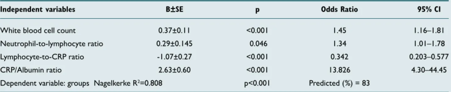
Based on the multivariate logistic regression analysis, high WBC level (OR:1.45; 95% CI:1.16–1.81; p=0.001), high CAR level (OR:13.826; 95% CI:4.30–44.45; p=0.001), high NLR level (OR:1.34; 95% CI:1.01–1.78; p=0.046) and low LCR lev-el (OR:0.001; 95% CI:3.642–0.001; p=0.001) were indepen-dent variables for AA diagnosis in pregnant patients (Table 2). In ROC curve analyses of these independent variables, AUC was above 0.600 for WBC, CAR, NLR and LCR (Fig. 1).
When a cutoff value of >11.965/mm3 was used for WBC,
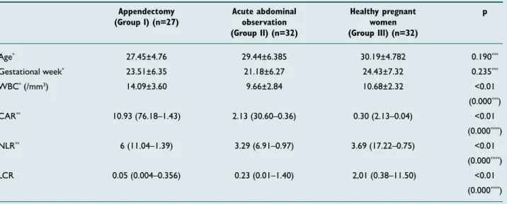
the sensitivity was 77% and the specificity was 81% (accuracy rate 79%, AUC ± SE = 0.828±0.055 and p<0.001) for AA diagnosis. For CAR variable to predict AA diagnosis, the sen-sitivity was 96% and the specificity was 80% (accuracy rate 88%, AUC±SE = 0.917±0.028, p<0.001) using a cutoff value of >2.473. For NLR, the sensitivity was 68% and the speci-ficity was 86% (accuracy rate 77%, AUC ± SE= 0.781±0.065 Table 1. Gestational age and hemogram parameters of the study groups
Appendectomy Acute abdominal Healthy pregnant p (Group I) (n=27) observation women
(Group II) (n=32) (Group III) (n=32)
Age* 27.45±4.76 29.44±6.385 30.19±4.782 0.190*** Gestational week* 23.51±6.35 21.18±6.27 24.43±7.32 0.235*** WBC* (/mm3) 14.09±3.60 9.66±2.84 10.68±2.32 <0.01 (0.000***) CAR** 10.93 (76.18–1.43) 2.13 (30.60–0.36) 0.30 (2.13–0.04) <0.01 (0.000****) NLR** 6 (11.04–1.39) 3.29 (6.91–0.97) 3.69 (17.22–0.75) <0.01 (0.000****) LCR 0.05 (0.004–0.356) 0.23 (0.01–1.40) 2,01 (0.38–11.50) <0.01 (0.000****)
*Mean±standard deviation; **Median (max-min); ***One-way ANOVA test; **** Kruskal Wallis Test.
and p<0.001) for the prediction of AA diagnosis when a cut-off value of >5.025 was used. When a cutcut-off value of <0.127 was used for LCR to predict AA diagnosis, the sensitivity was 73% and the specificity was 89% (accuracy rate 81%, AUC±SE = 0.895±0.033, p<0.001). Proposed cutoff values and performance characteristics for these variables were shown in Table 3.
DISCUSSION
AA is among the most common causes of emergency surgery in pregnant patients. Physical examination and anamnesis are important in diagnosing AA. There are difficulties in AA diag-nosis due to physiological and anatomical changes observed during pregnancy and due to the restrictions on the use of radiological methods. This increases the importance of using parameters involving acute phase reactants secondary to an inflammatory reaction in the body. In the present study, we found that inflammatory parameters, such as WBC, NLR, CAR, and LCR, could be considered to be statistically signifi-cant in AA diagnosis for pregnant patients.
WBC is a highly cost-effective and easily accessible laborato-ry parameter that is widely used in the diagnosis of AA. Ele-vated WBC level is not a pathognomonic finding in patients with AA, but is used as an auxiliary parameter for AA
diag-nosis.[14] Elevated WBC levels in peripheral blood are used
as an acute phase reactants secondary to inflammation.[15]
Keskek et al.[15] and Panagiotopoulou et al.[16] reported that
the WBC value of patients with AA was higher than the
nor-mal population. Yazar et al.,[4] on the other hand, found that
the average WBC value was 10.762±1.513/mm3 in healthy
pregnant women and 13.768±3.443/mm3 in patients
under-going an appendectomy. Yilmaz et al.[17] reported an average
WBC value of 12.702±4.180/mm3 in pregnant women who
were operated due to AA. In the present study, the average WBC value in healthy pregnant women was 10.680±2.32/
mm3, which was 14.090±3.60/mm3 in pregnant patients
op-Table 2. Results of the multinomial logistic regression analysis of white blood cell count, neutrophil-to-lymphocyte ratio, CRP/
albumin ratio and lymphocyte-to-CRP ratio to determine independent predictors of acute appendicitis in pregnant women
Independent variables B±SE p Odds Ratio 95% CI
White blood cell count 0.37±0.11 <0.001 1.45 1.16–1.81 Neutrophil-to-lymphocyte ratio 0.29±0.145 0.046 1.34 1.01–1.78 Lymphocyte-to-CRP ratio -1.07±0.27 <0.001 0.342 0.203–0.577 CRP/Albumin ratio 2.63±0.60 <0.001 13.826 4.30–44.45 Dependent variable: groups Nagelkerke R2=0.808 p<0.001 Predicted (%) = 83
Multinomial logistic regression. B: The set of coefficients estimated for the model; CI: Confidence interval; SE: Standard error; CRP: C-reactive protein.
Table 3. The results of ROC analysis
Parameter Cut-off values Accuracy rate (%) Sensitivity (%) Specificity (%) AUC ± SE p
WBC >11.965 0.79 0.77 0.81 0.828±0.055 <0.001 CAR >2.473 0.88 0.96 0.80 0.917±0.028 <0.001 NLR >5.025 0.77 0.68 0.86 0.781±0.065 <0.001 LCR <0.127 0.81 0.73 0.89 0.895±0.033 <0.001
AUC: Area under the receiver operating characteristic curve; SE: Standard error; WBC: White blood cells. CAR: CRP/Albumin ratio; NLR: Neutrophil-to-lymphocyte ratio; LCR: Lymphocyte-to-CRP ratio.
Figure 1. Receiver operating characteristic (ROC) curve analyses
of significant parameters for the diagnosis of acute appendicitis: CAR: CRP/Albumin ratio; WBC: White blood cell count; NLR: Neu-trophil-to-lymphocyte ratio; LCR: Lymphocyte-to-CRP ratio.
1.0 0.8 0.6 Sensitivity ROC Curve Source of the Curve CAR WBC NLR LCR Reference Line 1 - Specificity 0.4 0.2 0.0 0.0 0.2 0.4 0.6 0.8 1.0
erated due to AA. According to these results, WBC value was significantly higher in patients who were operated for AA compared to healthy pregnant women and pregnant women under acute abdominal observation (Table 1). Based on the multivariate logistic analysis (multinomial logistic regression), WBC was found to be an independent risk factor for the diagnosis of AA in pregnant patients (Table 2). Previous stud-ies reported sensitivity levels of 73.0–97.8%, specificity levels of 52.0–55.7%, PPV levels of 42.0–91.3%, and NPV levels of
25.2–82.0% for WBC in AA diagnosis.[14] Such large sensitivity
and specificity ranges could be due to the different cut-off values used in the diagnosis of AA. For example, Keskek et al.
[15] reported a cut-off value of 10.500/mm3, while Körner et
al.[18] mentioned a value of 12.300/mm3. Yazar et al.[4]
calculat-ed the sensitivity as 57.1% and specificity as 82.9% when they
used a cut-off level of 13.880, while Çınar et al.[19] obtained
a sensitivity level of 72.5% and a specificity level of 72.3% using a cut-off value of 10.300. Considering a cut-off value of >11.965/mm3, we calculated the sensitivity as 77%, specificity
as 81% (Table 3). Based on these findings, elevated WBC level could be used by clinicians as a parameter to support physical examination and anamnesis findings for the diagnosis of AA in pregnant women.
In AA cases, a characteristic shifting to the left is observed in
hemogram due to neutrophilia and lymphopenia.[20,21] Markar
et al.[22] and Yavuz et al.[23] reported that NLR had
statistical-ly higher diagnostic sensitivity for AA than WBC and CRP.
Eren et al.,[21] on the other hand, reported that the NLR
ra-tio was higher in patients with complicated appendicitis than in patients subjected to negative appendectomy. This finding was attributed to elevated neutrophilia severity secondary to increased inflammation level and a more evident decrease in
lymphopenia.[22] In the present study, the median NLR
val-ue observed in patients undergoing appendectomy was 6.00 (1.39–11.04), which was 3.69 (0.75–17.22) in healthy preg-nant women. According to these results, the NLR value was significantly higher in patients who were diagnosed with AA and who had appendectomy compared to healthy pregnant women and pregnant women under acute abdominal obser-vation (Table 1). Based on multivariate logistic regression analysis using the data obtained in the present study (mul-tinomial logistic regression), NLR was found to be an inde-pendent risk factor for the diagnosis of AA (Table 2). The most appropriate cut-off value for NLR was reported as >3.5 by Białas et al.[24] and as ≥4.5 by Eren et al.[21] Yavuz et al.[23]
calculated a sensitivity level of 92.5% and a specificity level of 59.3% for NLR in geriatric patients, when they considered a cut-off value of 3.95. In all three studies, it was stated that the elevated NLR value was associated with complicated ap-pendicitis. Yazar et al.[4] calculated that for AA diagnosis
accu-racy rate of NLR was 79.4% and AUC ± SE was 0.852±0.049 (p<0.001) when they used a cut-off value of >6.84. On the other hand, Çınar et al.[19] used a cut-off value of >5.50 and
calculated the sensitivity as 90%, specificity as 89.4%, accura-cy rate as 90.8% and AUC ± SE value as 0.920±0.034 for NLR
in AA diagnosis. In the present study, we used a cut-off value of >5.025 for NLR, and calculated sensitivity level as 68% and specificity level as 86% (Table 3). Similar to those reported by Yazar[4] and Çınar,[19] sensitivity and specificity values in the
present study were also significant. Based on our findings, el-evated NLR level could be used as a supportive parameter to physical examination and anamnesis findings for AA diagnosis in pregnant patients.
Bacterial infections, trauma, malignant neoplasms, burns, tissue infarctions, immunological and inflammatory events and birth are stimuli that cause acute phase response in the body. The purpose of the acute phase response is to neutral-ize pathogens by isolating them, to reduce tissue damage to a minimum by limiting them, to prevent the generalization of the events, to start the repair, thereby allowing the host hemostatic mechanisms to restore the normal physiological
function in a fast manner.[25] AA causes the initiation of an
inflammatory process secondary to bacterial infection in the body, resulting in the formation of an acute phase response by organism against the pathogen. Regardless of the localized or generalized nature of the disease, the acute phase response is a general host reaction. Proteins whose serum or plasma levels change during this response are called acute phase pro-teins (AFP). Synthesis of AFP propro-teins occurs in the liver as a result of cytokines released from tissue macrophages, and these proteins reflect nonspecifically the presence and
sever-ity of inflammation.[26,27] Proteins whose synthesis increase
due to AFY are called positive reactants, while those whose synthesis decrease is termed acute phase reactants. In acute phase response secondary to inflammation in the organism, the amount of CRP increases, whereas the amount of albu-min decreases.[26]
CRP is an acute phase reactant that starts to increase in the body within 8-12 hours due to the acute phase response caused by inflammation. Its increase is somewhat slower than that of WBC and reaches a maximum level within 24–48
hours.[28] CRP is an acute phase reactant used quite
frequent-ly in the diagnosis of AA, and its sensitivity ranges from 40.0 to 95.6%, and its specificity varies from 53 to 82%.[27] In many
studies examining the relationship between AA and CRP, the CRP level was reported to be especially high in complex ap-pendicitis cases, such as perforation and periapical abscess.
[27,29] Using a CRP cut-off level of 20 mg/L, Ayrık et al.[14]
re-ported a sensitivity level of 54.33% and a specificity level of 56.06% for CRP in AA cases. On the other hand, Yokoyama et al.[30] found a sensitivity level of 84.3% and specificity level
of 75.8% using a cut-off value of 49.5 mg/L for CRP. Yang et
al.[31] reported a CRP cut-off value of 24.1 mg/L for AA
cas-es. There are publications reporting that CRP value increases while the albumin level decreases in the acute phase response
secondary to inflammation in pregnant patients.[10,32]
Fair-clough[10] and Karasahin[11] reported that the combined use
of decreased albumin and elevated CRP levels improved the accuracy rate in detecting the acute infection. Both studies
reported that CAR elevation increased as parallel to the se-verity of the disease. There are reports showing that elevated CAR levels were associated with aggressive tumor behavior and poor prognosis in oncology patients.[33–35] Qin et al.[10]
and Gibson et al.[12] reported that high CAR values reflected
the severity of infection in the acute phase of inflammatory bowel disease. Both studies showed that elevated CAR val-ues were associated with the extent and severity of the
in-fection. Goulart[13] stated that increased CAR values could be
used as an early marker to determine surgical site infection in cases operated due to colorectal cancer. In the present study, the median CAR value was 10.93 (1.43–76.18) in patients who underwent appendectomy, which was 0.30 (0.04–2.13) in healthy pregnant women. The CAR value was significantly higher in patients who underwent appendectomy after AA di-agnosis compared to pregnant women who were under acute abdominal observation or healthy pregnant women (Table 1). Multivariate logistic regression analysis (multinomial logistic regression) in the present study indicated CAR as an inde-pendent predictor for AA diagnosis (Table 2). Goulart et al.[13]
calculated sensitivity and specificity of CAR as 77.3% and 66.2%, respectively, for identifying the surgical site infection when they used a cut-off value of 43 for CAR. Karaşahin et al.[11] studied infection vulnerability in geriatric patients using
a cut-off value of 1.70 for CAR, and they calculated the sensi-tivity as 74.3% and specificity 79.6% (p<0.001). In the present study, using a CAR cut-off value of >2.473 for AA diagnosis in pregnant patients, we determined the sensitivity and spec-ificity of CAR values to be 96 and 80%, respectively (Table 3). In the study conducted by Goulart et al., high CAR values were because their study included oncology patients and that the measurements were made in the postoperative period. According to these findings, elevated CAR levels could help physicians in the diagnosis of AA in pregnant patients as an additional parameter to support physical examination and an-amnesis.
Lymphocytes are involved in immune system regulation and
their number increases with inflammation.[36,37] Low
lympho-cyte number and high CRP level may indicate an infection in the body. Therefore, the combination of lymphocytes and CRP can be used as a biochemical marker to determine the severity of the infection. There are studies reporting that low lymphocyte count and elevated CRP level can be used as an infection marker in orthopedic prosthetic surgeries for an early onset of treatment for the infection.[38,39] Evaluating
the data from 554 gastric cancer patients, Okugawa et al.[40]
mentioned that low LCR values can be used as a marker to
determine surgical site infection. Yazar et al.[4] and Çınar et
al.[19] studied pregnant patients and reported that the number
of lymphocytes was lower, but CRP was higher in the group of patients who had appendectomy compared to healthy pregnant women. In both studies, the number of lymphocytes decreased while the CRP value increased depending upon the severity of the infection. In the present study, the median LCR value was 0.05 (0.004–0.356) in patients who underwent
appendectomy due to AA and 2.01 (0.38–11.50) in healthy pregnant women. Decrease in the rate of lymphocytes and an increase in CRP value secondary to infection resulted in a negative correlation between these two parameters. Thus, LCR value was significantly lower in patients who underwent appendectomy after AA diagnosis compared to pregnant women under acute abdominal observation and healthy preg-nant women (Table 1). Multivariate logistic analysis (multino-mial logistic regression) in the present study revealed that LCR was an independent risk factor for the diagnosis of AA in pregnant women patients (Table 2). Using a cut-off value of <0.127, LCR could predict AA in pregnant women with an accuracy of 81%, a sensitivity of 73% and a specificity of 89% (Table 3). Based on these result, low LCR values could be used by clinicians as support data to physical examination and anamnesis for AA diagnosis in pregnant patients.
Our study carries the limitations inherent in retrospective case studies. In addition, the scarcity of the patients who had appendectomy and exclusion of a small number of patients with complicated appendicitis from the study were the main limitations. Another limitation of this study is the lack of in-formation between the blood withdrawal and the operation time since the inflammatory values may change with time.
Conclusion
In pregnant patients with suspected AA, WBC, NLR, CAR and LCR could be used as support parameters to the findings from anamnesis, physical examination and imaging methods in the diagnosis. Such a practice could lower the maternal and fetal morbidity/mortality rates and negative laparotomy rates. Our results could contribute and provide valuable insights to limited literature associated with AA in pregnant women. Prospective studies with large cohorts in this area could be useful.
Ethics Committee Approval: Approved by the local
eth-ics committee.
Peer-review: Internally peer-reviewed.
Authorship Contributions: Concept: A.A., M.T., B.O.;
Design: A.A., N.A.H., B.O.; Supervision: A.A., Y.A., M.T.; Ma-terials: A.A., B.O.; Data: M.T., B.O.; Analysis: Z.A.K., H.Y.; Lit-erature search: Y.A., M.T., S.M., H.Y.; Writing: A.A., N.A.H.; Critical revision: S.M., H.Y.
Conflict of Interest: None declared.
Financial Disclosure: The authors declared that this study
has received no financial support.
REFERENCES
1. Zhang Y, Zhao YY, Qiao J, Ye RH. Diagnosis of appendicitis during pregnancy and perinatal outcome in the late pregnancy. Chin Med J (Engl) 2009;122:521−4.
2. Wray CJ, Kao LS, Millas SG, Tsao K, Ko TC. Acute appendicitis: contro-versies in diagnosis and management. Curr Probl Surg 2013;50:54−86.
3. Yagci MA, Sezer A, Hatipoglu AR, Coskun I, Hoscoskun Z. Acute ap-pendicitis in pregnancy. Dicle Tip Derg 2010;37:134−139.
4. Yazar FM, Bakacak M, Emre A, Urfalıoglu A, Serin S, Cengiz E, et al. Predictive role of neutrophil-to-lymphocyte and platelet-to-lymphocyte ratios for diagnosis of acute appendicitis during pregnancy. Kaohsiung J Med Sci 2015;31:591−6. [CrossRef ]
5. Abbassi-Ghanavati M, Greer LG, Cunningham FG. Pregnancy and laboratory studies: a reference table for clinicians. Obstet Gynecol 2009;114:1326−31. [CrossRef ]
6. Franca Neto AH, Amorim MM, Nóbrega BM. Acute appendicitis in pregnancy: literature review. Rev Assoc Med Bras (1992) 2015;61:170−7. 7. İlhan M, İlhan G, Gök AF, Bademler S, Verit Atmaca F, Ertekin C. Eval-uation of neutrophil-lymphocyte ratio, platelet-lymphocyte ratio and red blood cell distribution width-platelet ratio as early predictor of acute pan-creatitis in pregnancy. J Matern Fetal Neonatal Med 2016;29:1476−80. 8. Daldal E, Akbas A, Dasiran MF, Dagmura H, Bakir H, Okan I.
Prog-nostic importance of neutrophil/lymphocyte and lymphocyte/crp ratio in cases with malignant bowel obstruction. Med Science 2019;8:927−30. 9. Sevgisunar NS, Şahinduran Ş. Hayvanlarda akut faz proteinleri, kullanım
amaçları ve klinik önemi. MAKÜ Sag Bil Enst Derg 2014;2:50−72. 10. Qin G, Tu J, Liu L, Luo L, Wu J, Tao L, et al. Serum Albumin and
C-Re-active Protein/Albumin Ratio Are Useful Biomarkers of Crohn’s Disease Activity. Med Sci Monit 2016;22:4393−400. [CrossRef ]
11. Karaşahin Ö, Tosun Taşar P, Timur Ö, Baydar Ö, Yıldırım F, Yıldız F, et al. Palyatif Bakım Alan Geriatrik Hastalarda Enfeksiyon Tanı ve Prog-nozunda Laboratuvar Belirteçlerin Değeri. Tepecik Eğit ve Araşt Hast Dergisi 2016;26:238−42.
12. Gibson DJ, Hartery K, Doherty J, Nolan J, Keegan D, Byrne K, et al. CRP/Albumin Ratio: An Early Predictor of Steroid Responsiveness in Acute Severe Ulcerative Colitis. J Clin Gastroenterol 2018;52:e48−52. 13. Goulart A, Ferreira C, Estrada A, Nogueira F, Martins S,
Mesquita-Ro-drigues A, et al. Early inflammatory biomarkers as predictive factors for freedom from infection after colorectal cancer surgery: a prospective co-hort study. Surgical infections 2018;19:446−50. [CrossRef ]
14. Ayrık C, Karaaslan U, Dağ A, Bozkurt S, Toker İ, Demir F. Lökosit sayısı, yüzde nötrofil oranı ve C-reaktif protein konsantrasyonlarının “kesim değeri” düzeylerinde apandisit tanısındaki değerleri. Ulus Travma Acil Cerrahi Derg 2016;22:76−83.
15. Keskek M, Tez M, Yoldas O, Acar A, Akgul O, Gocmen E, Koc M. Re-ceiver operating characteristic analysis of leukocyte counts in operations for suspected appendicitis. Am J Emerg Med 2008;26:769−72. [CrossRef ]
16. Panagiotopoulou IG, Parashar D, Lin R, Antonowicz S, Wells AD, Bajwa FM, Krijgsman B. The diagnostic value of white cell count, C-reactive protein and bilirubin in acute appendicitis and its complications. Ann R Coll Surg Engl 2013;95:215−21. [CrossRef ]
17. Yilmaz HG, Akgun Y, Bac B, Celik Y. Acute appendicitis in pregnancy— risk factors associated with principal outcomes: a case control study. Int J Surg 2007;5:192−7. [CrossRef ]
18. Körner H, Söndenaa K, Söreide JA. Perforated and non-perforated acute appendicitis—one disease or two entities? Eur J Surg 2001;167:525−30. 19. Çınar H, Aygün A, Derebey M, Tarım İA, Akalın Ç, Büyükakıncak S, et al. Significance of hemogram on diagnosis of acute appendicitis during pregnancy. Ulus Travma Acil Cerrahi Derg 2018;24:423−8. [CrossRef ]
20. Andersson RE. Meta-analysis of the clinical and laboratory diagnosis of appendicitis. Br J Surg 2004;91:28−37. [CrossRef ]
21. Eren T, Tombalak E, Burcu B, Özdemir İA, Leblebici M, Ziyade S, et al. Akut Apandisit Olgularında Nötrofil/Lenfosit Oranının Tanıda ve
Hastalığın Şiddetini Belirlemedeki Prediktif Değeri. Dicle Tıp Derg 2016;43:279−84.
22. Markar S, Karthikesalingam A, Falzon A, Kan Y. The diagnostic value of neutrophil: lymphocyte ratio in adults with suspected acute appendicitis. Acta Chir Bel 2010;110:543−7. [CrossRef ]
23. Yavuz E, Erçetin C, Uysal E, Solak S, Biricik A, Yiğitbaş H, et al. Diag-nostic Value of Neutrophil/Lymphocyte Ration in Geriatric Cases With Appendicitis. Turkish J Geriatrics 2014;17:345−9.
24. Białas M, Taran K, Gryszkiewicz M, Modzelewski B. Evaluation of neutrophil-lymphocyte ratio usefulness in the diagnosis of appendicitis. Wiad Lek 2006;59:601−6.
25. Uluğ M, Can-Uluğ N, Selek Ş. Akut brusellozlu hastalarda akut faz reak-tanlarının düzeyi. Klimik Dergisi 2010;23:48−50. [CrossRef ]
26. Yaylı G. İnfeksiyon hastalıklarında C-reaktif protein, sedimantasyon ve lökositler. Ankem Derg 2005;19:80−4.
27. Soylu L, Aydin OU, Yıldız M. Diagnostic value of procalcitonin, C-re-active protein, and erythrocyte sedimentation rate for acute complicated appendicitis. J Clin Anal Medicine 2018;9:47−50. [CrossRef ]
28. Wu HP, Lin CY, Chang CF, Chang YJ, Huang CY. Predictive value of C-reactive protein at different cutoff levels in acute appendicitis. Am J Emerg Med 2005;23:449−53. [CrossRef ]
29. Shogilev DJ, Duus N, Odom SR, Shapiro NI. Diagnosing appendicitis: evidence-based review of the diagnostic approach in 2014. West J Emerg Med 2014;15:859−71. [CrossRef ]
30. Yokoyama S, Takifuji K, Hotta T, Matsuda K, Nasu T, Nakamori M, et al. C-Reactive protein is an independent surgical indication marker for appendicitis: a retrospective study. World J Emerg Surg 2009;4:36. 31. Yang HR, Wang YC, Chung PK, Chen WK, Jeng LB, Chen RJ.
Labora-tory tests in patients with acute appendicitis. ANZ J Surg 2006;76:71−4. 32. Beyazıt F, Pek E, Türkön H. Serum Ischemia-Modified Albumin Con-centration and Ischemia-Modified Albumin/Albumin Ratio in Hyper-emesis Gravidarum. Med Bull Haseki 2018;56:292−8. [CrossRef ]
33. Yu ST, Zhou Z, Cai Q, Liang F, Han P, Chen R, Huang XM. Prognostic value of the C-reactive protein/albumin ratio in patients with laryngeal squamous cell carcinoma. Onco Targets Ther 2017;10:879−84. [CrossRef ]
34. Liu Z, Jin K, Guo M, Long J, Liu L, Liu C, et al. Prognostic Value of the CRP/Alb Ratio, a Novel Inflammation-Based Score in Pancreatic Can-cer. Ann Surg Oncol 2017;24:561−8. [CrossRef ]
35. Akbas A, Bakir H, Dasiran M, Dagmura H, Ozmen Z, Celtek NY, et al. Significance of Gastric Wall Thickening Detected in Abdominal CT Scan to Predict Gastric Malignancy. J Oncol 2019;2019. [CrossRef ]
36. Maurizi G, Della Guardia L, Maurizi A, Poloni A. Adipocytes properties and crosstalk with immune system in obesity-related inflammation. J Cell Physiol 2018;233:88−97. [CrossRef ]
37. Ustundag Y, Huysal K, Gecgel SK, Unal D. Relationship between C-re-active protein, systemic immune-inflammation index, and routine he-mogram-related inflammatory markers in low-grade inflammation. Int J Med Bioch 2018;1:24−8. [CrossRef ]
38. Bekmez Ş, Çağlar Ö, Atilla B. Total kalça artroplastisi sonrası enfeksiyon. TOTBİD Derg 2013;12:268–75. [CrossRef ]
39. Smith T. Nutrition: its relationship to orthopedic infections. Orthop Clin North Am 1991;22:373−7.
40. Okugawa Y, Toiyama Y, Yamamoto A, Shigemori T, Ichikawa T, Yin C, et al. Lymphocyte-to-C-reactive protein ratio and score are clinically fea-sible nutrition-inflammation markers of outcome in patients with gastric cancer. Clin Nutr 2020 39:1209−17. [CrossRef ]
OLGU SUNUMU
Gebe hastalarda enflamatuvar belirteçlerin akut apandisit tanısı koymadaki değeri
Dr. Ahmet Akbaş,1 Dr. Zeliha Aydın Kasap,2 Dr. Nadir Adnan Hacım,1 Dr. Merve Tokoçin,1
Dr. Yüksel Altınel,1 Dr. Hakan Yigitbaş,1 Dr. Serhat Meriç1, Dr. Bakiye Okumuş3
1Sağlık Bilimleri Üniversitesi, İstanbul Bagcılar Eğitim ve Araştırma Hastanesi, Genel Cerrahi Kliniği, İstanbul 2Karadeniz Teknik Üniversitesi Tıp Fakültesi, Biyoistatistik ve Tip Bilişimi Anabilim Dalı, Trabzon
3İstinye Üniversitesi Tıp Fakültesi, Kadın Hastalıkları ve Doğum Anabilim Dalı, İzmir
AMAÇ: Gebelik esnasında en sık obstetrik dışı cerrahi müdahaleye neden olan hastalık akut apandisittir (AA). AA tanısı laboratuvar testleri eşli-ğinde anamnez ve fizik muayene ile birlikte konulmaktadır. Gebelikte gözlenen fizyolojik ve anatomik değişiklikler nedeni ile AA tanısı gebe olmayan hastalara göre daha zordur. Bu çalışmada, hamilelik esnasında AA tanısında beyaz küre (WBC), nötrofil/lenfosit oranı (NLR), CRP/albümin oranı (CAR) ve lenfosit/CRP oranı (LCR) önemini araştırdık.
GEREÇ VE YÖNTEM: Çalışmada, Eylül 2015–Aralık 2019 yıları arasında genel cerrahi kliniğinde AA ön tanısı ile yatışı yapılarak takibi yapılan gebe hastalar “International Statistical Classification of Diseases and Related Health Problems-10” (ICD-10) tanı kodu kullanılarak retrospektif olarak belirlendi. Hastalar iki gruba ayrılarak I. Gruba AA nedeni ile apendektomi uygulanan, patolojik değerlendirme sonucuna göre süpüratif apandisit tanısı konulan hastalar, II. Gruba AA ön tanısı ile yatırılan ve takiplerinde ameliyat edilmeden şifa ile taburcu edilen gebe hastalar dahil edildi. Kontrol grubuna (Grup III) ise hastanemiz kadın doğum polikliniğinde takibi yapılan çalışma kriterlerine uygun, rastgele seçilmiş, çalışmaya katılmayı kabul eden 32 sağlıklı gebe prospektif olarak belirlenerek dahil edildi.
BULGULAR: Çalışmaya, yaş ortalaması 29.20±4.47 olan 96 gebe hasta (32 sağlıklı gebe, 32 akut batın müşahede ile takip edilen gebe, 32 apendek-tomi uygulanmış gebe) alındı. Bu hastalardan ameliyat ve histopatolojik bulgulara göre süpüratif apandisit olmayan üç olgu (negatif apendekapendek-tomi) ile perfore apandisit tespit edilen iki olgu çalışma dışı bırakıldı. Yapılan değerlendirmelerde Grup I oluşturan hastaların WBC değeri (p=0.001), CAR değeri (p=0.001), NLR değeri (p=0.001) grup II ve III’den anlamlı düzeyde yüksek iken, LCR değerinin düşük olduğu gözlendi (p=0.001).Yapılan çok değişkenli lojistik regresyon analizine göre; WBC, CAR, NLR yüksekliği ile LCR düşüklüğü gebe hastalarda AA tanısında bağımsız değişken olduğu gözlendi.
TARTIŞMA: Tıbbi öykü, fizik muayene ve görüntüleme tekniklerine ek olarak, gebe kadınlarda AA tanısı için WBC, NLR, CAR ve LCR değerlerinin göz önünde bulundurulması klinisyene karar vermede kolaylık sağlayabilir.
Anahtar sözcükler: Akut apandisit; CRP albümin oranı; gebelik; lenfosit; nötrofil.
Ulus Travma Acil Cerrahi Derg 2020;26(5):769-776 doi: 10.14744/tjtes.2020.03456