Oxidative Stress Parameters, Selenium Levels, DNA
Damage, and Phthalate Levels in Plastic Workers
Gulru Gurdemir,a,b Pinar Erkekoglu,a Aylin Balci,a Unzile Sur,a,c Gizem Ozkemahlı,a,d Engin Tutkun,e Hinc Yılmaz,f Ali Asci,c & Belma Kocer-Gumuselg,*a Department of Toxicology, Faculty of Pharmacy, Hacettepe University Ankara, Turkey; bTurkish Medicines and
Medical Devices Agency, Ankara, Turkey; cDepartment of Toxicology, Faculty of Pharmacy, Atatürk University,
Erzurum, Turkey; dDepartment of Toxicology, Faculty of Pharmacy, Erzincan University, Erzincan, Turkey; e
-Department of Public Health, Bozok University, Yozgat, Turkey; fDepartment of Public Health, Faculty of Medicine,
Yıldırım Beyazıt University, Ankara, Turkey; gDepartment of Toxicology, Faculty of Pharmacy, Lokman Hekim
University Ankara, Turkey
* Address all correspondence to: Belma Kocer-Gumusel, Lokman Hekim University, Faculty of Pharmacy, Department of Toxicology, Ankara, Turkey; Tel.: +05325009533, E-mail: [email protected]
ABSTRACT: Di(2-ethylhexyl)phthalate (DEHP) is the most widely used phthalate. DEHP is highly used in PVC floor-ings and PVC windows and carpeting. The objective of this study was to determine sex hormone levels, oxidative stress parameters, selenium levels, DNA damage, and phthalate levels in plastics workers (n = 24, age = 20–58 years) working in the production of rubber mechanical goods and exposed to DEHP in workplace. The control group (n = 29, age = 25–54, all male) was selected from age-matched healthy adults. Antioxidant parameters and DNA damage were determined by spectrophotometry. Selenium levels were determined by atomic absorption spectroscopy. Plasma hormone levels were measured by chemiluminescence microparticle immunoassay. Plasma phthalate levels were determined by high-pressure liquid chromatography. Plastic workers had lower serum testosterone and free T4 levels and higher folli-cle-stimulating hormone levels vs. controls. Liver enzyme activities were markedly higher in workers vs. controls. There were also increases in plasma glutathione peroxidase levels and marked decreases in plasma selenium and erythrocyte to-tal glutathione levels in plastics workers (P < 0.05 vs. control). Plasma 8-hydroxy-2′-deoxyguanosine levels were 14-fold higher in plastics workers than in controls. Plasma DEHP and mono(2-ethylhexyl)phthalate were also markedly higher in workers vs. controls. The results of this study show that occupational exposure to DEHP may lead to disturbances in sex hormones, increased liver problems, higher oxidative stress and DNA damage levels, and lower trace element concentra-tions in workers. More comprehensive and mechanistic studies with higher numbers of subjects are needed to show the unwanted effects of occupational exposure to DEHP.
KEY WORDS: DNA damage, oxidative stress, phthalate, antioxidant system, plastics worker, selenium, sex hormones
I. INTRODUCTION
Humans are exposed to different kinds of phthalates in everyday life. These compounds increase the du-rability, flexibility, and pliability of polyvinyl chlo-ride (PVC) plastics.1–4 Di(2-ethylhexyl)phthalate (DEHP) is the most widely used phthalate. It is pres-ent in food, baby products, cosmetics, PVC floor-ings and undercarpets, PVC windows, and medical devices.4,5 DEHP is suggested to cause reproductive toxicity in both animals and humans.6–10 Moreover, DEHP is a peroxisome proliferator that is categorized as a group IIB carcinogen (possible carcinogenic to humans) by the International Agency for Research on Cancer (IARC).11 DEHP is an endocrine-disrupt-ing chemical, as shown by many in vitro and in vivo
studies, and it is suggested to cause antiandrogenic effects. DEHP was also shown to cause oxidative stress in different organs of rodents.12–17 Moreover, DEHP is suggested to lead to reprotoxicity, partic-ularly in males, and it was also shown to cause de-velopmental anomalies and neuroendocrine system toxicity. DEHP is associated with different diseases, like precocious puberty (PP), diabetes, gynecomas-tia, obesity and/or metabolic syndrome, asthma, and autism.18–24 Occupational exposure to DEHP is high in PVC film manufacturing, PVC compounding, rubber boot manufacturing, and PVC flooring, win-dow, and carpet manufacturing workplaces.25–27
Oxidative stress is an imbalance between cellular antioxidants and oxidants which favors the latter. High levels of intracellular reactive oxygen species (ROS)
can lead to the depletion of cellular thiols and antiox-idant enzymes and can cause high levels of intracel-lular oxidation. While celintracel-lular antioxidant enzymes, such as glutathione peroxidases (GPxs), thioredoxin reductases (TrxRs), superoxide dismutase (SOD) and catalase (CAT), can handle high levels of intracellu-lar ROS, such as hydrogen peroxide (H2O2), other peroxidases, superoxide, and hydroxyl radicals, they cannot always cope with the chronic oxidative stress caused by different chemicals, including phthalates. Therefore, occupational exposure to several indus-trial chemicals including phthalates can lead to high oxidant levels, lipid peroxidation, protein oxidation, and DNA base damage.28–32
As PVC workers can continuously be exposed to phthalates by different routes (oral, inhalation, dermal), the number of studies evaluating the toxic effects of DEHP and phthalates has been increas-ing, particularly in China and Taiwan. Most of these studies have focused on one or two param-eters. Pan et al. evaluated the association between urinary DEHP and dibutyl phthalate (DBP) and serum testosterone levels in Chinese plastics work-ers. The same working group later evaluated haz-ard indices (HIs) for DBP and DEHP and examined the relationship between the urinary levels of these hormones and urinary phthalate concentrations in phthalate-exposed and nonexposed Chinese men.33 Another study, also conducted in China, determined urinary 8-hydroxy-2′-deoxyguanosine (8-OHdG) levels, antioxidant enzyme activities (SOD), and lipid peroxidation in workers exposed to DEHP in a plastics-recycling site in China.34 Studies conducted on polyvinyl chloride workers in Taiwan also pointed out significant effects of DEHP exposure on reproductive hormones, sperm parameters, ROS generation, and apoptosis.35,36 However, no compre-hensive study has examined the many parameters and their correlations in PVC workers who are ex-posed to phthalates.
Concerning all available data and the information presented here, the aim of this study was to determine changes in plasma thyroid hormones, reproductive hormones, liver enzymes, plasma/erythrocyte and antioxidant/oxidant parameters as well DNA base damage and correlate them with levels of exposure to DEHP and its main metabolite mono(2-ethylhexyl)
phthalate (MEHP), and to evaluate the correlations between the measured parameters.
II. MATERIALS AND METHODS A. Subjects
The study group consisted of 24 plastics workers (age = 20–58 years, all male) who had been admit-ted to Turkish Ministry of Health Ankara Occupa-tional Diseases Hospital between 2013 and 2014. All of the workers had been working in plastics-pro-ducing factories or workplaces using PVC and DEHP as plasticizers for more than two years and had been periodically examined by physicians in the Ankara Occupational Diseases Hospital. During their admission to the hospital, they were asked to contribute to the study. The control group (n = 29, age = 25–54, all male) was selected from age-matched healthy adults without chronic, endocrine, or genetic disease not taking any medication. Blood samples were collected on Fridays (the last shift day of the week).
The study was approved by the Hacettepe Uni-versity Scientific Research Evaluation Commission Ethical Committee. Written informed consent was obtained from all subjects recruited to the study. A standard questionnaire was applied to determine ed-ucational status, cigarette smoking, alcohol use, di-etary habits, occupational information, and potential exposure route to phthalates.
B. Deplasticization of Glassware
Extreme caution was taken to prevent subject con-tact with plastic material throughout the study. All test tubes and vials were deplasticized on a heater for 4 h at 400οC. All other glassware (beakers, glass pipettes, etc.) were deplasticized with tetrahydrofu-ran:n-hexane [50:50 (v/v)] for 2 h and dried in an incubator for 2 h at 37οC.
C. Sample Preparation
For the determination of the hormones, liver func-tion parameters, plasma and erythrocyte antioxidant parameters, and selenium levels, heparinized blood
samples (∼ 10 mL) were drawn by a routine tech-nique. Samples were centrifuged immediately at 800 × g for 15 min in order to separate plasma and erythrocytes. Both plasma and erythrocyte samples were aliquoted and kept at −80°C until analysis. For the determination of plasma DEHP and MEHP levels, blood samples were drawn by the dropping method. Briefly, blood was drawn by a stainless steel needle from the left-arm cubital vein and allowed to drop into heparinized glass test tubes directly. The tube openings were covered by clean aluminum foil to protect the sample from contact with plastic material. Centrifugation was performed at 800 × g for 15 min, plasma were separated, and all samples were immedi-ately aliquoted into glass vials covered with aluminum foil and stored in a freezer at −80°C until analysis.
Spot urine samples (∼ 5 mL) were collected in beakers for determination of F2-isoprostane levels. The samples were aliquoted and kept at −80°C until analysis.
D. Chemicals and Kits
All chemicals and colorimetric assay kits for protein determination, GSH, and TrxR were obtained from Sigma-Aldrich (St. Louis, MO). MEHP was ob-tained from Cambridge Chemicals (Woburn, MA). Alanine aminotransferase (ALT), aspartate amino-transferase (AST), direct/total bilirubin, and total protein and blood glucose kits were obtained from Beckman Coulter (Brea, CA). GPx, CAT, SOD, and thiobarbituric acid reactive substance (TBARS) col-orimetric assay kits were obtained from Cayman Chemical (Ann Arbor, MI). Urinary isoprostane en-zyme immunoassay kits and 8-hydroxy-2′-deoygua-nosine quantitative assay kits were from obtained from Oxis International (Foster City, CA). All high-performance liquid chromatography (HPLC) equipment was obtained from Agilent (Santa Clara, CA).
E. Determination of Plasma Hormone Levels
Plasma fT4, fT3, TSH, FSH, LH, and testosterone were measured by chemiluminescence microparti-cle immunoassay (CLIA) on an Architect i200 SR analyzer (Stillwater, MN).
F. Determination of Plasma Liver Function Parameters and Fasting Blood Glucose
Liver function parameters were measured using ALT, AST, direct bilirubin, total bilirubin, total pro-tein, and fasting blood glucose kits on a Beckman Coulter AU analyzer.
G. Determination of Oxidant/Antioxidant Parameters
For spectrophotometric measurements, a SpectraMax M2 spectrophotometer (Molecular Devices, Sunny-vale, CA) was used. SoftMax Pro software (Molecu-lar Devices) was used for quantification.
Glutathione peroxidases (GPxs) are seleni-um-containing antioxidant enzymes that reduce hy-drogen peroxide to water in order to overcome its harmful effects. These selenoproteins are present in erythrocytes and plasma and in different tissues, particularly the liver.37,38 The commercial kit used in the study indirectly measures the GPx activity by a coupled reaction with glutathione reductase (GR). Oxidized glutathione (GSSG) upon reduction by a hydroperoxide (i.e., cumene hydroperoxide) is later recycled to its reduced state by GR and by NADPH oxidation. The oxidation of NADPH to NADP+ is accompanied by a decrease in absorbance at 340 nm that is directly proportional to the GPx activity in the erythrocyte sample. Measurement was con-ducted for 5 min, plasma samples were diluted to 1:5, and erythrocyte samples were diluted to 1:200 with sample buffer. Erythrocyte GPx1 activity was expressed as nmol/min/mg Hb; plasma GPx (GPx3) activity was expressed as U/mL.
Thioredoxin reductases (TrxRs) are a family of selenoenzymes categorized as pyridine nucleo-tide-disulphide oxidoreductases. They are the only known enzymes that can reduce thioredoxin, which serves as a general protein disulfide oxido-reduc-tase.39,40 Plasma TrxR activity was measured based on the reduction of substrate 5,5′-dithiobis(2-nitro-benzoic) acid (DTNB) into 5-thio-2-nitrobenzoic acid (TNB). TNB concentration was measured at 412 nm by a kinetic assay. GPx1 and GR activity is also a contributor to DTNB reduction and can be estimated using a specific TrxR inhibitor. In order
to determine DTNB reduction due only to TrxR ac-tivity, two consecutive assays were performed: total DTNB reduction and DTNB reduction in the pres-ence of TrxR inhibitor solution. The differpres-ence be-tween the two was the DTNB reduction due to TrxR activity, expressed as µmol/min/mL.
Superoxide anion is dismutated by SODs to H2O2. SOD1 is the major intracellular SOD (cytosolic Cu/ ZnSOD).41 Total erythrocyte SOD activity is mea-sured by a colorimetric assay. Xantine oxidase (XO) causes superoxide ion production while converting xanthine and water to uric acid and H2O2. A radical detector (tetrazolium salt solution) produces a wa-ter-soluble formazan dye upon reduction with a super-oxide anion. Supersuper-oxide can be dismutated by SOD (within the erythrocyte sample, dilution: 1:2,000 by sample buffer) to oxygen and H2O2 in the same cycle. The reduction rate of superoxide is linearly associated with XO activity and can be inhibited by SOD. The 50% inhibition activity of SOD (IC50) was determined by this colorimetric, end-point reading method. Since absorbance at 440 nm was proportional to the amount of superoxide anion, SOD inhibition was quantified by measuring the decrease in color development at 440 nm. One unit of SOD is defined as the amount of enzyme needed to exhibit 50% dismutation of the superoxide radical, expressed as U/mg Hb.
Hydrogen peroxide is catalyzed to H2O by CAT, which is an important peroxisomal enzyme.42 Eryth-rocyte CAT activity is measured based on its per-oxidative potential. CAT reacts with methanol in the presence of H2O2 and the end-product, formal-dehyde, reacts with the chromogen 4-amino-3-hy-drazino-5-mercapto-1,2,4-triazole to produce a purple-colored complex. The intensity of the purple is measured at 540 nm and is directly proportional to the amount of formaldehyde produced. Erythrocyte samples were diluted to 1:1,250 with sample buffer during measurements. The erythrocyte CAT activity was expressed as nmol/min/mg Hb.
H. Determination of Erythrocyte Total Glutathione Levels
Glutathione, an atypical tripeptide (γ-glu-cys-gly), is the most important thiol in many living organisms. In the presence of an oxidant, reduced GSH
mol-ecules are oxidized and generate a cystine bridge between the two glutathione molecules, producing GSSG.43 First, the protein in the erythrocyte samples were denatured by the addition of 5-sulfosalicylic acid (SSA) 5% solution and centrifuged at 10,000 rpm. The precipitate was used as the main sample. The method employed relies on the reaction of the sulfhydryl group of GSH with DTNB to produce TNB; in the same cycle, GSH is simultaneously converted to GS-TNB. GS-TNB can be reduced to produce GSH and TNB by GR. The TNB level is di-rectly proportional to the GSH concentration in the sample. Absorbance was measured at 405 nm for 5 min in a kinetic assay; an increase in absorbance indicated the GSH level. The results were expressed as nmol/mg Hb.
I. Determination of Plasma Malondialdehyde Levels
Lipid peroxidation is a process by which oxidants such as free radicals or nonradical species attack lipids containing carbon-carbon double bond(s), especially polyunsaturated fatty acids (PUFAs) that involve hydrogen abstraction from a carbon, with oxygen insertion resulting in lipid peroxyl radicals and hydroperoxides. Glycolipids, phospholipids, and cholesterol are also important targets of dam-aging and potentially lethal peroxidative modifica-tion.44 Oxidation of lipids has many consequences, including protein oxidation. Plasma lipid peroxida-tion levels were quantified using a TBARS assay kit that measures the concentration of malondialdyde (MDA) by spectrophotometric assay. MDA is an important indicator of lipid peroxidation. It forms a complex with thiobarbituric acid (TBA) under high temperature (90–100οC). Acidic conditions and the color intensity of MDA-TBA complex were mea-sured at 540 nm spectrophotometrically. The amount of MDA was measured using MDA standards (0, 0,5, 5, 10, 20, 30, and 50 µM) and expressed as µM.
J. Determination of Hemoglobin Levels
Hemoglobin (Hb) levels in erythrocytes were deter-mined according to Fairbanks et al.45 Optical den-sities were read at 546 nm spectrophotometrically.
K. Determination of Urinary 8-Epi- Prostaglandin (F2α) Levels
The predominant n-6 fatty acid is arachidonic acid, which can be reduced to prostaglandins, leukot-rienes, thromboxanes, and other cyclooxygenase, lipoxygenase, or cytochrome P450-derived prod-ucts via enzymatic peroxidation; or it can be re-duced to MDA, 4-hydroxynonenal, isoprostanes, and other lipid peroxidation end-products (more stable and toxic than hydroperoxides) via nonenzy-matic peroxidation through oxygen radical–depen-dent oxidative routes.44
Another biomarker for lipid peroxidation is 8-epi-prostaglandin (F2α).46 Urinary F2α-prostane levels were determined by ELISA. F2α-prostane in the samples or by standards (0.05, 1, 5, 10, 20, 30, and 50.0 ng/mL) competes with F2α-prostane con-jugated to horseradish peroxidase (HRP) for binding to a polyclonal antibody specific for F2α-prostane coated on the microplate. HRP activity results in color development when the substrate is added, with the intensity of the color proportional to the amount of bound F2α-prostane and inversely proportional to the amount of unconjugated F2α-prostane in the sam-ples or standards at 450 nm. A four-parameter logistic regression curve was used, and urinary F2α-pros-tane levels in the samples were calculated using RIDASOFT Win software (Darmstadt, Germany). F2α-prostane levels were expressed as ng/mL.
L. Determination of Plasma 8-Hydroxy- 2′-Deoyguanosine Levels
Oxidative stress can permanently cause damage to lipids of cellular membranes, proteins, and DNA. In both nuclear and mitochondrial DNA, 8-OHdG is the most commonly observed ROS-induced oxi-dative DNA lesion. It is widely being used as a bio-marker of oxidative DNA damage, carcinogenesis, and degenerative diseases.47 Plasma 8-OHdG lev-els were determined by sandwich ELISA. Briefly, 8-OHdG monoclonal antibodies, by sample or standards, was added to 8-OHdG-coated wells; the anti body was competitively bound to these wells, so higher concentrations of 8-OHdG in the sample solution led to reduced binding of antibodies to
the 8-OHdG on the plate. The antibodies bound to the 8-OHdG in the sample were washed out of the well, while those bound to the 8-OHdG coated on the plate remained. The enzyme-labeled sec-ondary antibodies were then added to each well and were bound to the monoclonal antibodies that remained on the plate. Unbound enzyme-labeled secondary antibodies were removed by a wash step. The addition of a chromogen resulted in the development of color in proportion to the amount of antibody bound to the plate. Finally, the color reaction was terminated by a stop solution and absorbance of the samples or standards was mea-sured at 450 nm. A four-parameter logistic regres-sion curve was used, and plasma 8-OHdG levels in the samples, expressed as pg/mL, were calculated using RIDA®SOFT Win.
M. Determination of Erythrocyte Selenium Levels
Selenium (Se) is a trace element with roles in a variety of metabolic processes, including thyroid hormone metabolism, protection against oxida-tive stress, and immunity. It is the integral com-ponent of many GPxs (including GPx1 and GPx3) and TrxRs.48 A single-beam atomic absorption spectrometer (PerkinElmer, Waltham, MA) with a Zeeman background correction equipped with an Fs-go plus furnace auto-sampler was used in the determination of plasma Se levels. A stock solution of 1,000 mg/L Na2SeO3 was used on a daily basis to prepare appropriate concentrations of standard selenium solutions (10, 20, 50, and 100 µg/L). Plasma samples (0.2 mL) were diluted with 0.5 mL 0.2% HNO3 (v/v in deionized water) and 0.3 mL 0.2% Triton X-100 (v/v in deionized water). Samples (20 μL) and standard solutions (20 μL) along with matrix modifier (5 μL) were wet-injected three times in a graphite furnace at 400οC. Lamp current was 290 mA; bandwidth was 2 nm. Optical densities were read at 196 nm and mean concentrations of three readings were calculated. Selenium concentration in the sam-ples was calculated from the calibration curve ob-tained from the peak areas of selenium standards and expressed as µg/L.49
N. Measurement of Plasma Di-(2- Ethylhexyl) Phthalate and Mono- (2-Ethylhexyl) Phthalate Levels
Plasma DEHP and MEHP levels were detected by HPLC according to Paris et al. with modifications.50 Briefly, 200 μL of plasma sample was spiked with 20 μL 20-ppm DEHP and 20 μL 20-ppm MEHP (1 ppm in the last volume, both). After extraction us-ing 1-N NaOH (400 μL), 50% H3PO4 (100 μL), and acetonitrile (800 μL), samples were vortexed and the mixture was centrifuged at 5,000 rpm for 10 min. Su-pernatants (600 μL) were placed in another tube and evaporated under nitrogen stream. The residues were kept at −20οC until analysis. On the day of analy-sis, residues were dissolved in 60% (v/v) acetonitrile (300 μL) and 100 μL of the resultants were injected to HPLC (Hewlett Packard Agilent 1200 Series, Vi-enna, Austria; equipped with an auto sampler and a UV detector). A Spherisorb C18 ODS2 column (25 cm × 5 m × 4.6 mm i.d.) (Waters, Milford, MA) and an ODS C18 precolumn (4 cm) (Waters) were used for analysis. The mobile phase was 0.1% phosphoric acid and acetonitrile [pH 3.0, 20:80 (v/v)], and the flow rate was 1 mL/min. Retention times for DEHP and MEHP were 39.3 and 4.7 min, respectively. With-in-day precisions were 1.12 ± 0.56% CV for DEHP and 4.15 ± 1.73% CV for MEHP. Between-run preci-sions were 10.31 ± 6.09% CV for DEHP and 8.42 ± 4.42% CV for MEHP. DEHP and MEHP concentra-tions in the samples were calculated from standards and the peak-area calibration curve was used. Limit of detections (LODs) for both DEHP and MEHP were 0.05 g/mL.
O. Statistical Analysis
Statistical analysis was performed using SPSS 22.0 (IBM, Armonk, NY). The distribution of values was analyzed by Levene test. For nonparametric
compari-sons, the Mann–Whitney U test was used. Categorical variables were compared using the Fisher chi-square test. Correlations between values were analyzed us-ing either Spearman rank-order correlation or Pear-son’s correlation (according to whether parametric or nonparametric). Data were expressed as mean ± stan-dard deviation, and p values < 0.05 were considered statistically significant.
III. RESULTS
A. Characteristics of the Study Groups
Subject ages, body weights, and working periods in the plastics industry are given Table 1. The two
groups were not statistically different from each other in age and body weight. For the workers, mean working period in the plastics industry was 8.17 ± 2.94 years and ranged 2–36 years. Seven workers had more than 10 years. Three workers refused to declare their working period.
In the control group, 10.71% of the subjects graduated from primary school and 10.71% gradu-ated from secondary school; 50% were high school graduates and 32.14% had a university degree or higher. In the workers group, 20.83% graduated from primary school and 41.66% graduated from second-ary school; 25% were high school graduates and only 12.5% had a university degree or higher. Of the controls, 55.2% were regular smokers while 58.3% of the plastics workers smoked regularly; 13.8% of controls consumed alcohol, while 4.2% of the work-ers were drinking only to socialize. There were no heavy alcohol drinkers in the two study groups.
B. Questionnaire Related to Plastics Exposure from Different Sources
In Table 2, answers obtained from the questionnaire are given. This questionnaire evaluated exposure to plastic TABLE 1: Age, body weight, and working period in the plastics industry
Controls (n = 29) Plastic workers (n = 24)
Age (year) 35.86 ± 1.67 30.71 ± 2.41
Weight (kg) 73.83 ± 2.72 81.34 ± 2.57
Working period (year) — 8.17 ± 2.94
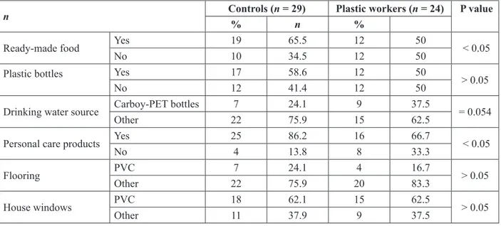
material from other sources (not from the working en-vironment). Controls had significantly higher consump-tion of ready-made food vs. plastics workers (P < 0.05). They also had an insignificantly higher consumption of soda from plastic bottles vs. plastics workers (P > 0.05); 86.2% of controls used personal care products, while this rate was 66.7% for the plastics workers. Rates of exposure to PVC house flooring and windows were not significantly different (P > 0.05, both).
C. Hormone Levels in the Study Groups
Hormone levels in the study groups are given in Table 3. TSH, fT3, and LH levels were not significantly different in controls and plastics workers (p > 0.05,
all). However, plasma fT4 levels were markedly lower in plastics workers vs. controls (P < 0.05). Moreover, plasma FSH levels were 56% higher in plastics work-ers when compared to controls (P < 0.05). In addition, plasma testosterone levels were, significantly, 22% lower in plastics workers vs. controls (P < 0.05).
D. Liver Function Parameters and Fasting Blood Glucose Levels in the Study Groups
Liver function parameters and fasting blood glu-cose levels are given in Table 4. Both ALT (49%) and AST (36.6%) levels were significantly higher in plastics workers vs. controls. However, neither the plastics workers nor the controls had ALT or AST TABLE 2: Evaluation of exposure to plastics from other sources
n Controls (n = 29)% Plastic workers (n = 24) P value
n %
Ready-made food Yes 19 65.5 12 50 < 0.05
No 10 34.5 12 50
Plastic bottles Yes 17 58.6 12 50
> 0.05
No 12 41.4 12 50
Drinking water source Carboy-PET bottles 7 24.1 9 37.5 = 0.054
Other 22 75.9 15 62.5
Personal care products Yes 25 86.2 16 66.7 < 0.05
No 4 13.8 8 33.3
Flooring PVC 7 24.1 4 16.7 > 0.05
Other 22 75.9 20 83.3
House windows PVC 18 62.1 15 62.5 > 0.05
Other 11 37.9 9 37.5
TABLE 3: Hormone levels in the study groups
Normal range Controls (n = 29) Plastic workers (n = 24)
TSH (mIU/L) 0.4–4.0 1.33 ± 0.05 1.28 ± 0.12 fT3 (ng/d) 2.6–4.8 3.46 ± 0.07 3.47 ± 0.07 fT4 (ng/dL) 0.7–2.0 1.97 ± 0.16 1.19 ± 0.03* FSH (mIU/L) 1.3–19.3 2.83 ± 0.26 4.41 ± 0.57* LH (IU/L) 1.8–8.6 4.83 ± 0.23 5.14 ± 0.32 Testosterone (ng/dL) 300–1000 442.94 ± 17.16 344.10 ± 21.97*
levels above normal ranges. There were no marked differences in direct and total bilirubin levels (P > 0.05, both), and none of the study subjects showed in direct and total bilirubin levels above normal ranges. In addition, total protein levels were not dif-ferent. However, total protein levels in six plastics workers and five controls were slightly higher than the normal range (6–8.3 g/dL), which may be a sign of mild inflammation. Fasting blood glucose levels were higher in plastics workers vs. controls (P < 0.05), and 11 workers had blood glucose levels ≥ 100 mg/dL (normal range 70–100 mg/dL).
E. Selenium Levels and Antioxidant Enzyme Activities in the Study Groups
Selenium levels and antioxidant enzyme activities in the study groups are shown in Table 5. There were no significant differences in erythrocyte GPx1 activ-ity, SOD activactiv-ity, CAT activactiv-ity, or plasma TrxR ac-tivity in plastics workers vs. controls (P > 0.05, all). However, selenium levels were significantly lower (17%) in plastics workers (P < 0.05). In addition,
plasma GPx3 activity was Markedly Higher (48%) in Plastics Workers (P < 0.05).
F. Erythrocyte Glutathione, Plasma MDA, Urinary F2α Isoprostane, and Plasma 8-Ohdg Levels
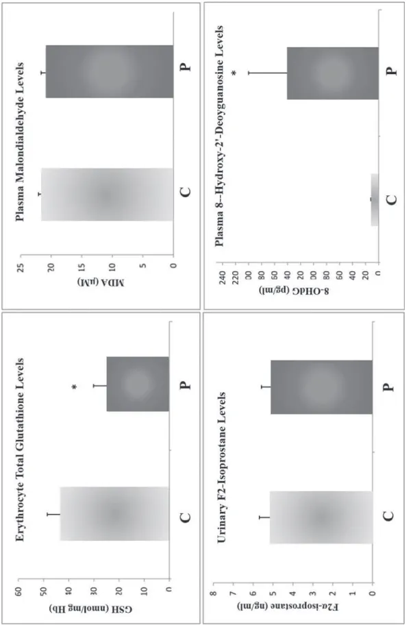
Erythrocyte total GSH levels were significantly lower (43%) in plastics workers vs. controls (24.51 ± 5.61 nmol/mg Hb vs. 42.82 ± 5.70 nmol/ mg Hb, P < 0.05). Plasma MDA levels did not mark-edly differ in plastics workers vs. controls; nor did urinary F2α isoprostane levels. However, 8-OHdG levels were ∼14-fold higher in plastics workers vs. controls (140 ± 60 pg/mL vs. 10 ± 2 pg/mL, P < 0.05) (Fig. 1).
G. Plasma DEHP and MEHP Levels
Plasma DEHP and MEHP levels were measured in only 17 samples as some plastics workers refused to give blood by the dropping method; for the same reason, only 26 control samples were measured. TABLE 4: Liver function parameters and fasting blood glucose levels in the study groups
Normal range Controls (n = 29) Plastic workers (n = 24)
ALT (U/L) 7–55 16.51 ± 1.39 24.70 ± 2.42*
AST (U/L) 8–48 16.96 ± 0.72 23.16 ± 1.23*
Direct bilirubin (mg/dL) 0–0.4 0.31 ± 0.03 0.29 ± 0.02
Total bilirubin (mg/dL) 0.3–1.0 0.69 ± 0.15 0.68 ± 0.13
Total protein (g/dL) 6–8.3 7.84 ± 0.54 8.00 ± 0.54
Fasting blood glucose (mg/dL) 70–100 80.69 ± 8.11 91.00 ± 12.77*
*P < 0.05 vs. control
TABLE 5: Selenium levels and antioxidant enzyme activity in the study groups Controls (n = 29) Plastic workers (n = 24)
Selenium (µg/L) 178.44 ± 6.07 148.73 ± 3.51* GPx1 (nmol/min/mg Hb) 0.56 ± 0.08 0.58 ± 0.09 GPx3 (U/mL) 23.27 ± 2.32 34.64 ± 2.22* SOD (U/mg Hb) 13.90 ± 0.36 15.57 ± 0.91 CAT (nmol/min/mg Hb) 363.00 ± 18.21 368.50 ± 20.89 TrxR (µmol/min/mL) 0.21 ± 0.06 0.17 ± 0.02
Note: GPx1, SOD, and CAT activity and selenium levels were measured in erythrocytes; GPx3 and TrxR activity was measured in plasma. *P < 0.05
FIG. 1: Erythrocyte tota l glutath ione, plasma malondialdehyde, urinary F2α-isoporstane, and plasma 8-hydroxy-2′-deoxyguanosine levels in the study groups. *
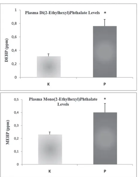
Plasma DEHP levels were significantly higher (2.5-fold) in plastics workers vs. controls (0.76 ± 0.10 ppm vs. 0.31 ± 0.04 ppm, P < 0.05), as expected. Plasma MEHP levels also were markedly increased (1.7-fold) in plastics workers vs. controls (0.40 ± 0.07 ppm vs. 0.23 ± 0.02 ppm, P < 0.05) (Fig. 2).
H. Plasma Correlations between Measured Parameters in Plastics Workers
Correlations between measured parameters in plas-tics workers are given in Table 6. GPx3 activity was
inversely correlated with working period in the plas-tics industry (ρ = −0.440, P < 0.05). Direct bilirubin levels were positively correlated with working pe-riod (ρ = 0.477, P < 0.05). Plasma ALT levels were positively correlated with plasma AST levels, as ex-pected (ρ = 0.466, P < 0.05).
Plasma testosterone levels were inversely cor-related with MEHP levels (ρ = −0.520, P < 0.01); plasma DEHP levels were positively correlated with plasma MEHP levels, as expected (ρ = 0.583, P < 0.05). Erythrocyte GSH levels were positively correlated with plasma LH (ρ = 0.495, P < 0.05) and plasma TrxR
lev-FIG. 2: Plasma di(2-ethylhexyl)phthalate and mono(2-ethylhexyl)phthalate levels in the study groups. *P < 0.05 vs. controls.
els (ρ = 0.513, P < 0.05). Urinary F2α isoprostane levels were inversely correlated with erythrocyte CAT activity (ρ = −0.469, P < 0.05) and positively correlated with plasma 8-OHdG levels (ρ = 0.413, P < 0.05). Eryth-rocyte SOD activity was positively correlated with erythrocyte GPx1 (ρ = 0.447, P < 0.05) and CAT (ρ = 0.724, P < 0.01) activities and inversely correlated with 8-OHdG levels (ρ = −0.541, P < 0.01).
I. Reconsideration of Measured Parameters According to Working Period in Plastics Workers
When working period was taken into account, plastics workers were divided into two groups:
P > 10 consisted of seven subjects who had worked in the plastics industry for more than 10 years; P < 10 consisted of ten subjects who had worked in the plastics industry for less than 10 years. In P >10, testosterone levels were sig-nificantly lower (18%) and ALT levels (34.6%) were significantly higher than those in P < 10. In addition, erythrocyte SOD activity (33%) and erythrocyte CAT activity (22.5%) were higher in P > 10 than in P < 10. Plasma DEHP (51%) and MEHP (76%) levels were also higher in P > 10 than in P < 10; however, due to low number of subjects within the subgroups, the differences in plasma phthalate levels were not statistically sig-nificant (Table 7).
TABLE 6: Correlations between measured parameters in plastics workers
Parameters Correlation coefficient
Working period GPx3 −0.440*,a
Direct bilirubin 0.477*,a
ALT AST 0.466*,b
Testosterone MDA −0.520**,a
DEHP MEHP 0.583*,b
GSH LH 0.495*,a
TrxR 0.518*,a
F2α-isoprostane CAT −0.469*,a
8-OHdG 0.413*,b SOD 8-OHdG −0.541**,a GPx1 0.447*,b CAT 0.724**,b *P < 0.05 ; **P < 0.01. aPearson; bSpearman
TABLE 7: Reconsideration of measured parameters according to working period in plastic workers Controls (n = 26) Plastic workers p < 10 (n = 10) Plastic Workersp > 10 (n = 7)
Testosterone (ng/dL) 442.94 ± 17.16 350.05 ± 29.8* 286 ± 30.0*.# ALT (U/L) 16.51 ± 1.39 23.18 ± 2* 31.2 ± 5.6*.# SOD (U/mg Hb) 13.90 ± 0.36 14.66 ± 0.91 19.51 ± 2.8*.# CAT (U/mg Hb) 363.00 ± 18.21 356.97 ± 22.9 437.2 ± 75.4*.# DEHP (ppm) 0.31 ± 0.04 0.7 ± 0.12* 1.06 ± 0.17* MEHP (ppm) 0.23 ± 0.02 0.33 ± 0.07* 0.58 ± 0.19* *P < 0.05 vs. controls; # p < 0.05 vs. P < 10 group
IV. DISCUSSION
Exposure to plasticizers from various sources is inevitable in today’s life. Phthalates are the most commonly used plasticizers in the PVC industry. Though phthalates have many advantages such as increasing the flexibility and durability of plastics, they are suggested to cause endocrine disruption and other toxic effects. DEHP is the most widely used phthalate derivative as it is highly effective in shaping PVC material.4,51–54 PVC workers are highly exposed to phthalates.26,32,33,54 All of the plastics workers in this study had been working in PVC in-dustry for more than two years (mean 8.17 ± 2.94 years) and were mainly exposed to DEHP. Although they may have been exposed to plasticizers through different routes, their main exposure seems to be the workplace.
Phthalates are suggested to cause liver toxicity in both rodents (peroxisome proliferation and finally hepatocarcinogenesis) and humans.12,17,55 However, it is not clear whether they cause peroxisome pro-liferation in the human liver given that the conse-quence of molecular events in the rodent liver after phthalate exposure does not seem to be operative in humans.11 We have observed that DEHP affects hepatic (ALT, AST) biomarkers in rodents.16 In the current study, we observed significant increases in both ALT and AST activities, which are important biomarkers of liver damage, in plastics workers vs. controls. However, levels of these hepatic bio-markers were in the normal range (ALT: 7–55 U/L; AST: 8–48 U/L), so we could not determine if liver damage was present. In addition, neither total/direct bilirubin nor total protein concentrations were sig-nificantly different between the two study groups. Nevertheless, fasting blood glucose levels were markedly higher in plastics workers vs. controls and ∼ 46% of the plastics workers had fasting blood glu-cose ≥ 100 mg/dL.
Wang et al. investigated the effects of occupa-tional DEHP exposure on plasma cholinesterase and renal and hepatic biochemical markers in 352 ex-posed workers and 104 controls. Regression anal-yses showed that plasma cholinesterase activity decreased markedly with increasing plasma DEHP concentrations. Serum ALT, AST, creatinine, urea,
gamma glutamyltransferase (GGT), MDA, total an-tioxidant, and C-reactive protein levels were signifi-cantly higher in workers vs. controls: 116 exposed workers (33.0%) had a daily DEHP intake of 22.7 μg/kg b.w./day, which was more than the 20 μg/kg b.w./day specified by the US Environmental Protec-tion Agency. The researchers suggested that DEHP could cause health hazards due to its effects on dif-ferent hepatic and renal biomarkers.55 It has been posited that there is a relationship between phthal-ates and type 2 diabetes56; our results indicate that high phthalate exposure could lead to high levels of fasting blood glucose, the importance of which needs to be evaluated.
Both plasma DEHP and MEHP levels in plastics workers were significantly higher when compared to controls in this study (2.5-and 1.7-fold, respec-tively). Though we did not measure oxidized me-tabolites of DEHP other than MEHP, the correlation between plasma DEHP and MEHP levels suggests that occupational exposure to high levels of DEHP can lead to high production of its metabolites, which are suggested to be more toxic than the parent com-pound. Fong et al. measured oxidized metabolites of DEHP [MEHP, mono(2-ethyl-5-oxohexyl) phthal-ate (MEOHP), and mono(2-ethyl-5-hydroxyhexyl) phthalate (MEHHP)] in preshift and postshift urine samples of 89 PVC workers (high-exposure group: 66 raw-materials workers; low-exposure group: 23 administrative workers). They also measured DEHP air concentrations. The geometric means of airborne concentrations of DEHP were 5.3 μg/m3 (low-ex-posure group) and 32.7 μg/m3 (high-exposure group) (P < 0.01). Analysis showed a consistent and marked correlation between airborne DEHP con-centrations and urinary DEHP metabolite concen-trations in the high-exposure group. The researchers suggested that absorbed airborne DEHP inhalation in the workplace caused a significant increase in to-tal-body DEHP burden in PVC workers.57
Phthalate exposure may affect serum hor-mone levels, although the results of various stud-ies are contradictory. In this study, plasma fT4 and testosterone levels were found to be significantly decreased in plastics workers when compared to controls. Plasma FSH levels were markedly lower as well. Grasso et al. suggested that both DEHP and
MEHP lead to disruption in FSH binding of Sertoli cell membranes in vitro. This phenomenon may cause unresponsiveness of Sertoli cells to FSH and finally higher circulating FSH levels.58 Agarwal et al. determined that benzyl butyl phthalate (BBzP) causes a significant decrease in plasma testosterone and a significant dose-dependent increase in LH and FSH in male rats. However, they observed no marked degenerative decreases in Leydig cells and therefore suggested that decreases in testosterone and increases in LH and FSH were due to unrespon-siveness in a “negative feedback system” with no apparent changes in the hypothalamus-pituitary-tes-tis (HPT) axis.58 Erkekoglu et al. also observed a marked increase in FSH in DEHP-exposed rats, most likely for the same reason.8
Pan et al. examined testosterone levels and uri-nary phthalate monoester concentrations in male workers (n = 74) exposed to DBP and DEHP at a factory producing unfoamed PVC flooring, and compared them with concentrations in male workers (n = 63) in a construction company group-matched for age and smoking status. Compared to the unex-posed workers, the exunex-posed workers had substan-tially and significantly elevated concentrations of the DBP main metabolite mono-n-butyl phthalate (MBP) (644.3 vs. 129.6 µg/g creatinine, P < 0.001) and MEHP (565.7 vs. 5.7 µg/g creatinine, P < 0.001). Free testosterone levels were significantly lower (8.4 µg/g creatinine vs. 9.7 µg/g creatinine, P = 0.019) in exposed workers than in unexposed workers and were negatively correlated with both MBP (r = −0.25, P = 0.03) and MEHP (r = −0.19, P = 0.095) in the phthalate-exposed workers. In re-gression analyses, free testosterone was found to decrease markedly with increasing total phthalate ester score (the sum of quartiles of MBP and MEHP; r = −0.26, P = 0.002). Pan et al. reported a modest but significant reduction in serum free testosterone in workers with higher levels of urinary MBP and MEHP compared to unexposed workers.33
In another study, Pan et al. examined associa-tions between HIs of DEP and DEHP exposure and serum reproductive hormone levels (free testoster-one, estradiol, LH, FSH) among occupationally ex-posed workers (n = 74) and unexex-posed construction workers (n = 63, matched for age and smoking
sta-tus) in China. The median HI value was 5.30 for the plastics workers, which was almost 53-fold higher when compared to the unexposed construction workers. The researchers found a significantly neg-ative association between HIs and free testosterone in exposed workers (r = −0.195, P = 0.096). How-ever, there was no association between HIs and free testosterone in the unexposed construction work-ers. The exposed workers had inverted long-tailed J-shaped free testosterone and FSH curves and small changes in the LH curve, whereas the unexposed construction workers showed inverted and flattened S-shaped free testosterone and mirror S-shaped LH and FSH curves. Both testosterone production and the hypothalamo-pituitary-testis (HPT) axis func-tion were found to be damaged in workers with high HIs of phthalate exposure.27
In a recent study, Fong et al. investigated the re-lationship between urinary metabolites of DEHP and reproductive hormones in male PVC plant workers (n = 82). There were marked positive relationships between urinary levels of DEHP metabolites and estradiol (P < 0.01) and in the ratio of estradiol to testosterone (P < 0.05) in multiple regression mod-els adjusted for potential confounders. In addition, quartile analysis showed significant positive rela-tionships between total urinary DEHP metabolite levels and estradiol (P trend = 0.024) and in the ratio of estradiol to testosterone (p trend = 0.031). Asso-ciations between reproductive hormones and total urinary DEHP levels in the PVC plant workers were markedly positive. Fong et al. suggested that these associations were a sign of incremental aromatase activity in male workers exposed to DEHP.35
The results of Kasai’s study on flexible-PVC workers (n = 69) showed that urinary concentrations of the oxidative metabolites of DEHP vary pre- and postshift [12.6 and 28.7 μg/L for MEHP, 38.6 and 84.4 μg/L for (5-carboxy-2-ethylpentyl) phthalate (5cx-MEPP) and 20.4 and 70.6 μg/L for 2-ethyl-hexanoic acid (2-EHA)]. These concentrations were also significantly higher than those in controls.61
Hines et al. recruited 156 workers in 2003–2005 from eight industry sectors in the United States to analyze occupational contributions to urinary phthalate metabolite levels and compare end-of-shift metabolite concentrations in workers to those
in the general population. Occupational exposure to DEHP was the highest in PVC film manufacturing, PVC compounding, and rubber boot manufacturing. End-of-shift concentrations of oxidative metabolites of DEHP were several-fold higher in the workers than in the general population. Occupational expo-sure to DBP was most evident in rubber gasket and phthalate (raw material) and rubber hose manufac-turing, with DBP metabolite concentrations highly exceeding those in the general population. Con-centrations of DEP and dimethylphthalate (DMP) metabolites in workers in phthalate manufacturing were also several folds higher than in the general population. Hines et al. suggested that urinary me-tabolite concentrations are good identifiers of work-places causing phthalate exposure.62
Another study by Hines et al. on workers in PVC film–processing (n = 25) and PVC custom com-pounding (n = 12) exposed to DiNP reported that urinary concentrations of mono(carboxy-isooctyl) phthalate (MCiOP, the primary metabolite of DiNP) ranged 0.42–80 μg/g creatinine in the film workers and 1.11–13.4 μg/g creatinine in the compounding workers. PVC film workers directly exposed to DiNP (n = 7) had the highest urinary MCiOP end-of-shift concentrations (25.2 μg/g creatinine) followed by those who worked on a shift where DiNP was used (n = 11) (17.7 μg/g creatinine). These concen-trations were significantly higher when compared to controls (2.92 μg/g) or to workers who had shift ex-posure to DiNP (2.08 μg/g creatinine).26
We observed a change in the intracellular redox state, as evidenced by increases in plasma GPx3 activity and decreases in erythrocyte GSH levels. GPx3, the main plasma GPx, has many important functions. It contains a selenocysteine (Sec) residue at its active site. This enzyme is a major scavenger of ROS in plasma and acts as a redox signal modula-tor. It mainly catalyzes reductions in H2O2, lipid per-oxides, and organic hydroperoxide using GSH. In different cancers, higher aberrant methylation of the GPx3 gene has been observed, leading to silencing, down-regulation of GPx3 expression, and incre-mental intracellular oxidation.37,38,43 The increases in GPx3 activity and decreases in GSH levels were possibly caused by higher ROS levels in plastics workers vs. controls. Although phthalates are not
considered oxidants, several pathways triggered by those substances as well as their oxidative metab-olites may lead to oxidative stress and may cause increments in liver function parameters.
One of the most important aspects of this study is the approximate 14-fold increase in 8-OHdG levels in plastics workers that we found. 8-OHdG has been widely used as a biomarker for oxidative stress.46,60,61,63 In a study conducted in Hunan Prov-ince, China, two study sites were chosen: an exposed site with a history of over 20 years of waste plastic recycling and a reference site without known DEHP pollution (about 50 km from the exposed site). TSH, MDA, and 8-OHdG levels as well as SOD activi-ties were measured in male recycling workers (n = 181) and gender- and age-matched farmers (n = 160). DEHP concentrations in water and cultivated soil samples and micronuclei frequency in human capillary blood lymphocytes were also determined. As expected, in different sources of water and soil, DEHP levels were greater at the recycling site than at the reference site (P < 0.05, all). The recycling workers had higher median levels of MDA compared with the farmers (3.80 vs. 3.14 nmol/mL) and lower SOD activity (112.15 vs. 123.82 U/mL; P < 0.01, all)—contrary to our findings. In addition, urinary 8-OHdG levels in the recycling workers were sig-nificantly higher than those in the farmers (340.37 vs. 268.18 μmol/mol creatinine)—in agreement with our findings. Multivariate analysis revealed that working in plastic recycling is an independent risk factor for increased urinary 8-OHdG concentrations in male workers (P < 0.01), in which, the researchers suggested, occupational DEHP exposure might con-tribute to oxidative DNA base damage.34
Testicular dysgenesis syndrome and cancer are highly related to phthalate exposure, which is also postulated to be responsible for early-onset testic-ular cancers by epigenetic and genetic effects.64–67 In a case-control study, Ohlson and Hardell exam-ined 148 cases of testicular cancer and 314 healthy controls. Among plastics workers exposed to PVC, there was a six-fold increase in the risk for semi-noma, one type of testicular cancer.68 It is well-known that DNA base modifications and damages are highly mutagenic and carcinogenic, so we can postulate that an underlying factor in testicular
can-cer caused by xenoestrogens, particularly phthalates, may be the increase in DNA damage. Previously, we determined that DEHP and MEHP can lead to DNA damage in human prostate cancer cells and mouse Leydig cells, most probably due to increased intra-cellular ROS production.13,69,70
In a study of PVC workers (n = 47) exposed to DEHP, sperm concentration and motility were markedly decreased and sperm ROS generation and apoptosis were significantly increased vs. controls (n = 15) and were positively correlated with MEHHP and MEOHP concentrations, respectively.37
An interaction between selenium and phthal-ates has been suggested in both in vitro and in vivo studies. The bioavailability of selenium has been observed to be decreased by MEHP, and selenium supplementation has been proposed to ameliorate the toxic effects of DEHP and MEHP both in vitro and in vivo.13–17,69–71 In this study, selenium levels in plastics workers were 17% lower than in controls, suggesting that DEHP exposure may deplete eryth-rocyte selenium, which is a good indicator of long-term selenium availability.72
We found several significant correlations be-tween the parameters measured in plastics work-ers. As expected, plasma ALT and AST levels (ρ = 0.466) as well as plasma DEHP and MEHP levels were highly correlated (ρ = 0.583). On the other hand, the positive correlation between plasma F2α-isoprostane and 8-OHdG levels and the nega-tive correlation between SOD activity and 8-OHdG levels indicate that as oxidative stress increases, 8-OHdG may increase. In addition, SOD and GPx1 activity and SOD and CAT activity were positively correlated. These correlations suggest that important components of the antioxidant system have highly interactive relationships.
We observed that plastics working years signifi-cantly affect testosterone and ALT levels as well as SOD and CAT activity. Decreasing testosterone lev-els can be a sign of declining fertility. However, more hormone levels (including dehydroepiandrosterone and its sulfate form DHEA/S) and sperm parame-ters should be measured in order to confirm this. The marked increases in SOD and CAT activity in plas-tics workers with more than 10 years in the industry mainly suggest increasing oxidative stress. Because
ALT levels were significantly higher in these workers as well (when compared to workers with less than 10 years in the industry), we suggest that chronic plas-ticizer exposure can also affect the liver, leading to changes in liver biochemistry if not liver damage.
Our study has limitations. For one, the sam-ple size was relatively low. For another, DEHP and MEHP levels were not measured in all plastics workers because some were unwilling to have blood drawn by the dropping method and others were un-willing to state their working period in the plastics industry. In spite of its limitations, this study is so far the most comprehensive investigation of the effects of DEHP in exposed plastics workers. More work should be done to explicate workers’ deteriorating health throughout their working years as a result of DEHP exposure. Case-control studies with large numbers of subjects are needed to show the undesir-able effects of phthalates used in the PVC industry. They should be mechanistic, particularly regarding the interaction between phthalates and DNA, which is one of the most important findings of our work here—that DNA base damage levels were higher in plastics workers than in controls.
Because mutations and cancer mainly originate from DNA base damage, prospective studies should highlight the relationship between chronic exposure to plasticizers and carcinogenicity. Our aim is to study different phthalate derivatives and compare their chronic toxic effects on plastics workers.
REFERENCES
1. Wormuth M, Scheringer M, Vollenweider M, Hunger-buhler K. What are the sources of exposure to eight frequently used phthalic acid esters in Europeans? Risk Anal. 2006;26:803e24.
2. Ticker JA, Schettler T, Guidotti T, McCally M, Rossi M. Health risks posed by use of di-ethylhexyl phthalate, DEHP, in PVC medical devices, a critical review. Am J Ind Med. 2001;39:100e11.
3. Taiwan Food and Drug Administration. Report. May 28. 2011. Available from: http//www.fda.gov.tw/news.aspx-?newssnZ7641&key_
4. Hauser R, Calafat AM. Phthalates and human health. Oc-cup Environ Med. 2005;62:806–18.
5. Kay VR, Chambers C, Foster WG. Reproductive and de-velopmental effects of phthalate diesters in females. Crit Rev Toxicol. 2013; 43:200–19.
6. Balbuena P, Campbell J Jr, Clewell HJ 3rd, Clewell RA. Evaluation of a predictive in vitro Leydig cell assay for anti-androgenicity of phthalate esters in the rat. Toxicol in vitro. 2013; 27:1711–18.
7. Christiansen S, Boberg J, Axelstad M, Dalgaard M, Ving-gaard AM, Metzdorff SB, Hass U. Low-dose perinatal exposure to di, 2-ethylhexyl. phthalate induces anti-an-drogenic effects in male rats. Reprod Toxicol. 2010;30: 313–21.
8. Erkekoglu P, Zeybek ND, Giray B, Asan E, Arnaud J, Hincal F. Reproductive toxicity of di-(2-ethylhexyl) phthalate in selenium-supplemented and selenium-defi-cient rats. Drug Chem Toxicol. 2011;34:379–89. 9. Kortenkamp A, Faust M. Combined exposures to
an-ti-androgenic chemicals, steps towards cumulative risk assessment. Int J Androl. 2010;33:463–74.
10. Svechnikov K, Svechnikova I, Söder O. Inhibitory ef-fects of mono-ethylhexyl phthalate on steroidogenesis in immature and adult rat Leydig cells in vitro. Reprod Toxicol. 2008;25:485–90.
11. IARC (International Agency for Research on Cancer). Some chemicals present in industrial and consumer prod-ucts, food and drinking-water [monograph]. In: IARC monographs on the evaluation of carcinogenic risks to humans. Lyon, France: International Agency for Research on Cancer; 2013. Available from: http://monographs.iarc. fr/ENG/Monographs/vol101/mono101-006.pdf
12. Rusyn I, Peters JM, Cunningham ML. Modes of action and species-specific effects of di-, 2-ethylhexyl.phthalate in the liver. Crit Rev Toxicol. 2006;36:459–79.
13. Erkekoglu P, Rachidi W, Yüzügüllü OG, Giray B, Oz-türk M, Favier A, Hıncal F. Induction of ROS, p53, p21 in DEHP- and MEHP-exposed LNCaP cells—pro-tection by selenium compounds. Food Chem Toxicol. 2011;49:1565–71.
14. Erkekoglu P, Zeybek ND, Giray BK, Rachidi W, Kızıl-gün M, Hininger-Favier I, Favier A, Asan E, Hincal F. The effects of di, 2-ethylhexyl.phthalate exposure and selenium nutrition on Sertoli cell vimentin structure and germ-cell apoptosis in rat testis. Arch Environ Contam Toxicol. 2012;62:539–47.
15. Erkekoglu P, Giray BK, Kizilgun M, Hininger-Favier I, Rachidi W, Roussel AM, Favier A, Hincal F. Thyroi-dal effects of di(2-ethylhexyl) phthalate in rats of dif-ferent selenium status. J Environ Pathol Toxicol Oncol. 2012;31:143–53.
16. Erkekoglu P, Giray BK, Kızilgün M, Rachidi W, Hininger-Favier I, Roussel AM, Favier A, Hincal F. Di, 2-ethylhexyl.phthalate-induced renal oxidative stress in rats and protective effect of selenium. Toxicol Mech Methods. 2012;22:415–23.
17. Erkekoglu P, Zeybek ND, Giray BK, Rachidi W, Kızılgün M, Hininger-Favier I, Favier A, Asan E, Hincal F. The ef-fects of di, 2-ethylhexyl.phthalate on rat liver in relation to selenium status. Int J Exp Pathol. 2014;95:64–77.
18. Buluş AD, Aşci A, Erkekoglu P, Balci A, Andiran N, Koçer-Gümüşel B. The evaluation of possible role of endocrine disruptors in central and peripheral preco-cious puberty. Toxicol Mech Methods. 2016;26:493– 500.
19. Dong R, Zhao S, Zhang H, Chen J, Zhang M, Wang M, Wu M, Li S, Chen B. Sex differences in the asso-ciation of urinary concentrations of phthalate metab-olites with self-reported diabetes and cardiovascular diseases in Shanghai adults. Int J Environ Res Public. 2017;14(6):598. doi: 10.3390/ijerph14060598.
20. Durmaz E, Aşçı A, Erkekoğlu P, Akçurin S, Gümüşel BK, Bircan I. Urinary bisphenol A levels in girls with idiopathic central precocious puberty. J Clin Res Pediatr Endocrinol. 2014;6:16–21.
21. Durmaz E, Ozmert EN, Erkekoglu P, Giray B, Derman O, Hincal F, Yurdakök K. Phthalate-induced oxidative stress and association with asthma-related airway inflam-mation in adolescents. Int J Hyg Environ Health. 2017; 220:468–77.
22. Franken C, Lambrechts N, Govarts E, Koppen G, Den Hond E, Ooms D, Voorspoels S, Bruckers L, Loots I, Nelen V, Sioen I, Nawrot TS, Baeyens W, Van Larebeke N, Schoeters G. Phthalate-induced oxidative stress and association with asthma-related airway inflammation in adolescents. Int J Hyg Environ Health. 2017; 220: 468–77.
23. Kondolot M, Ozmert EN, Ascı A, Erkekoglu P, Oztop DB, Gumus H, Kocer-Gumusel B, Yurdakok K. Plasma phthalate and bisphenol A levels and oxidant-antioxidant status in autistic children. Environ Toxicol Pharmacol. 2016;43:149–58.
24. Stojanoska MM, Milosevic N, Milic N, Abenavoli L.The influence of phthalates and bisphenol A on the obesity development and glucose metabolism disorders. Endo-crine. 2017;55:666–81.
25. Allsopp M, Santillo D, Johnston P. Hazardous chemicals in carpets. Exeter, UK: Greenpeace Research Labora-tories; 2001. Technical Note 01/2001. Available from: http//www.greenpeace.to/publications/carpet.pdf 26. Hines CJ, Nilsen Hopf NB, Deddens JA, Calafat AM,
Silva MJ, Grote AA, Sammons DL. Occupational expo-sure to diisononyl phthalate, DiNP, in polyvinyl chloride processing operations. Int Arch Occup Environ Health. 2012;85: 317–25.
27. Pan G, Hanaoka T, Yu L, Na J, Yamano Y, Hara K, Ichiba M, Nakadate T, Kishi R, Wang P, Yin H, Zhang S, Feng Y. Associations between hazard indices of di-n-butyl-phthalate and di-2-ethylhexyldi-n-butyl-phthalate exposure and serum reproductive hormone levels among occupation-ally exposed and unexposed Chinese men. Int J Androl. 2011;34:e397–e406.
28. Ansari KN. The free radicals—the hidden culprits—an update. Indian J Med Sci. 1997;51: 319–36.
29. Gutteridge JM, Halliwell B. Free radicals and antioxi-dants in the year 2000. A historical look to the future. Ann NY Acad Sci. 2000;899:136–47.
30. Halliwell B. Biochemistry of oxidative stress. Biochem Soc Trans. 2007;35:1147–50.
31. Halliwell B. Free radicals, proteins and DNA, oxidative damage versus redox regulation. Biochem Soc Trans. 1996;24:1023–27.
32. Hayes JD, McLellan LI. Glutathione and glutathi-one-dependent enzymes represent a co-ordinately regu-lated defence against oxidative stress. Free Radic Res. 1999;31:273–300.
33. Pan G, Hanaoka T, Yoshimura M, Zhang S, Wang P, Tsukino H, Inoue K, Nakazawa H, Tsugane S, Taka-hashi K. Decreased serum free testosterone in workers exposed to high levels of di-n-butyl phthalate, DBP and di-2-ethylhexyl phthalate, DEHP, a cross-sectional study in China. Environ Health Perspect. 2006;114: 1643–8.
34. Wang Q, Wang L, Chen X, Rao KM, Lu SY, Ma ST, Jiang P, Zheng D, Xu SQ, Zheng HY, Wang JS, Yu ZQ, Zhang R, Tao Y, Yuan J. Increased urinary 8–hydroxy-2′-deox-yguanosine levels in workers exposed to di-, 2-ethyl-hexyl.phthalate in a waste plastic recycling site in China. Environ Sci Pollut Res Int. 2011;18:987–96.
35. Fong JP, Lee FJ, Lu IS, Uang SN, Lee CC. Relation-ship between urinary concentrations of di(2-ethylhexyl. phthalate, DEHP, metabolites and reproductive hor-mones in polyvinyl chloride production workers. Occup Environ Med. 2015;72:346–53.
36. Huang LP, Lee CC, Fan JP, Kuo PH, Shih TS, Hsu PC. Urinary metabolites of di,2-ethylhexyl.phthalate relation to sperm motility, reactive oxygen species generation, and apoptosis in polyvinyl chloride workers. Int Arch Occup Environ Health. 2014;87:635–46.
37. Lubos E, Loscalzo J, Handy DE. Glutathione perox-idase-1 in health and disease, from molecular mech-anisms to therapeutic opportunities. Antioxid Redox Signal. 2011;15:1957–97.
38. Takahashi K, Cohen HJ. Selenium-dependent gluta-thione peroxidase protein and activity, immunological investigations on cellular and plasma enzymes. Blood. 1986;68:640–5.
39. Mustacich D, Powis G. Thioredoxin reductase. Biochem J. 2000;346(Pt 1):1–8.
40. Holmgren A. Thioredoxin structure and mechanism, con-formational changes on oxidation of the active-site sulf-hydryls to a disulfide. Structure. 1995;3:239–43. 41. Fukai T, Ushio-Fukai M. Superoxide dismutases, role in
redox signaling, vascular function, and diseases. Anti-oxid Redox Signal. 2011;15: 1583–606.
42. Zamocky M, Furtmüller PG, Obinger C. Evolution of catalases from bacteria to humans. Antioxid Redox Sig-nal. 2008;10:1527–48.
43. Lemire J, Alhasawi A, Appanna VP, Tharmalingam S, Appanna VD. Metabolic defense against oxidative stress, the road less travelled—so far. J Appl Microbiol. 2017 Oct;123(4):798–809. doi: 10.1111/jam.13509
44. Ayala A, Muñoz MF, Argüelles S. Lipid peroxidation, production, metabolism, and signaling mechanisms of malondialdehyde and 4-hydroxy-2-nonenal. Oxid Med Cell Longev. 2014;2014:360438.
45. Fairbanks V, Klee GG. Biochemical aspects of hematol-ogy. In: Tietz NW, editor. Textbook of clinical chemistry. Philadelphia: W.B. Saunders; 1986. p. 1532–4.
46. Milne GL, Yin H, Morrow JD. Human biochemistry of the isoprostane pathway. J Biol Chem. 2008;283:15533–7. 47. Valavanidis A, Vlachogianni T, Fiotakis C. 8-hydroxy-2′
-deoxyguanosine (8-OHdG), a critical biomarker of oxidative stress and carcinogenesis. J Environ Sci Health C Environ Carcinog Ecotoxicol Rev. 2009; 27:120–39. 48. Baltaci AK, Mogulkoc R, Akil M, Bicer M. Review:
se-lenium—its metabolism and relation to exercise. Pak J Pharm Sci. 2016;29:1719–25.
49. Kirkbright GE. Atomic absorption spectroscopy. In: Ele-mental analysis of biological materials, current problems and techniques with special reference to trace elements. Vienna Technical Report Series 197. Vienna: Interna-tional Atomic Energy Agency; 1980. pp 141–65. 50. Paris I, Ruggieri F, Mazzeo P, Carlucci G.
Simultane-ous determination of dimethylhexyl phthalate and mono ethyhlhexyl phthalate in human plasma by HPLC. Anal Lett. 2003;36:2649–58.
51. Blass CR. PVC as a biomedical polymer—plasticizer and stabilizer toxicity. Med Device Technol. 1992;3:32–40. 52. Bowman JD, Choudhury M. Phthalates in neonatal health,
friend or foe? J Dev Orig Health Dis. 2016;7:652–64. 53. Shea KM, American Academy of Pediatrics
Commit-tee on Environmental Health. Pediatric exposure and potential toxicity of phthalate plasticizers. Pediatrics. 2003;111:1467–74.
54. Skinner MK. Endocrine disruptors in 2015, epigene-tic transgenerational inheritance. Nat Rev Endocrinol. 2016;12: 68–70.
55. Wang W, Xu X, Fan CQ. Health hazard assessment of occupationally di-2-ethylhexyl.phthalate-exposed work-ers in China. Chemosphere. 2015;120: 37–44.
56. Song Y, Chou EL, Baecker A, You NC, Song Y, Sun Q, Liu S. Endocrine-disrupting chemicals, risk of type 2 dia-betes, and diabetes-related metabolic traits. A systematic review and meta-analysis. J Diabetes. 2016;8:516–32. 57. Fong JP, Lee FJ, Lu IS, Uang SN, Lee CC. Estimating the
contribution of inhalation exposure to di-2-ethylhexyl phthalate, DEHP, for PVC production workers, using personal air sampling and urinary metabolite monitoring. Int J Hyg Environ Health. 2014;217:102–9.
58. Grasso P, Heindel JJ, Powell CJ, Reichert LE Jr. Effects of mono(2-ethylhexyl) phthalate, a testicular toxicant, on
follicle-stimulating hormone binding to membranes from cultured rat Sertoli cells. Biol Reprod. 1993;48:454–9. 59. Agarwal DK, Maronpot RR, Lamb JC 4th, Kluwe WM.
Adverse effects of butyl benzyl phthalate on the repro-ductive and hematopoietic systems of male rats. Toxicol-ogy. 1985;35:189–206.
60. Gaudin R, Marsan P, Ndaw S, Robert A, Ducos P. Bio-logical monitoring of exposure to di,2-ethylhexyl.phthal-ate in six French factories, a field study. Int Arch Occup Environ Health. 2011;84: 523–31.
61. Kasai H. What causes human cancer? Approaches from the chemistry of DNA damage. Genes Environ. 2016;38:19. 62. Hines CJ, Nilsen Hopf NB, Deddens JA, Calafat AM, Silva
MJ, Grote AA, Sammons DL. Urinary phthalate metabolite concentrations among workers in selected industries, a pi-lot biomonitoring study. Ann Occup Hyg. 2009;53: 1–17. 63. Poulsen HE, Nadal LL, Broedbaek K, Nielsen PE,
Wei-mann A. Detection and interpretation of 8-oxodG and 8-oxoGua in urine, plasma and cerebrospinal fluid. Bio-chim Biophys Acta. 2014;1840:801–8.
64. Singh S, Li SS. Epigenetic effects of environmental chemicals bisphenol A and phthalates. Int J Mol Sci. 2012;13:10143–53.
65. Rusyn I, Corton JC. Mechanistic considerations for human relevance of cancer hazard of di,2-ethylhexyl. phthalate. Mutat Res. 2012;750:141–58.
66. Erkekoglu P, Kocer Giray B. Unpredictable effects of chemical mixtures on liver in health and disease. J Liver Dis Transplant. 2012;1:1.
67. Hu GX, Lian QQ, Ge RS, Hardy DO, Li XK. Phthal-ate-induced testicular dysgenesis syndrome, Leydig cell influence. Trends Endocrinol Metab. 2009; 20: 139–40.
68. Ohlson CG, Hardell L. Testicular cancer and occu-pational exposures with a focus on xenoestrogens in polyvinyl chloride plastics. Chemosphere. 2000;40: 1277–82.
69. Erkekoğlu P, Rachidi W, De Rosa V, Giray B, Favier A, Hincal F. Protective effect of selenium supplementation on the genotoxicity of di,2-ethylhexyl.phthalate and mono,2-ethylhexyl.phthalate treatment in LNCaP cells. Free Radic Biol Med. 2010;49:559–66.
70. Erkekoglu P, Rachidi W, Yuzugullu OG, Giray B, Favier A, Ozturk M, Hincal F. Evaluation of cy-totoxicity and oxidative DNA damaging effects of di-(2-ethylhexyl) phthalate, DEHP and mono-(2-eth-ylhexyl-phthalate), MEHP on MA-10 Leydig cells and protection by selenium. Toxicol Appl Pharmacol. 2010; 248:52–62.
71. Garberg P, Högberg J. Selenium metabolism in isolated hepatocytes, inhibition of incorporation in proteins by mono,2-ethylhexyl.phthalate, a metabolite of the peroxi-some proliferator di,2-ethylhexyl.phthalate. Carcinogen-esis. 1991;12:7–12.
72. Stefanowicz FA, Talwar D, O’Reilly DS, Dickinson N, Atkinson J, Hursthouse AS, Rankin J, Duncan A. Eryth-rocyte selenium concentration as a marker of selenium status. Clin Nutr. 2013;32: 837–42.