The Analysis of Glutathione Level and Superoxide Dismutase Activity in Normal and
Preeclampsia Pregnant
NAİM UZUN1*
1Agri Ibrahim Cecen University, Faculty of Pharmacy, Department of Clinical Pharmacy, Agri, Turkey
Abstract
Preeclampsia is a disease characterized by hypertension and significant proteinuria that occurs after the twentieth week of pregnancy. The presence of mechanisms that detoxify free oxygen radicals associated with increased oxygen consumption in preeclampsia is very important. In mitochondrial and microsomal systems, molecular oxygen is reduced by taking an electron and superoxide radical is formed which is an unstable structure. Superoxide Dismutase is a metalloenzyme which converts superoxide radical to Hydrogen Peroxide. Glutathione Peroxidase degrades hydrogen peroxide by converting it into water and molecular oxygen using glutathione. Twenty-seven preeclampsia patients and 32 healthy pregnant control groups were selected at 20-28 weeks of gestation. There was a significant difference (p <0.05) between Superoxide dismutase and glutathione levels in patients with preeclampsia and control group. We aimed to investigate the effect of two important components of the antioxidant system, superoxide dismutase and glutathione, on preeclampsia causing fetal and maternal mortality.
Keywords: Preeclampsia, Superoxide Dismutase,
Glutathione.
Received: 09.01.2019 Revised: 21.01.2019 Accepted:06.03.2019
*Corresponding author: Naim Uzun, PhD
Agri Ibrahim Cecen University, Faculty of Pharmacy Clinical Pharmacy, Agrı Turkey
E-mail: [email protected]
Cite this article as: N. Uzun, The Analysis of Glutathione Level and Superoxide Dismutase Activity in Normal and Preeclampsia Pregnant, Eastern Anatolian Journal of Science, Vol. 5, Issue 1, 23-29,2019
Introduction
Preeclampsia is an early and definite manifestation of hypertension, proteinuria and edema (Azman 2009), pregnancy-specific, mainly young and first child pregnant women, after 20 weeks of pregnancy, 5-10% of pregnancies, intrauterine growth retardation, premature birth, fetal and maternal mortality disease (Berköz 2009, Altunhan 2011). In the clinic this condition is also called 'pregnancy toxemia'. (Berköz 2009).
Proteinuria is an indicator of glomerular damage in preeclampsia. Detection of 300 mg or more protein in 24-hour urine; In the case of at least two urine samples taken at 6 hours or more, more than one proteinuria is sufficient for the diagnosis of pathological proteinuria (Azman 2009). The first symptom in preeclampsia is edema. Edema is mostly seen on the face and extremities. However, edema has no definitive diagnostic value (Çelik 2012).
Preeclampsia is the most common cause of perinatal and maternal morbidity and mortality in many parts of the world (Sağol; Özkınay 2000). Preeclampsia is generally known as the disease of young and non-nulliparous women. A woman who had preeclampsia in a pregnancy had a recurrence risk of 3% in subsequent pregnancies (Azman 2009).
Preeclampsia is more common in women with some risk factors (Azman 2009). Risk factors for preeclampsia; null parity, maternal age> 35 years, black race, family history, history of preeclampsia in previous pregnancies, chronic hypertension, obesity, renal diseases, connective tissue diseases, vascular diseases, antiphospholipid syndrome, thrombophilia, pregestational diabetes mellitus, molar (plural) pregnancy, in pregnancy trophoblastic disease, fetal hydrops, polyhydramnios, smoking. Preeclampsia is less common in smoking women than in non-smokers, but fetal risk is higher if pregnant women with preeclampsia smoke (Altunhan 2011).
Complications depend on the week of pregnancy and other medical problems. Complications can be divided into two main categories: fetal and maternal. Fetal Complications; fetal growth retardation, perinatal death, premature labor, oligohydramnios, fetal asphyxia. Maternal Complications; convulsions, acute renal failure, heart failure, pulmonary edema, intracranial hemorrhage, blindness, liver subcapsular hematoma and rupture, thrombocytopenia, disseminated intravascular coagulation, HELLP syndrome (Azman 2009). HELLP (Hemolysis; Elevated Liver Enzymes; Low Platelets) syndrome is classified as a subgroup of severe preeclampsia with high mortality and morbidity (Baha 2002).
Pathophysiology often includes inadequate trophoblastic invasion, placental ischemia, generalized vasospasm, abnormal hemostasis associated with activation of the coagulation system, vascular endothelial dysfunction, abnormal nitric oxide and abnormal lipid metabolism, abnormal leukocyte activation, changes in cytokines, and insulin resistance (Dekker 1998).
Increased oxygen demand during pregnancy causes an increase in the production of free oxygen radicals. Major antioxidant enzymes in the placenta are Superoxide Dismutase, Catalase, Glutathione Peroxidase, Glutathione Reductase and Glucose-6-Phosphate Dehydrogenase. It has been shown that production of lipid peroxides increases in preeclamptic pregnancies, enzymatic (superoxide dismutase, glutathione peroxidase) and non-enzymatic (Vitamin C and E) antioxidants decrease (Berköz 2009). The cause of the increase of oxidative stress caused by the placenta was evaluated as hypoxia and ischemia during spinal artery structuring (Saito et al 1990). Free radicals cause damage to membranes, proteins and DNA, and increase cell death in trophoblasts. Increased cell death in placentas of preeclamptic pregnants has been shown (Hazar 2012).
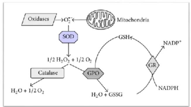
Reduction of molecular oxygen in mitochondrial and microsomal systems creates a superoxide radical which is unstable. SOD is a metalloenzyme which converts superoxide to hydrogen peroxide. (Memişoğulları 2005; Rencüzoğulları 2006). Superoxide is converted by SOD into Hydrogen Peroxide; Hydrogen Peroxide is converted into water and molecular oxygen by Catalase and Glutathione Peroxidase (Figure 1) (Koch et al. 2014).
Oxidant-Antioxidant System consists of production of superoxide radicals, production of hydrogen peroxide, production of hydroxyl radical, inactivation of hydrogen peroxide.
Figure 1. Schematic representation of the endogenous
defense system against oxidative stress.SOD, Superoxide Dismutase; GPO, Glutathione Peroxidase; GR, Glutathione Reductase; GSH / GSSG, reduced / oxidized glutathione.
Glutathione (GSH) is a tripeptide known as γ-L-glutamyl-L-cysteinyl-glycine. Glutathione contains two important biological structures (thiol group and γ-glutamine base) (Figure 2). Glutathione, an important antioxidant due to the thiol group and high cell internal concentration, is present in more than 99% reduced form. The ability to keep in this form is dependent on Glutathione Reductase and NADPH produced in the Pentose phosphate pathway (Thorburn 1985). Total glutathione is present in the cells in free form or depending on the proteins. Free glutathione is present in reduced state, in the case of oxidative stress, it is converted to oxidized form and then reduced again by the enzyme glutathione reductase. The redox state depends on the relative amounts of the reduced and oxidized forms of glutathione (GSH / GSSG), a critical finding for the cell. GSH / GSSG ratio in the cell in resting state exceeds 100, this rate falls between 1 and 10 in case of exposure to oxidative stress (Kasap 2010).
Figure 2. Structure of reduced glutathione; glutamate
is linked in an isopeptide bond (via its γ-carboxyl group) to cysteine, which in turn forms a peptide linkage with glycine (Ziglari T and Allameh A. 2013).
Glutathione has been shown to play an important role in the pathogenesis and progression of diseases such as HIV infection, cirrhosis, respiratory diseases, gastrointestinal and pancreatic inflammation, diabetes, neurological diseases, aging, preterm delivery, intrauterine growth retardation, preeclampsia (Wu et al. 2004). In addition, glutathione plays a role in the detoxification of hydrogen peroxide, other peroxides, free radicals and a wide variety of xenobiotics (Pastore et al. 2003). Glutathione is not only responsible for protecting cells from oxidative damage but also allows the sulfydryl groups required for normal functions of proteins to be reduced (Bharath et al. 2002).
Materials and Methods
Pregnant women with preeclampsia who were diagnosed with preeclampsia and healthy pregnant women with the same gestational week were included in the study. Pregnant women who had a systemic
disease, consumed cigarette and alcohol, used drugs for different reasons and had gestational diabetes were not included in the study.
Venous blood samples were collected from 26 preeclampsia patients and 32 healthy pregnant women with age ranges between 22 and 38 years. 5 ml of peripheral venous blood was collected from the two groups of subjects (patient group and control group) after hospitalization and before delivery. Samples were allowed to stand at room temperature for 30 minutes and then were separated by centrifugation at 3000 r / min for 20 min. It was noted that the selected samples were not hemolyzed. Serums were maintained at -20 ° C until the working time. All samples and test kits were brought to room temperature before working. Protein analyzes were performed with manual Micro Lowry Sigma kit and superoxide dismutase and gluttayon ELISA method. Statistical analysis was performed using the SPSS package program (version 22.0) (SPSS Inc. Chicago, USA) and Microsoft Excel (Windows 10, Office 2013). Analysis of variance was determined by Kruskal-Wallis. Mann-Whitney test was used for nonparametric analysis. Data were calculated according to p˂0.05 significance values according to mean and standard deviations.
Before starting the study, approval was obtained from Atatürk University Medical Faculty Ethics Committee.
Results
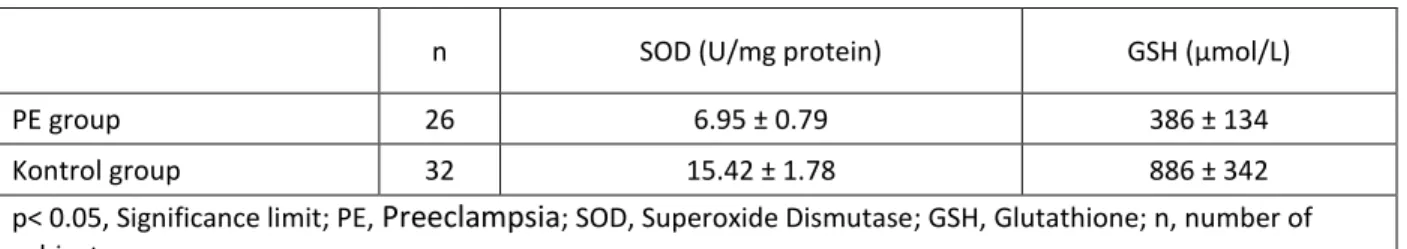
The results of the analysis of the research are given in Table 1.
Table 1: Preeclampsia and control group analysis results.
n SOD (U/mg protein) GSH (μmol/L)
PE group 26 6.95 ± 0.79 386 ± 134
Kontrol group 32 15.42 ± 1.78 886 ± 342
p< 0.05, Significance limit; PE,
Preeclampsia
; SOD, Superoxide Dismutase; GSH, Glutathione; n, number of subjects.There was a significant difference between serum Superoxide Dismutase levels in patients with Preeclampsia and control group; there was a significant difference between serum Glutathione levels in patients with Preeclampsia and control group (p <0.05).
Discussion
Oxidative damage progresses according to the balance between reactive oxygen species produced and antioxidant species (Murray 2009). Antioxidant molecules can be divided into two groups as enzymatic and non-enzymatic. Superoxide Dismutase (SOD), Glutathione Peroxidase (GPO) and Catalase enzymatic; Glutathione (GSH), vitamin C, vitamin E, carotenes, etc. are non-enzymatic basic antioxidants (Murray 2009, Taşcan 2014). While SOD plays an important role in the detoxification of O2•- , GSH is the first protection against endogenous and exogenous reactive oxygen species. GSH is a molecule that plays a major role in the reduction of cellular proteins (Taşcan 2014).
The oxidative balance continues during pregnancy. Preeclamptic pregnant women fail to develop this compensatory mechanism. Shanklin et al. (1989) showed that preeclamptic patients had higher levels of membrane lipids than healthy pregnant patients, and this damage is more pronounced in the severe preeclampsia group than in the mild preeclampsia group, and there is a close relationship between the severity of lipid injury and the clinical severity of preeclampsia. In preeclamptic women, especially in severe preeclampsia, antioxidant capacity is reduced (Chamy et al. 2006). It has been reported that antioxidant vitamin (Vitamin C and E) (Murray 2009) supplementation with protective roles against oxidative damage of the cell membrane can prevent
high endothelial dysfunction and reduce the incidence of preeclampsia (Chappell 1999).
The normal process in pregnancy is that it can be compensated by an increase in antioxidant defenses parallel to an increase in free radical production, but lipid-peroxidation is significantly increased in patients with preeclampsia (Altunhan 2011). Prostaglandin H2 synthesis in endothelial cells (Hoffbrand 2016) increases the oxygen radicals resulting from peroxidation and combines with the oxygen radicals formed by the polyunsaturated fatty acids to stimulate lipid peroxidation (Wang et al. 1991). Placental Glutathione Peroxidase inhibits prostaglandin H2 in normal pregnancy but increases the levels of lipid peroxidation in placental Glutathione peroxidase deficiency (Walsh; Wang 1993).
The need for selenium, the cofactor of glutathione peroxidase, increases during pregnancy and lactation. In most body fluids, selenium levels decrease with the progression of pregnancy. Depending on the level of decreased selenium, the amount of plasma Glutathione and Glutathione Peroxidase activity is particularly reduced from the middle of the second trimester of pregnancy (Mihailovic 2000). Glutathione in Plasma Reduction in peroxidase activity is fast, while erythrocytes are slow. Because plasma thiols may be reduced during pregnancy due to increased plasma volume; Erythrocyte lysate thiols increase with pregnancy. Plasma and erythrocyte thiols have important functions as non-specific oxidative stress buffers. GSH (glutathione) is the most important erythrocyte lysate thiol. Erythrocyte glutathione levels do not change in the first two trimesters, but in late pregnancy, erythrocyte lysate thiol levels decrease in pregnant women with preeclampsia. The erythrocyte Glutathione peroxidase activity during pregnancy has been reported to remain unchanged and the change in glutathione level has
been reported to be independent of Glutathione Peroxidase (Walsh 2000).
Superoxide dismutase (SOD) has been observed to increase in normal pregnancy. The increase in serum SOD activity begins from the first months of pregnancy. SOD has the function of protecting embryos against lipid peroxidation, especially in the early stages of gestation. In women with preeclampsia, serum SOD activity is lower than in normal pregnant women. In placental homogenization, the SOD level is low in the early stages of pregnancy but may increase by 2-3 times with the progression of pregnancy. The reason for the change in placental SOD levels during gestational period is due to the change in placental oxygen requirement during pregnancy. The placental SOD activity in placenta-related pathologies was lower than in normal pregnant women (Walsh 2000). In the study performed by Taşcan (2014), serum SOD activity in preeclampsia groups was found to be statistically significantly higher compared to the control group. However, the literature also reported that SOD activity in preeclampsia was higher, lower and no significant differences than control group. In a clinical study of 21 patients diagnosed with preeclampsia, antioxidant enzyme (SOD, CAT, GPO) activities and MDA levels were increased; there is no contradiction between the increase in both MDA growth and its inhibitory enzymes. Because all antioxidant enzymes, all isoforms of these enzymes and all non-enzymatic antioxidants should be taken into consideration. As a result, it is stated that simultaneous rise in both SOD and GPO and MDA values is predictable (Alaçam 2008).
Gheorghe et al. (2007) found a significant decrease in erythrocyte MDA and SOD and GPO activities in patients with preeclampsia and a significant decrease in erythrocyte GSH, ascorbic acid, plasma vitamin E levels and plasma CAT activities. The researchers evaluated the increase in antioxidant enzyme activity as a compensatory reaction to oxidative stress. As a result, they suggested that excessive intrauterine hypoxia would cause a reperfusion oxidative damage, which may cause abnormalities in trophoblast development. Hoffman et al. (2008) predicted that increased amounts of reactive oxygen species would result in placental abnormalities in pregnancy and this would result in maternal symptoms in preeclampsia.
In studies investigating the status of lipid peroxidation and antioxidant enzymes in pregnancy-induced hypertension patients, thiobarbutiric acid (TBARS) level was significantly increased and SOD, CAT, GPO and glutathione reductase activities decreased significantly. With these findings, the researchers suggested that oxidative stress in preeclampsia increases with no doubt (Patil et al. 2007).
In our study, there was a significant difference between serum Superoxide Dismutase levels in patients with Preeclampsia (6.95 ± 0.79 U/mg protein) and control group (15.42 ± 1.78 U/mg protein); there was a significant difference between serum Glutathione levels in patients with Preeclampsia (386 ± 134 μmol/L) and control group (886 ± 342 μmol/L) (p <0.05).
Bainbridge et al. (2007) in a study of patients with preeclampsia hydrogen peroxide, nitrite, peroxinitrite, lipid peroxides and glutathione S-transferase activity is high; superoxide anion, SOD, CAT and Glutathione Reductase activities are low; there was no difference in vitamin E, reduced glutathione and oxidized glutathione concentrations have achieved. Based on these results, it has been claimed that patients with preeclampsia are under an oxidative stress caused by the inability of the oxidant-antioxidant system to stabilize.
It has been hypothesized that low glutathione concentrations and antioxidant vitamin status, lipid peroxidation are important causal factors in the pathogenesis of preeclampsia in patients with hypertension due to pregnancy (Krishna and Venkataramana 2007). The intracellular enzyme SOD levels are suggested to be similar in healthy pregnancies and in complicated pregnancies; it was reported that there was no difference in plasma levels of SOD activity between pregnancies that did not develop complications and pregnancies that resulted in preterm delivery (Hsieh et al. 2012).
Preeclampsia can cause morbidity and mortality in maternal and neonatal. It is often emphasized that oxidative stress is present in its pathogenesis. Increased oxygen demand, especially in pregnant women, causes an increase in free oxygen radicals and therefore more severe oxidative damage in preeclamptic pregnants. Further clinical and biochemical research is needed to better understand the relationship between oxidative stress and perinatal
disease. There is a need for a sensitive biochemical marker to prevent PE in the early stages and allow for clinical interventions.
References
ALAÇAM, H. , 2008. Preeklampside Asimetrik Di
Metil Arjinin (Adma) Ve
Oksidan/Antioksidan Sistemin Rolü. Hacettepe Üniversitesi Tıp Fakültesi Biyokimya Anabilim Dalı. Uzmanlık Tezi 2008: 51-
ALTUNHAN, H. , 2011. Preeklamptik Anne Bebeklerinde Total Oksidan Seviye, Total Antioksidan Seviye ve Paraoksonaz Düzeyleri. Selçuk Üniversitesi Meram Tıp Fakültesi Çocuk Sağlığı Ve Hastalıkları Anabilim Dalı Neonatoloji Bilim Dalı, Uzmanlık Tezi 2011: 28-32
AZMAN, P. G., Haziran 2009. Kliniğimizde 2004– 2009 Yılları Arasında Doğum Yapmış Olan Preeklampsi Vakalarının Retrospektif Değerlendirilmesi Ve Mpv Değerinin Preeklampsinin Şiddetini Öngörmedeki Yeri. Haydarpaşa Numune Eğitim Ve Araştırma Hastanesi Kadın Hastalıkları Ve Doğum Kliniği. Uzmanlık Tezi 2009: 5
BAHA M.SIBAI. Hypertension. (In): Gabbe SG, Niebly JR, Simpson JL, eds. Obstetrics Normal and problem pregnancies 4 th ed.Churcill Livingstone, 2002:945- 1004 BAINBRIDGE SA, ROBERTS JM. Uric Acid as a
Pathogenic Factor in Preeclampsia. Placenta. 2008; 29:S:67-72.
BERKÖZ, M. VE YALIN, S. , 2009. Normal ve Preeklamptik Gebelerde Lipid Peroksidasyonu Ve Antioksidan Aktivite. Mersin Üniversitesi Eczacılık Fakültesi, Biyokimya Anabilim Dalı, Mersin, Türkiye. ADÜ Tıp Fakültesi Dergisi 2009; 10(2) : 53 - 58
BHARATH, S. , HSU, M., KAUR, D., RAJAGOPALAN, S., ANDERSEN, J.K. (2002). Glutathione, iron and Parkinson’s disease. Biochemical Pharmacology, 64: 1037-1048.
CHAMY VM, LEPE J, CATALÁN A, RETAMAL D, ESCOBAR JA, MADRID EM. 2006. Oxidative stress is closely related to clinical
severity of pre-eclampsia. Biol Res. 2006;39(2):229-36.
CHAPPELL LC, SEED PT, BRILEY AL, KELLY FJ, LEE R, HUNT BJ, PARMAR K, BEWLEY SJ, SHENNAN AH, STEER PJ, POSTON L (1999) Effect of antioxidants on the occurrence of pre-eclampsia in women at increased risk: A randomised trial. Lancet 354: 810-816
ÇELIK, E. KARAER, A. YILMAZ, E. TÜRKÇÜOĞLU, I. ÇELIK, Ö. FILMLEK, Y. KIRICI, P. ÖZEROL, E. TANBEK, K. , 2012.İkinci trimesterde amniyotik sıvıda oksidatif stres belirteçlerinin düzeyleri ile preeklampsi gelişimi ve erken doğum arasındaki ilişkinin araştırılması. İnönü Üniversitesi Tıp Fakültesi Kadın Hastalıkları ve Doğum Anabilim Dalı, Malatya. Perinatoloji Dergisi 2012;20(3):140-145. DEKKER GA, SIBAI BM. Pathogenesis and etiology
of preeklampsia. Am J Obstet Gynecol 1998; 179: 1359.
GHEORGHE CP, MOHAN S, OBERG KC. 2007. Gene expression patterns in the hypoxic murine placenta: a role in epigenesis? Reproductive Sciences. 2007; 14, 3.
HAZAR, D. , 2012. Ağır Preeklampside Vasküler Endotelyal Büyüme Faktörü, Solubl Fms Benzeri Tirozin Kinaz-1 Ve Endotelin Düzeyleri Ve Bunların Birbirleri İle Olan İlişkileri. E Üniversitesi Tıp Fakültesi Kadın Hastalıkları Ve Doğum Anabilim Dalı. Uzmanlık Tezi 2012: 19-26.
HOFFBRAND A. V., MOSS P. A. H. 2016. Hoffbrand’s Essential Haematology. Seventh Edition. John Wiley & Sons Ltd. USA. HSIEH TT, CHEN SF, LO LM, LI MJ, YEH YL,
HUNG TH. The association between maternal oxidative stress at midgestation and subsequent pregnancy complications. Reprod Sci 2012; 19: 505-12.
KASAP, Y. , 2010. Glutatyon (Gsh) Düzeyinin Plasentada Araştırılması. Ankara Üniversitesi Sağlık Bölümleri Enstitüsü. Yüksek Lisans Tezi 2010; 1-29.
KOCH, K. HAVERMANN, S. BÜCHTER, C. , 2014. Caenorhabditis Elegans As Model System. İn Pharmacology And Toxicology: Effects Of Flavonoids On Redox-Sensitive
Signalling Pathways And Ageing, nstitute of Agricultural and Nutritional Sciences, Martin-Luther-Universität Halle-Wittenberg, Weinbergweg 22 (Biozentrum), 06120 Halle, Germany. Figure 2
KRISHNA MOHAN, S. AND
VENKATARAMANA, G. Status of lipid peroxidation, glutathione, ascorbic acid, vitamin E and antioxidant enzymes in patients with pregnancy--induced hypertension. Indian J Physiol Pharmacol. 2007; 51: 284-288.
MEMIŞOĞULLARI, R. , 2005. Diyabette Serbest Radikallerin Rolü ve Antioksidanların Etkisi. AİBÜ Düzce Tıp Fakültesi. Düzce Tıp Fakültesi Dergisi 2005; 3: 30-39
MIHAILOVIC M, CVETKOVIC M, LJUBIC A, et al. Selenium and malondialdehyde content and glutathione peroxidase activity in maternal and umbilical cord blood and amniotic fluid. Biol Trace Elem Res 2000; 73: 47-54.
MURRAY R. K., BENDER D. A., BOTHAM K. M., KENNELLY P. J., RODWELL V. W., 2009. Harper’s Illustrated Biochemistry. Twenty-Eighth Edition. a LANGE medical book. The McGraw-Hill Companies.
PASTORE A, FEDERICI G, BERTINI E, PIEMONTE F. 2003. Analysis of glutathione: implication in redox and detoxification. Clin Chim Acta. 2003 Jul 1;333(1):19-39.
PATIL SB, KODLIWADMATH MV,
KODLIWADMATH SM. Role of lipid peroxidation and enzymatic antioxidants in pregnancy-induced hypertension. Clin Exp Obstet Gynecol. 2007; 34: 239-41.
RENCÜZOĞULLARI N. Ratlarda deneysel olarak oluşturulan kadmiyum toksikasyonu üzerine likopenin etkilerinin araştırılması. Hatay, Mustafa Kemal Üniversitesi Sağlık Bilimleri Enstitüsü Biyokimya (Vet) Anabilim Dalı, 2006
SAĞOL S, ÖZKINAY E. Preeklampsi etyopatogenezinde lipid peroksidasyonu. Türkiye Klinikleri Jinekolojik Obstetrik Dergisi 2000; 10: 7-15.2002:112-9 (7-9) SAITO S, SSHIOZAKI A, NAKASHIMA A, SAKAI
M, SASAKI Y.The rol of immune system in
preeclampsia. Mol Aspec Med. 1990; 28: 192-209 34.Zusterzeel PL, Rutten H, Roelofs HM, Peters WH, Steegers EA. Protein carbonyls in decidua and placenta of pre-eclamptic women as markers for oxidative stree. Placenta. 2001; 22: 213-219(5-32) TAŞCAN A. , 2014. Preeklampsili Gebelerde
Oksidatif/Nitrozatif Stresin Araştırılması. Gaziantep Üniversitesi Sağlık Bilimleri Enstitüsü. Yüksek Lisans Tezi 2014; 1-40. THORBURN DR, KUCHEL PW. Regulation of the
Human Erythrocyte Hexose Monophosphate Shunt Under Conditions of the Oxidative Stress. Eur J Biochem 1985; 150: 371-86 WALSH S. W., VAUGHAN J. E., WANG Y.,
ROBERTS L. J.. 2000. Placental isoprostane is significantly increased in preeclampsia. 2000; 14; 10.
WALSH SW, WANG Y. 1993. Deficient glutathione peroxidase activity in preeclampsia is associated with increased placental production of thromboxane and lipid peroxides. Am J Obstet Gynecol;169:1456-61.
WANG Y, WALSH SW, GUO J, ZHANG J. 1991. The imbalance between thromboxane and prostacyclin in preeclampsia is associated with an imbalance between lipid peroxides and vitamin E in maternal blood. Am J Obstet Gynecol;165:1695-700.
WU, G. FANG, Y.Z. , YANG, S. , LUPTON, J.R. , TURNER, N.D. (2004).Glutathione metabolism and its implications for health. J Nutr;134(3):489-92.
ZIGLARI T, ALLAMEH A. (2013) The significance of glutathione conjugation in aflatoxin metabolism. - Aflatoxins-Recent Advances and Future. intechopen.com.