NUMERICAL ANALYSIS OF SDS-PAGE PROTEIN PATTERNS
OF FACULTATIVE ALKALIPHILIC BACILLUS SPECIES
ISOLATED FROM LAKE VAN, TURKEY
İsmet Berber1 and Şerife Berber21Department of Biology, Faculty of Arts and Science, Yüzüncü Yıl University, 65080 Van, Turkey 2Departments of Biometrics and Genetics, The Institute of Science, Yüzüncü Yıl University, 65080 Van, Turkey
SUMMARY
In the present study, seven reference Bacillus species and a total of eighteen new facultative alkaliphilic
Bacil-lus strains, isolated from the water of Lake Van and its soil
surroundings, were identified using phenotypic charac-teristics and the numerical analysis of whole-cell protein profiles. According to morphological, biochemical and physiological characteristics, it was found that the new isolated strains belonged to Bacillus genus. In addition, the research indicated that SDS-PAGE of polypeptides of whole-cell extracts can provide more valuable taxonomic information than conventional tests based on the pheno-typic characteristics at the species and subspecies levels. Numerical analysis of whole-cell protein profiles of test strains revealed 4 basic clusters (I-IV) at dissimilarity val-ues of 18.9 % or above. The results of numerical analysis confirmed that each cluster had characteristic and distinc-tive protein profiles. Indeed, this study showed that the application of numerical analysis, coupled with the utiliza-tion of a standardized identificautiliza-tion system instead of sim-ple quantitative comparison of protein patterns, greatly enhanced the utilization of whole-cell protein profiles for identification of the facultative alkaliphilic Bacillus species.
KEYWORDS: Lake Van, Bacillus spp., numerical analysis,
protein profiles, phenotypic characterization, SDS-PAGE.
INTRODUCTION
Lake Van, the world’s largest soda lake (volume 607 km3, area 3570 km2, maximum depth 450 m, lake level
1648 m above sea level, continental climate) is located on the Anatolian high plateaus in eastern Turkey (38.5ºN and 43ºE). It has a pH of 9.7-9.8, and a salinity of 21.7‰ con-tributed to in equal shares by NaCl and sodium carbon-ates, and with minor contributions from sulfate, potassium
and magnesium [1]. Soda lakes are highly alkaline aquatic environments containing a number of alkaliphilic bacte-ria [2]. Up to now, many of the microorganisms character-ized from soda lakes have relatives in salt lakes, except those being alkaliphilic or, at least, highly alkali-tolerant [3]. It was reported that Bacillus genera consist of alkaliphilic strains, some of which are Gram-positive, endospore- form-ing, aerobic and facultative anaerobic [4]. In the last dec-ade, the taxonomical studies on alkaliphilic Bacillus strains are rising due to possessing valuable and commercially interesting enzymes [5].
The conventional identification and classification meth-ods based on morphological, physiological and biochemi-cal properties can clearly lead to misclassification in some bacterial taxa. Due to this fact, the development and use of new molecular methods for improving the identification and detection of microorganisms are advisable [6, 7].
Some researchers have studied identification and char-acterization of alkaliphilic Bacillus strains based on the phenotypic characteristics, DNA-DNA relatedness data, and phylogenetic analysis of the 16S rRNA sequence [8-11]. Although these methods have been used for classifi-cation of alkaliphilic Bacillus species, the characterization of these microorganisms is still considered to be compli- cated [6, 12].
Protein electrophoresis has been of great value for the delineation of numerous bacterial taxa. The primary level of information for identification of bacteria is given by nucleo-tide base sequencing. Second level information is given by the cellular proteins, and different types of electrophoresis are used to explore relationship at this level [13]. SDS elec-trophoresis in a discontinuous system is, by far, the most widely used electrophoretic technique in bacterial systemat-ics. The protein profiles produced by SDS-PAGE of whole cells and extra-cellularly by bacteria have been observed to correlate closely with DNA-DNA hybridization results, and to be suitable for rapid bacterial identification [14-17].
Computer-aided numerical analysis of protein patterns has a high potential in microbial systematics. High resolu-tion polyacrylamide gel electrophoresis (PAGE) of proteins with computerized analysis of profiles provided an effec-tive approach to the investigation of taxonomic relation-ships among many bacterial species [18, 19]. The aim of the present study is to identify seven reference Bacillus species and eighteen new alkaliphilic Bacillus strains, isolated from the water of Lake Van and the soil of its surroundings. The strains were identified, based on phenotypic characteris-tics and the numerical analysis of whole-cell protein pro-files.
MATERIALS AND METHODS Bacteria and growth conditions
The reference bacteria used in this study were provided from various researchers: B. megaterium GCM 1842, B.
megaterium DSM 32 and B. cereus ATCC 7064 from Prof.
Dr. Cumhur Cokmus (Department of Biology, Faculty of Sciences, Ankara University, Tandoğan 06100 Ankara, Turkey), B. sphaericus MRS 400 from Prof. Dr. Allan A. Yousten (VPI & State University, Blacksburg, VA, USA), and B. firmus ATCC 14573, B. pseudofirmus OF4 and B.
alcalophilus ATCC 27647 from Dr. Arthur A. Guffantı and
Dr. Terry A. Krulwich (Department of Biochemistry, Mont Sinai School of Medicine of the City University of New York, 10029 New York, USA). The nonalkaliphilic strains were cultivated at 30 °C and pH 7.5 for 24 hrs in Nutrient Yeast Salt Broth (NYSM) medium consisting of 0.8% nu-trient broth (Difco), 0.05% yeast extract (Difco), CaCl2 x
2H2O - 7.0 x 10⁻⁴ M, MnCl2 x 4 H2O - 5.0 x 10⁻⁵ M, and
MgCl2 x 6 H2O - 1.0 x 10⁻³ M, whereas the alkaliphilic
strains were propagated at 30 °C and pH 7.5 for 24 hrs in Peptone Yeast Alkali Broth (PYA) medium consisting of 1% soluble starch (Difco), 0.5% polypeptone (Difco), 0.5% yeast extract (Difco), 0.1% K2HPO4, 0.02% MgSO4 × 7H2O,
and 0.5% Na2CO3. The solution of Na2CO3 were autoclaved
separately and added to the medium. Isolation of alkaliphilic Bacillus spp.
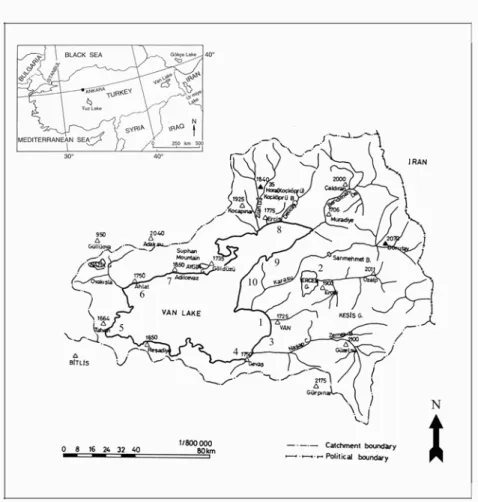
New facultative alkaliphilic Bacillus strains were iso-lated from the water of Lake Van and the soil of its sur-roundings (Figure 1) by using the isolation procedure of Horikoshi and Akiba [20], and PYA Agar medium (1% soluble starch (Difco), 0.5% polypeptone (Difco), 0.5% yeast extract (Difco), 0.1% K2HPO4, 0.02% MgSO4 × 7H2O,
0.5 % Na2CO3, 2% Agar, pH 10.5).
FIGURE 1 - Location of Lake Van, Turkey and map of the sampling points.
Physiological and morphological properties of the strains For the phenotypic test studies, the following refer-ence strains were used: B. megaterium GCM 1842, B.
megaterium DSM 32, B. sphaericus MRS 400, B. cereus
ATCC 7064, B. firmus ATCC 14573, B. pseudofirmus OF4, and B. alcalophilus ATCC 27647. New isolated strains in the present study were identified by conventional micro-biological methods [20-22]. The morphology of vegetative cells and sporangia, and the shape and position of spores were observed under a phase contrast microscope (Nikon, Japan). In addition, the following phenotypic tests were performed: motility, catalase and oxidase test; anaerobic growth; Voges-Proskauer test; methyl red test; gas produc-tion from glucose; degradaproduc-tion of starch, urea and casein; acid from D-glucose, L-arabinose, D-xylose, D-mannitol, fructose, galactose, maltose, lactose, inulin and sucrose; de-gradation of tyrosine; deamination of phenylalanine; egg-yolk lecithinase; nitrate reduced to nitrite; formation of indole; H2S production; DNase test; utilization of citrate;
NaCl and KCl required; growth at pHs 6.8 and 5.5 of nutri-ent broth; growth in NaCl 2%, 5%, 7% and 10%; and growth at 10, 30, 40, 45 and 50 ºC.
Antibiotic sensitivity
Sensitivity to antibiotics was determined by using the routine diffusion plate technique. Reference Bacillus strains (Nutrient Yeast Salt Agar medium at pH 7.5) and alkaliphilic strains (Peptone Yeast Alkali Broth medium at pH 7.5) were grown overnight at 30 °C, and used to prepare suspensions with optical density of 0.5 McFarland Standard (1.5 x 10⁸
cells per mL). 100 µL of each test bacterium was plated onto the agar, and disks containing antibiotics were placed onto the surface of the medium. After overnight-incubation at 30°C, the diameters of the zones of growth inhibition were measured. The following antibiotics were used (µg/ disk): imipenem (10 µg), ceftizoximine (30 µg), ciproflox-acine (5 µg), cefodizime (30 µg), ofloxciproflox-acine (5 µg), amox-icillin and clavulanic acid (10 µg), and sulbactam/ ampicil-lin (20 µg).
Extraction of whole-cell proteins from bacteria
All test strains were, at least, propagated in duplicate to prepare the synchronous culture. For each synchronous culture, 100 µL was inoculated into 15 mL NYSM or PYA broth, and incubated under rotation for 48 hrs (at 30 °C, 150 rpm). Each sample was centrifuged for 5 min at 12, 100 rpm, and the pellet collected was resuspended in 200 µL of CelLyticTM B-II Bacterial Cell Lysis/Extraction Reagent
(Sigma) [23]. The suspension was incubated for 30 min at room temperature. Afterwards, the sample was again cen-trifuged and 80 µL from each sample was transferred into a new 1.5 mL Eppendorf tube. Then, 25 µL of SDS-samples` buffer (0.06 M Tris-HCl, 2.5% glycerol, 0.5% SDS, 1.25% β-mercaptoethanol) was added and the whole mixture was vortexed to ensure good homogenization. The prepared samples were kept on a boiling water bath for 5 min and denaturated proteins stored at -70 °C until electrophoresis.
SDS-PAGE
Solubilized proteins were subjected to SDS-PAGE in gel slabs of 1 mm thickness (3.5 cm, 4% stacking and 15.5 cm, 12% resolving gels) as described by Laemmli [24]. Electrophoresis was performed with a discontinuous buffer system in an UVP Vertical Electrophoresis Unit (Cam-bridge, UK). The gel was run at 30 mA, until the bromo-phenol blue marker had reached the bottom of the gel. Protein molecular masses were calculated on the basis by comparison with the following standards (Prestained SDS-PAGE Standards, High Range, BIO-RAD): myosin (201 kDa), β-galactosidase (118 kDa), bovine serum al-bumin (83 kDa), and ovalal-bumin (48 kDa). After electro-phoresis, the gels were rinsed out for 20 min in an isopro-panol-acetic acid-water (1:3:6) solution, then for 5 min in methanol-acetic acid-water (3:1:6) solution. Then, the gels were stained for 6 hrs in 0.01 % (w/v) Coomassie Bril-liant Blue R-250. Afterwards, the gels were destained in a methanol-acetic acid-water (3:1:6) mixture, until protein bands became clearly visible.
Statistical analysis of protein profiles
The gels were scanned via densitometer (Desaga CD-60 Densitometer, Germany), and the molecular weight of each band was determined by one-dimensional analysis software (Lab Image Version 2.6, Halle, Germany). Data were coded as 0 (absent) and 1 (present). A hierarchical cluster analysis was performed using the average linkage method with the squared Euclidean distances [25]. The dendrogram, based on the whole-cell protein patterns of the test strains, was constructed by the program SPSS for Windows, version 12.0 (Chicago, Ill., USA).
RESULTS AND DISCUSSION
In the present research, eighteen facultative alkaliphilic
Bacillus strains isolated from the water of Lake Van and
its surrounding soil were examined. Morphological and physiological characteristics revealed that the new strains were all members of Bacillus genus. Morphological, physio-logical and biochemical properties of newly isolated strains are presented in Table 1. The data show that all strains are Gram-positive, aerobic rod, endospore-forming, facultative alkaliphilic, peritrichously flagellate, motility and catalase- positive. Spores were generally ellipsoidal without paraspo-ral crystals, and located centparaspo-ral and terminal, swelling in the young sporangium, except two strains numbered by 57 and 107. The strains were cultivated in nutrient broth at pHs between 6.8 and 10.5, and optimum pH was found to be 9. This case indicated that all strains were faculta-tive alkaliphilic. All the new strains were able to grow at temperature from 20 °C to 40 °C. The optimum growth temperature was 30 °C, but no growth was observed under 10 °C and above 50 °C. The native strains showed salt tol-erance at NaCl concentrations upto 5%. But, the strains numbered by 70, 74, 103 and 105, did not grow at 5% NaCl salinity. Other phenotypic characteristics of the strains were
determined to have the particular ability to reduce nitrate, utilization of citrate, acid formation from various carbo-hydrates, hydrolysis of casein, starch and urea, H2S
pro-duction, gas production from glucose, methyl red and Vo-ges-Proskauer tests, and DNase and lecithinase activities (listed in Table 1). The results of antibiotic tests indicated
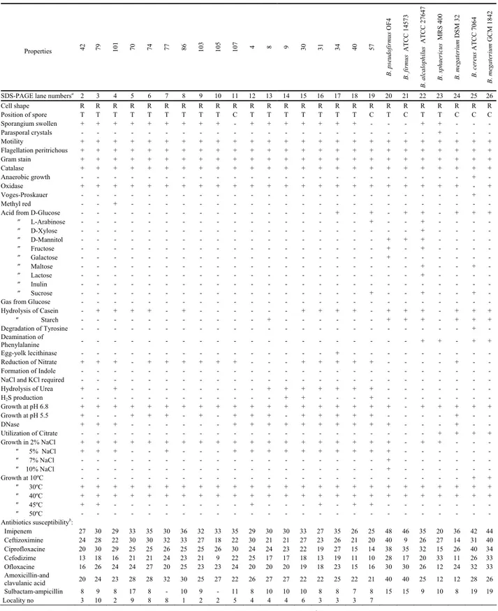
TABLE 1 - Morphological and phenotypical properties of the native and reference Bacillus strains.
Properties 42 79 101 70 74 77 86 103 105 107 4 8 9 30 31 34 40 57 B. pseudofi rmus O F 4 B. firmus ATCC 1457 3 B. alcaloph ilus A T CC 27647 B. sphaeric us MR S 40 0 B. megateri um DS M 3 2 B. cereus A T CC 70 64 B. megateri um GC M 1 842
SDS-PAGE lane numbersa 2 3 4 5 6 7 8 9 10 11 12 13 14 15 16 17 18 19 20 21 22 23 24 25 26
Cell shape R R R R R R R R R R R R R R R R R R R R R R R R R Position of spore T T T T T T T T T C T T T T T T T C T C T T C C C Sporangium swollen + + + + + + + + + - + + + + + + + - - - + + - - - Parasporal crystals - - - + - - - Motility + + + + + + + + + + + + + + + + + + + + + + + + + Flagellation peritrichous + + + + + + + + + + + + + + + + + + + + + + + + + Gram stain + + + + + + + + + + + + + + + + + + + + + + + + + Catalase + + + + + + + + + + + + + + + + + + + + + + + + + Anaerobic growth - - - + - Oxidase + + + + + + + + + + + + + + + + + + + + + + + - + Voges-Proskauer - - - + - Methyl red - - + - - -
Acid from D-Glucose - - - + - + - + + - + + +
″ L-Arabinose - - - + - - + - - - - ″ D-Xylose - - - + - - - - ″ D-Mannitol - - - + + + - - - - ″ Fructose - - - + - + - - - - ″ Galactose - - - + - - - ″ Maltose - - - + - - + - ″ Lactose - - - + - - - - ″ Inulin - - - ″ Sucrose - - - + - - + - - + -
Gas from Glucose - - -
Hydrolysis of Casein - + + + + - + - - - - + + + + - + + + - + + + ″ Starch - - - + - - - + + + - + + + Degradation of Tyrosine - - - + - Deamination of Phenylalanine - - - + + - + + Egg-yolk lecithinase - - - + - - - Reduction of Nitrate + + + - + + + + + + - - - + + + + + - - - - + - - Formation of Indole - - -
NaCl and KCl required - - -
Hydrolysis of Urea + - + - - - + + + + + + + - - - H2S production - - - + + - - - + - - - Growth at pH 6.8 + + + + + + + + + + + + + + + + + + + - + + + + + Growth at pH 5.5 + - - + + + - + - + + + - + + + + + - - - - + - - DNase + + + - - - + + + + + + + + + + - - - + - - Utilization of Citrate - - - + + + + Growth in 2% NaCl + + + + + + + + + + + + + + + + + + + - + + - - - ″ 5% NaCl + + + - - + - - - + + + + + + + + + + - - - - - - ″ 7% NaCl - - - + - - - ″ 10% NaCl - - - + - - - Growth at 10ºC - - - + + ″ 30ºC + + + + + + + + + + + + + + + + + + + + + + + + + ″ 40ºC + + + + + + + + + + + + + + + + + + + + + + + + + ″ 45ºC + + - - - + - - - + - + + - - - ″ 50ºC - - - Antibiotics susceptibilityb: Imipenem 27 30 29 33 35 30 36 32 33 35 29 30 30 33 27 35 26 25 48 46 35 20 36 42 44 Ceftizoximine 24 28 22 30 30 32 33 27 18 22 30 21 21 27 23 26 21 20 40 9 26 27 14 31 40 Ciprofloxacine 20 30 29 25 25 26 25 25 26 30 24 24 23 22 19 27 15 14 38 35 32 15 26 40 34 Cefodizime 13 18 16 21 21 24 23 21 9 22 25 17 17 18 13 19 11 10 28 17 20 33 11 26 33 Ofloxacine 16 26 24 24 27 20 25 23 23 24 20 20 20 19 18 23 15 16 30 30 26 12 24 32 33 Amoxicillin-and clavulanic acid 20 24 23 28 28 32 30 25 27 22 26 27 27 22 22 25 22 21 40 40 25 12 12 28 26 Sulbactam-ampicillin 8 9 8 17 8 - 10 9 - 11 8 10 10 10 8 8 7 8 15 15 9 10 8 19 19 Locality no 3 10 2 9 8 8 1 2 2 5 4 4 4 6 3 3 3 7
that reference Bacillus strains and native isolates had al-most equal sensitivity against the tested antibiotics. Never-theless, native strains were fairly resistant against sulbac-tam-ampicillin antibiotic.
The analysis of whole-cell protein profiles of the ref-erence and new facultative alkaliphilic Bacillus strains, obtained by one-dimensional denaturing gel electrophore-sis, are shown in Figure 2. The protein profiles of tested bacterial strains were inspected visually and compared with each others. The protein profiles of the test strains
revealed considerable differences; however, there were some similar protein bands between new strains and refer-ence Bacillus strains. The test strains have curiously dis-tinctive protein bands in the range of molecular weights be-tween 83-201kDa.
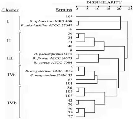
The numerical analysis of the whole-cell protein pro-files used for average linkage method with the squared Euclidean distances yielded a dendrogram, consisting of four basic clusters (I-IV) at dissimilarity values of 18.9 % or above (Figure 3). Cluster I comprised B. sphaericus
FIGURE 2 - SDS-PAGE of whole-cell protein profiles of the reference and the native Bacillus strains. Lane1, molecular
weight standards (kDa); lanes 2-19, the native strains; lines 20-26, the references strains. Lanes are identified in Table 1a.
FIGURE 3 - Grouping of the reference and the native Bacillus strains studied using cluster analysis (the squared Euclidean distances and average linkage clustering method) based on whole-cell protein profiles.
MRS 400, B. alcalophilus ATCC 27647 and two new facultative alkaliphilic strains, numbered as 4 and 107, at dissimilarity level between 0.8 % and 20 %. Cluster II had 5 facultative alkaliphilic strains numbered as 9, 30, 31, 34 and 40. The dissimilarity levels of the members of this group changed between 0.9 % and 12.4 %. The third clus-ter included three reference strains (B. pseudofirmus OF4,
B. firmus ATCC 14573, and B. cereus ATCC 7064), which
had 10 % of maximum distance. Cluster IV was divided in two subclusters (IVa and IVb) including B. megaterium GCM 1842, B. megaterium DSM 32 and 11 new strains (8, 42, 57, 70, 74, 77, 79, 86, 101, 103 and 105). Those strains were similar to B. megaterium GCM 1842 or B. megaterium DSM 32. Besides, the members of this cluster had the high-est similarity of protein profiles.
A number of researchers have reported that the strains of the genus Bacillus are recognized as being more pheno-typically heterogeneous than most other bacterial genera [26]. Truly, this genus exhibits a wide diversity of physio-logical ability, since the members of the genus are able to live in very different kinds of habitats. There is a diverse group of Bacillus species living in highly-alkaline terres-trial and aquatic environments. In the past decade, a full revision of alkaliphilic Bacillus classification was marked according to their phylogenetic and phenotypic character-istics [12, 21, 27]. The species of alkaliphilic and nonalka-liphilic Bacillus genera are notoriously difficult to be iden-tified by traditional methods, based on their physiological and morphological properties [6, 7, 12].
According to the present study, morphological, bio-chemical and physiological characteristics indicated that the new isolated strains are closely related to Bacillus ge-nus. The strains had almost similar biochemical characteris-tics. For instance, they did not utilize much carbohydrate for growth, except for glucose. Although the new isolated strains have been extensively investigated as a representa-tive strain of facultarepresenta-tive Bacillus, their taxonomic position is not yet known. As a result, it was determined that the conventional tests based on the phenotypic characteristics were insufficient for the differentiation of the native iso-lates.
Great attention has been given to protein electropho-resis, for the identification of a number of bacterial genera [14]. It is also widely acknowledged that the electropho-retic separation of cellular proteins is a sensitive technique, which mainly provides information on the similarity of the strains at and below the species level. The results of this study obviously showed that the electrophoretic method can provide more valuable information than phenotypic meth-ods at and below the species levels, and may be used for the discrimination of some new isolated and other Bacillus strains. The results are in good agreement with previous studies, which observed that some Bacillus species displayed distinct band variation in their SDS-PAGE whole-cell pro-tein patterns at the species and subspecies level [28, 29].
Analysis of whole-cell protein profiles of the Bacillus strains is useful for characterizing these microorganisms at the species and below the species level. However, com-puter-aided numerical analysis of protein patterns has pro-vided more useful information at subspecies level [14, 17]. In the present study, SPSS program was used to analyze the data because of the difficulties in the visual interpreta-tion of the bands, obtained in SDS-PAGE of whole-cell proteins. According to the results of the numerical analy-sis of whole-cell protein extracts, the new Bacillus isolates scattered distinctly within the reference Bacillus species. The results of numerical analysis confirmed that each clus-ter had characclus-teristical and distinctive protein profiles. For example, the members of clusters IVa and IVb had the high-est similarity of protein profiles related to B. megaterium DSM 32 strain (Fig. 2). The members of Cluster II had very similar protein profiles. However, there were some minor band variations. Besides, protein profiles from particular strains of the B. alcalophilus ATCC 27647 and B.
sphaeri-cus MRS 400 species were put into a distinct cluster (Fig. 3).
Cluster III included three reference strains (B.
pseudofir-mus OF4, B. firpseudofir-mus ATCC 14573, and B. cereus ATCC
7064), and it was slightly differed from Clusters IVa and IVb. The data presented here led the author to make four main clusters of new alkaliphilic Bacillus strains, which are similar to different reference Bacillus strains. Indeed, nu-merical analysis of one-dimensional SDS-PAGE of protein profiles of the native and reference strains was concluded to provide more useful approach towards clarifying rela-tionship within Bacillus species, compared to visual inspec-tion of protein bands.
In conclusion, this study showed that the application of numerical analysis, coupled with the utilization of a stan-dardized identification system, instead of simple quantita-tive comparison of protein patterns, greatly enhanced the utilization of whole-cell protein profiles for identification of the native and reference Bacillus species. In addition, the protein patterns of the isolated facultative alkaliphilic
Bacillus strains possibly represent new species of this
ge-nus. The numerical analysis of whole-cell protein profiles obtained by SDS-PAGE can provide valuable taxonomic position about new facultative alkaliphilic Bacillus strains.
ACKNOWLEDGMENTS
This research was supported by Scientific and Techni-cal Research Council of Turkey (TBAG-2103 (101T146). We would like to thank Prof Dr. Cumhur Cokmus, Dr. Ar-thur A. Guffantı, Dr. Terry A. Krulwich and Prof. Dr. Allan A. Yousten for reference Bacillus strains.
REFERENCES
[1] Kempe, S., Kazmierczak, J., Landmann, G., Konuk, T., Reimer, A. and Lipp, A. (1991). Largest known Microbialites discovered in Lake Van Turkey. Macmillan Magazines 349: 605-608.
[2] Grant, W.D., Jones, B.E. and Mwatha, W.E. (1990). Alka-liphiles: ecology, diversity and applications. FEMS Microbiol. Rev. 75:255-270.
[3] Jones, B.E., Granth, W.D., Collins, N.C. and Mwatha, W.E. (1994). Alkaliphiles: diversity and identification. In: Priest, F.G., Ramos-Carmenzana, A. and Tindall, B.J. (eds) Bacte-rial Diversity and Syst. Plenum Press, New York, 195-229. [4] Krulwich, T. and Guffanti, A.A. (1989). Alkalophilic
Bacte-ria. Ann. Rev. Microbiol. 43:435-463.
[5] Saeki, K., Hitomi, J., Okuda, M., Hataka, J., Kageyama, Y., Takaiwa, M., Kubota, H., Hagihara, H., Kobayashi, T., Kawai, S. and Ito, S. (2002). A novel species of alkaliphilic Bacillus that produces an oxydatively stable alkaline serine protease. Extremophiles 6: 65-72.
[6] Ash, C., Farrow, J.A.E., Wallbanks, S. and Collins, M.D. (1991). Phylogenetic heterogeneity of the genus Bacillus re-vealed by comparative analysis of small-subunit-ribosomal RNA sequences. Lett. Appl. Microbiol. 13:2002-2006. [7] Ivanova, P.E., Vysotskii, M.V., Svetashev, V.I.,
Ne-dashkovskaya, O.I., Gorskova, N.M., Mikhailov, V.V., Yu-moto, N., Shigeri, Y., Taguchi, T. and Yoshikawa, S. (1999). Characterization of Bacillus strains of marine origin. Int. Mi-crobiol. 2:267-271.
[8] Spanka, R. and Fritre, D. (1993). Bacillus cohnii sp. nov., a new, obligately alkaliphilic, oval-spore-forming Bacillus spe-cies with ornithine and aspartic acid instead of diaminopima-lic acid in the cell wall. Int. J Syst. Bacteriol. 43:150-156. [9] Nielsen, P., Fritre, D. and Priets, F.G. (1995). Phenetic
diver-sity of alkaliphilic Bacillus strains. Proposal for nine new species. Microbiology 141:1745-1761.
[10] Fritre, D. (1996). Bacillus haloalkaliphilus sp. nov. Int. J. Syst. Bacteriol. 46.98-101.
[11] Yumoto, I., Yamazaki, K., Hishinuma, M., Nodasaka, Y., Inoue, N. and Kawasaki, K. (2000). Identification of faculta-tively alkaliphilic Bacillus sp. Strain YN-2000 and its fatty acid composition and cell surface aspects depending on cul-ture pH. Extremophiles 4:285-290.
[12] Muntayan, M.S., Tourava, T.P., Lysenko, A.M., Kolganova, T.V., Fritze, D. and Skulachev, V. (2002). Molecular identi-fication of alkaliphilic and halotolerant strain Bacillus sp. FTU as Bacillus pseudofirmus. Extremophiles 6:195-199.
[13] Vauterin, L., Swings, J. and Kersters, K. (1993). Protein Elec-trophoresis and Classification. In: Goodfellow, M. and O’ Donnell, A.G. (Eds.) A Handbook of New Bacterial Systemat-ics, Academic Press Limited, London, pp.251-272.
[14] Vauterin, L., Vantomme, R., Pot, B., Hoste, B., Swings, J. and Kersters, K. (1990). Taxonomic analysis of
Xhantomo-nas campestris pv. begonidae and X. campestris pv. pelargo-nii by means of phythopathological, phenotypic, protein
elec-trophoretic and DNA hybridization methods. Syst and Appl. Bacteriol. 13:166-167.
[15] Nıemi, R.M., Nıemela, S.I., Bamford, D.H., Hantula, J., Hy-varınen, T., Forsten, T. and Raateland, A. (1993). Presum-tive Fecal Streptococci in Environmental Samples charac-terized by One- Dimensional Sodium Dodecyl Sulfate-Poly- acrilamide Gel Electrophoresis. App. and Env. Microbiol. 59: 2190-2196.
[16] Berber, I., Cokmus, C. and Atalan, E. (2003). Characteriza-tion of Staphylococcus species by SDS-PAGE of Whole-Cell and Extracellular Proteins. Microbiology 72:42-47.
[17] Berber, I. (2004). Characterization of Bacillus species by Nu-merical Analysis of their SDS-PAGE Protein Profiles. J. Cell and Mol. Biol. 3:33-37.
[18] Costas, M., Holmes, B., Frith, K.A., Riddle, C. and Hawkey, P.M. (1993). Identification and typing of Proteus penneri and
Proteus vulgaris biogroups 2 and 3, from clinical sources, by
computerized analysis of electrophoretic protein patterns. J. Appl. Bacteriol. 75:489-498.
[19] Zarnowski, R., Eichel, J., Lewicka, T., Rozyckı, H. and Pietr, S.J. (2001). Protein fingerprinting as a complementary tool for the classification of Pseudomonas bacteria. Cell & Mol. Biol. Lett. 6:913-923.
[20] Horikoshi, K. and Akiba, T. (1982). Alkalophilic Microbiolo-organisms. Japan Scientific Societies Press, Tokyo, Japan. [21] Fritze, D., Flossdorf, J. and Clause, D. (1990). Taxonomy of
alkaliphilic strains. Int. J. Syst. Bacteriol. 40:92-97. [22] Slepecky, R.A. and Hemphill, H.E. (1992). The Genus
Bacil-lus Nonmedical. In: Balows, A., Trüper, H.G., Dworkin, M.
Harder, W. and Schleifer, K.H. (Eds.) The Prokaryotes. Springer-Verlag, London pp.1662-1695.
[23] Mehigh, R. (2001). Lysis of E. coli prufication of soluble re-combinant proteins using CelLytic-ATM and CelLytic-BTM II.
Sigma Origins 3:15-16.
[24] Laemmli, U.K. (1970). Cleavage of structural proteins during the assembly of the head of bacteriophage T4. Nature (Lon-don) 227:680-685.
[25] Ten Bosch, J.J., Wan der Mei, H.C. and Busscher, H.J. (1991). Statistical analysis of bacterial species based on physico-chemical surface properties. Biofouling 4:141-150.
[26] Claus, D. and Berkeley, C.W. (1986). The Genus Bacillus. In: Sneath, P.H.A. (Ed.) Bergey’s Manual of Systematic Bacte-riology. Williams and Wilkins, Baltimore pp.1105-1139. [27] Takami, H., Han, C., Takakı, Y. and Ohtsubo, E. (2001).
Iden-tification and Distribution of New Insertion Sequences in the Genome of Alkaliphilic Bacillus halodurans C-125. J. Bacte-riol. 183:4345-4356.
[28] Zheng, G. and Slavik, M.F. (1999). Isolation, partial purifica-tion and characterizapurifica-tion of a bacteriocin produced by a newly isolated Bacillus subtilis strain. Lett. Appl. Microbiol. 28: 363-367.
[29] Berber, I. and Cokmus, C. (2001). Characterization of Bacillus
sphaericus strains by Native-PAGE. Bull. Pure and Appl Sci.
20:17-21. Received: September 12, 2005 Accepted: November 07, 2005 CORRESPONDING AUTHOR İsmet Berber Department of Biology Faculty of Arts and Science Yüzüncü Yıl University 65080 Van
Turkey
e-mail: [email protected]