Ankara Üniv Vet Fak Derg
42: ı39 - 141, 1995
MASS/VE FAT NECROS/S
/N A COW
Yılmaz Aydın
*
M. Yavuz Gülbahar**Massİve Fat Necrosis in a Cow
Özet: Bu raporda, 8yaşındaki Güneydoğu Anadolu Kırmızısı (GAK) bir inekte rastlanan masif yağ nekrozu tanımlanmıştır. Klinik olarak, rektal muaye-nede rektum ve distal kolonu çevreleyen sert, tümör benzeri yapılar belirlenmiş ve tanı patolojik incelemeler sonucunda konmuştur.
Summary: In this report, massive fat necrosis in a 8 year-old Southem
Anatolian Red cow is deseribed. ClinicaLLy, it was observed the presence of
hard, tumour-Like structures surrounding the distal part of the colon and rectum
during rectal examination. The diagnosis was made by post mortem and
histo-pathological examinations.
Introduction
Fat necrosis is a frequent finding at
au-topsy of the bovine (6). The etiologyand
patho-genesis is incompletely understood. Three
forms are recorded (I). Pancreatic necrosis (2).
Widespread or isolated focal necrosis of
abdo-minal and retroperitoneal fat (3). Massive fat
necrosis in cattle (6, 8). The third form is not uncommon and perhaps the most curious form in cattle as different from the other two forms (6, 8). In this form, the pathologic process oc-curs in any portion or all of the omental,
mesen-teric, and retroperitoneal fat. The lesion is
rea-dily recognized by the chalky white, lumpy to granular nodules of altered fat scattered within fat tissue (2, 3, 5, 8, 13).
Massive fat necrosis is confusing due to
the various names used for appearently the
same condition. In the previous reports, this
form was named as bovine lipomatosis (L, 2, 3).
Later, the essentially non-neoplastic character
of these lesions has been agreed upon and the term bovine fat necrosis has come into usage (4, 5, 12, 13). More recently, the term of massi-ve fat necrosis or diffuse lipogranulomatosis has been used (6).
In this report, clinical, gross and
microsco-pic observations have a very c10se similarity
with this form of fat necrosis in cattle.
Materials and Methods
Material of this study were constituted in
iO % neutral formalin fixed tissue samples from
a cow. The animal had been slaughtered,
nec-ropsied, and submitted for cause of condition
with its tissue samples and clinical and
nec-ropsy findings from Ceylanpınar Stud Farm to
Department of Pathology, Faculty of Veterinary
Medicine, University of Ankara, Turkey, on
July 24, i993. The tissues submitted were pro-cessed through alcohols and xylene, embedded in paraffın, sectioned at 5 to 6 micrometer, and
stained with haematoxylin and eosin. Frozen
sections from areas of the lesion were also pre-pared and stained with Oil red O in propylene glycoL.
Results
According to the report about the animal that had been examined by a veterinary surge-on, the animal was belonged to a herd of regis-tered Southem Anatolian Red cattle in Ceylan-pınar Stud Farm in Turkey. It was 8-year-old.
There was a history of long-standing infertility.
Lastly, the animal had been calved normally the years previously, and six months later, a double
injection of prostaglandin F2 had been
admi-nistrated with 11 days intervals and the n had
been inseminated by artificial insemination at
the observed estrus, but the pregnancy had not been achieved whereas this method had been
re-*Dr. Arş. Görevlisi, !\.Ü. Veteriner Fak. Patoloji Anabilim Dalı, Ankara.
140 Y. AYDIN - Y. GÜLBAHAR ::,. ~:-', ••• ,.. ••.•• -'$':.~~,l...., .,...__f'''".;;''~&,*,.\" -: ,,- •.~t. . r,I..
,..-.
.,.
••..."(
~",.~.'g".;
'J\"). ~ .•.• t'.'1~¥ IJL, " -~ • _~ hlJ•• •••••••••• •••'r .-.
.-
/'
.•..
~.\":.i. ~~ ". '.~L_,-'ı.
"'T.~.>~?/~~v1;'
- ",l" ._, ••~.y ., .~ .•'1~"""':"I~ ~. ıJ.~~..."•. ~ ';. ~~ ..~,~~.••.:..{ .... .ı'~ ~." ,,", ••••,-f!! ••. '...N"" ..
\lı, fI!.--w. •• ,...' , ~ ~ .. '". -... _ .,-:,.-. •. "';" ,.~ .• :..:i,: .•' •• ,•. ~'.'~.:. ';:. >..~-~~ "ıı- ..."".~.... ~;~-'~ (ii '1".;;'~_~::~""•..ı.~'\ "'f-' .~••••• ~.. ",1"''" ••••_""' •.{ . ~.f ~ •'- ••\~. .,~ı,Jif. ,;o ..••.••••.•'. - ~."" .•,.••..,''\''''';''::-'-~'''''''''f''''''fo_'i -.,> ."J.• ..__ ----_
-=.-::it_:-:~:~,..;,"'
..' •..- .' . o, • .,. ..." ~ .••. ,t •.•. • ''ii - ~ •l'...
, ...
~
':~'A."ı,.; •• ~~.:') . •\' • ..~i. ~ ,.," •• ~~ "."--" .•.•• , •.to. • ~ ,•...•.• ~. fo.ti~
""_ :1. ~,;..~;.>('L , .• ;;;~.,r-~ .", ~)."'...•.• :".~:,:••.•~" -.~'."JI>'~.!;.
ı;",!~~~.,"'"",,"1i:-.~
.1ıi1
ti
~t;,
".I •.• ~,...J:' .:~o>,i"
•... ~•• 'f':..••.•_~"'1-.~-C-f' •• ~74 •.•••~1'"....:T.~ .••!;,.
ı...
r- ,4'1-.... .rr.-
l"
. ...•..."
.I:, _,o._,' ! '. ~••• ;;..~ '.0 ? • '\ • :". , .,;,/. #~ •• .,. -~ •••...••..• ,,. ¥,."",''', -...:'? 1;~',".•..-,~. "" __".: "..ro ; •••••••• ,~~ J ~ •.(:".~:J
';,'\l;.. .•.,; - ~ -~
..1t .1';'~ ' ~-
•..~ .•••.:.'~-'~-"~""";"'i..~
..•,. .• •. •.~t\~'O::'.l:., '. --1
'l.- L:~""""'~"'" f' V •• ~ .,. • • ..,.••.~. ~,' :!.-:, ı" ".." t~ .. t.... • • ., ""-~ t, ';.," ~tf~ ••' 'i. ,j':. .ı"'--+-' !.. ••=" ••.• 1 i6'
:iJf
'f"
~~"'.~\
L ~.;", .
7~"::-.r.;!. •. .t#" I." ~,---'. ~ ._~;.~ •.••• '; <: r-...(:'Si ••
;t)...
J,
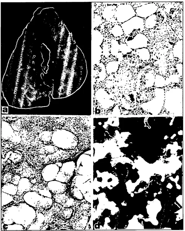
Figure ıa. Gross appearance of fat necrosis in cross-section of the colon that had been in fixative solution for several days. Notice connective lissue septums and the constricted lumen which is almost encircling by lesion.
b. Infiltration of Iymphocytes, plasma cells, macrophages, and mullinucleated foreign body giant cells around the lesions. H.E. x130.
c. Proliferation of fibrous lissue around the lesions. H.E. x 130.
d. Crysı.aJs aggregated in star-shaped dusters and fat droplets in fat cells. Frozen section, Dil red O, x i30. peated after eight months. During this time,
rec-tal palpation reyealed neither cross abnormaliti-es nor pregnancy findings. Eleyen months Iater, the condition was found when the animal was
examined for the cause of infertility. At this
time, clinical examination reyealed that
respira-tion, pul se and temperature were all normaL.
Rectal examination showed that the distal colon
and rectum were completely surrounded by a
large, hard. lobulated tumour-like stmcture. It
was difficult to palpate the utems and impossib-le to palpate the oyaries. A diagnosis of intesti-nal obstmction due to tumorous structures was made and it was decided to have the cow be sla-ughtered.
r
---MASSIVE FAT NECROSIS IN A COW
Post mortem exarrtination showed that the distal colon and rectum were enveloped in
mas-ses of lipomatous tissue. Areas of the lesion
were coarse, hard, almost encireling the
intesti-nal waıı, opaque appearance and whitish in
color. They were surrounded by a thick fibrous membrane. The cut surface of these lesions sho-wed lobules of fat with areas of a fibrous tissue (Fig. la). Lumens of the colon and rectum were constricted by this massiye fatty lesions. There
weren't sirrtilar lesions see n in other areas of
body fat. Aıı the organs and viscera were
nor-mal in appearance, with the exception of the
ovaries of which contained eysts.
Histopathologieaııy, the lesions were
SUf-rounded by thiekened conneetive tissue whieh
infiltrated deep into the lesions and divided
them into many irregular lobules. Each lobule eontained large fat ceııs. Inside the enlarged fat ceııs, several foam eeııs were lined up with a
cytoplasrrtic range. They had slightly stained
round or oval nuelei and frequently formed a syncytium. Around the lesions there were foeal
haemorrhages and an inflammatory infiltration
of lymphoeytes, plasma eeııs, macrophages and
multinueleated foreign body giant eeııs (Fig.
ib). In some areas there was fibroplasia with extensive coııagen formed into narrow septums or broad sheets (Fig. le). Most of the necrotie
eeııs contained a fine eosinophilie, crystaııine
mass which eaused the ceııs to be distended and
aggregated in star-shaped elusters. These
crystals were black stained in frozen sections with OH red O (Fig. Id).
Ovarian cysts observed in macroscopieal
examination were histopathologieaııy found to
be as luteinized cysts. The eavity of the cyst
was spherieal, fiııed by eosinophilie material,
and lined by a thick layer of fibrous tissue adja-cent to the zone of luteinized theca ceııs.
Discussion
A review of many reports which have been published on the disease indicates that identical lesions were present mainly in the adipose tis-sues (2, 8- iO). Most of such cases have been detected at the slaughterhouse or during routine autopsy or reetal exarrtination. In this cow, the condition was found around the intestinal waıı during rectal exarrtination, and was confırmed
by post-mortem and histopathological
examina-tions. The basic lesion was evidently identical with those of reported by several investigators
(5, 6, 8,
ı
2,ı
3). it is probable that the lesionshad been growing for some time without cau-sing symptoms.
141
In this cow the cause of infertility may be considered that was due to fat necrosis or ovari-an cysts or both. Because it is weıı known that both cause infertility in cattle (6, 7).
As a result, it must be remembered that
most of the cattle affected with elinical fat nec-rosis ineluding this cow, were died or condem-ned, because the etiology of this disease was unknown and there was no effective treatmenL
More recently, however, attempt to establish
the therapeutic effect of isoprothiolane on Japa-nese Black Cattle affected with subelinical fat necrosis was indieated satisfactory results (I I).
Acknowledgement
We thank to Dr. Sait Bulmuş for providing the material of this study.
Referenees
1. Arbuekle, B. (1962). Diffuse lipomatosis in a heifer. Vet.
Rec .. 74: 768.
2. Brldge, F.S., and Spratling, F.R. (1962). Bovine
Iipo-matosis. Vet Rec, 74: 1357-1362.
3. Edgson, F.A. (1952). Bovine lipomatosis. Vet. Rec., 64:
449-454.
4. Forney, M.M., Williams, D.J., Papp, E.M. and
Tyler, D.E. (1969). Limited survey of Georgia canlefor fat
necrosis. J Amcr Vet Med Ass, 154: 1603-1604.
5. ıto, T., Mlura, S., Ohshlma, K., and Numakunai,
S. (1968). Pathological studies on fat necrosis (lipomatosis)
in cattle. Jap J Vet S, 30: 141-150.
6. Julian, R.J. (1985). The peritoneum, Retroperitoneum. and
Mesentery. In: Pathology of Domestie Animals, 3rd Edit. K.V.F. Jupp, P.c. Kennedy. and N. Palmer, Eds. Aeademie Press, Ine., New York.
7. JuPP, K.V.F., Kennedy, P.C, and Palmer, N.
(1985). "Pathology of Domestic Animals" 3rd ed. Aeademie Press, Ine., New York.
8. Papp, E., and Williams, D.J. (1970). Bovine
lipomato-sis. Zentralbl Veterinaermed Rcihe A, 17: 735.742.
9. Ribelin, W.E. and neEds, F. (1960). Fat necrosis in
man and animals. J Amer Vet Med Ass, 136: 135-139.
LO. Rumsey, T.S., Stuedemann, J.A., Wilkinson, S.R.
and Williams, n.J. (ı 979). Chemical composifion of
ne-crotic fat lesions in beef cows grazing fertilized "KenlUcy-31" tallfescue. J Anİm Sei, 48:673.682.
11. Shlmada, Y., Katarnato, H., Ishlda, S., Kobaya.
shl, K. and Tohzyoh, H. (1988). Therapeutic effect of
Isoprothio/ane on bovine fat necrosis. Jap J Vet S, 50:
1017-1024.
12. Vitovee, J., Proks, C., and Valvoda, V. (1975).
Up-omatosis (fat necrosis) in cattle and pigs. J Comp Pathol, 84:
53-59.
13. Williams, n.J., Tyler, n.E., and Papp, E. (1969).
Abdominal fat necrosis as a herd problem in Georgia cartle.