Ankara Üniv Vet Fak Derg, 53, 2006 149 Ankara Üniv Vet Fak Derg, 53, 149-151, 2006
Short Communication / Kısa Bilimsel Çalışma
Pleuritis and pneumonia associated with nocardiosis and aspergillosis
in a domestic short haired cat
Rıfkı HAZIROĞLU1, Mehmet ŞAHAL2, Recai TUNCA3, Tolga GUVENC4,
SibelYasa DURU2, Lale ATASEVEN5
1Department of Pathology,Veterinary Medicine, Ankara University, 06110 Diskapi, Ankara, Turkey; 2Department of Internal Medicine, Veterinary Medicine, Ankara University, 06110 Diskapi, Ankara, Turkey; 3Department of Pathology,Veterinary Medicine, University of Kafkas, 36100 Pasacayiri, Kars, Turkey. 4Department of Pathology,Veterinary Medicine, University of 19
Mayis, 55139 Kurupelit, Samsun, Turkey. 5Department of Microbiology, Provincial Control Laboratory, Van, Turkey.
Summary: A severe, pyogranulomatous inflammation of the pleura and pnömonia associated with Nocardia spp. and
Aspergillus fumigatus was described in 9 month-old, domestic short haired male cat to the respect of clinical, microbiological and
pathological aspects. Severe abdominal respiration, open-mouthed breathing, weakness, dehydration and fever 39.6 °C were observed clinically. There was yellowish brown fluid in the thoracic cavity. Thick pleura and multiple adhesions were detected at necropsy. Many necrotic and pyogranulomatous lesions were observed histologically.
Key words: Aspergillosis, cat, nocardiosis, pleuritis, pneumonia
Evcil kısa tüylü bir kedide plöritis ve pnömoni ile karakterize nokardiozis ve aspergillozis
Özet: Erkek, kısa tüylü, 9 aylık, evcil bir kedide Nocardia spp. ve Aspergillus fumigatus tarafından oluşturulan, şiddetli pyogranülomatöz plöritis ve pnömoni olgusu klinik, patolojik ve mikrobiyolojik yönleri ile tanımlandı. Klinik olarak şiddetli abdominal solunum, ağız açık vaziyette nefes alma, güçsüzlük, dehidrasyon ve 39.6 °C ateş gözlendi. Nekropside, göğüs boşluğunda sarımtırak kahve renkli sıvı mevcuttu. Plöra kalınlaşmış ve çok sayıda adezyonlar bulunmaktaydı. Histolojik olarak nekrotik, pyogranülomatöz lezyonlar dikkati çekti.
Anahtar sözcükler: Aspergillozis, kedi, nokardiozis, plöritis, pnömoni.
Nocardia is a gram positive, aerobic and branching filamentous bacteria (5). Nocardiosis is acute or chronic infectious disease with granulomatous or suppurative in nature (4-6). Nocardia asteroides are most commonly isolated from the nocardiosis cases. Aspergillus is a fungal genus, which is widespread in the environment (3). Aspergillosis is either a granulomatous or necrotizing disease in cats, usually affecting lungs and intestines (7,9). These agents are widespread in the environment, although, infections caused by Nocardia spp. or Aspergillus spp. are more rare in cats (2,3,5,8). Infections caused by individual Aspergillus spp. and
Nocardia spp. have been described by several authors
(2,5,7-10), however, there has been no report about dual infection of these agents in cats.
A 9 month-old, domestic short haired male cat, with the complains of respiratoric difficulties and anorexia was brought to the Ankara University, Veterinary Faculty, Internal Medicine Department. The cat was died
after attending the clinic that performed complete clinical examination one week later. The cat was necropsied systemically. Tissue samples were fixed in 10 % neutral buffered formalin, embedded in paraffin wax, sectioned at 5-7 µm. and stained with haematoxylin and eosin. Selected sections were also stained with periodic acid – Schiff (PAS), Brown and Brenn (BB) and Gomori’s methenamine silver (GMS).
Severe abdominal respiration, open-mouthed breathing, weakness, dehydration and fever 39.6 °C were observed clinically. The sound of lungs and heart was not clear at the auscultation. There was decreasing in the auscultation area of lungs. The results of hemogram showed that there was an increasing in the white blood
cells (44530 n/µl) while erytrocyte (4.23 106µl),
hemotocrit (24.0 %) and hemoglobin (5.9 g/dl) levels were detected. The venal blood gases were as follows; blood pH 7.068, base insufficiency –15,8 mmol/l, bicarbonate amount 11,9 mmol/l and pO2 % 24.3 mmHg.
Rıfkı Hazıroğlu - Mehmet Şahal - Recai Tunca - Tolga Guvenc - Sibel Yasa Duru - Lale Ataseven 150
In radiographic examination, the cranial lobes of the lungs and heart were not clearly seen due to retention of a large amount of an exudate. From the thoracic cavity 50 ml of purulent and red-brownish liquid for bacteriological examination was taken. Oxygen therapy was applied to the cat. Intravenously Ringer lactate, dextrose % 5 and NaCl % 0.9, NaHCO3 % 1.3 were used to rehidrate and the cat to alleviate the acidosis. Additional antibiotic (lyncomycin-spectinomycin,
Linco-spectin ® flk.), B-complex (Berovit-B12 ® flk.) and C
(Redoxone ® flk.) vitamins were applied. There were no improvements in the patient condition after seven days of treatment. Unfortunately the patient was died and immediately necropsied.
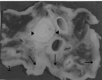
Macroscopically, the thoracic cavity contained approximately 300 ml of purulent yellowish-brown fluid that clotted after the thorax was opened. The pleura was thickened and there were multiple adhesions. The lungs were almost consolidated (Fig 1). The pericardial sac was thickened and adhered to the epicardium.
Figure 1: Macroscopic view of the cut surfaces of lungs and oesophagus (arrow heads), showing thickening of the pleura and consolidation of lungs (arrows).
Histopathologically, lesions always involved the serous membranes and extended into the parenchyma; cellular reaction was remarkably similar in each parts of the pleura. Lesions characterized large areas of granulomatous tissue containing several small, irregular and necrotic foci frequently confluent. Necrotic areas were surrounded by fibroblasts, neocapillaries, and mixture of polymorph leukocytes (PML) lymphocytes, plasma cells and macrophages. Numerous interspersed focal and larger accumulations of PML were often surrounded by areas of fibrosis, and many contained organisms colonized in the form of granules (Fig 2 a). When the sections stained with BB, these granules
containing numerous gram-positive, beaded branching bacilli that clearly resemble the Nocardia spp. were observed. The filaments were also stained GMS method, however, results of acid-fast staining were negative. Moreover, in the some center of the necrotic areas, septate and branching typical Aspergillus spp. hyphae were stained with PAS and GMS. In these areas, the pleural lesions extended into the lungs. The lesions in lungs were appeared to be circumscribed and well demarcated clearly by bands of compressed alveolar tissue and infiltrated with PML. The lesions were comprised with a typical a central zone of acidophilic necrotic debris. A band of tissue not only infiltrated mainly PML, but also plasma and epithelioid cells were detected surrounding this necrotic zone.
Bacteriologically, the specimens from the exudate, pleura, mediastinal lymph nodes and lungs were inoculated on 7% sheep blood agar, Sabouraoud dextrose agar (SDA) and Loewenstein Jensen agar. At the end of the incubation period, only Aspergillus fumigatus grew on blood agar and SDA (Fig 2 b). There were no
Nocardia spp. grown in same materials.
Figure 2: a-A nocardial nodule consisting of masses of organisms, surrounded by polymophnuclear leucocytes, mononuclear cells and fibroblasts. HE x 120. b- Insert preparations of the SDA, showing head of the conidia of A.
fumigatus. Native examination X 320.
Pleuritis and pneumonia due to dual infection with
Aspergillus spp. and Nocardia spp. have not been
described previously. Both infection caused by these agents are often thought to be a complication of some other debilitating disease (3,5,8). The histologic changes in this cat was similar to previous reports (3,5,8,10).
In nocardiosis, prognosis is usually good if the lesion localized but poor to guarded if is systemic or thoracic involvement (1,8). The nocardial agents are enter to the body via inhalation, food or skin wound (1). They have not been usually isolated from the exudates
Ankara Üniv Vet Fak Derg, 53, 2006 151
(8). In this case, the nocardial agents were probably entered to body via inhalation and/or food, since mark/s of the skin wound were not be able to see on the macroscopical examination. As a result, entering route of nocardial agents in this case was still obscure. No bacterial agents were isolated the exudates taken from thoracic cavity on the clinical examination. However,
Aspergillus fumigatus was isolated from the necropsy
materials, but Nocardia spp. was not. On the other hand, nocardial agents were identified using histochemical staining methods. This situation could be explained that broad spectrum antibiotics during one week period.
References
1. Edwards DF. (1998): Actinomycosis and nocardiosis. In Greene. Infectious diseases of the dog and cat. Saunders, Philadelphia, pp. 308-313.
2. Fox JG, Murphy JC, Shalev M. (1978): Systemic fungal
infections in cats. JAVMA, 173, 1191-1195.
3. Hazıroğlu R (2001): Solunum Sistemi. In: Veteriner Patoloji, (Haziroglu, R., and U. H. Milli, Eds.), Medipres Ankara, pp. 106-107.
4. Lobetti RG, M, Collett G, Leisewitz A. (1993): Acute
fibrinoprulent pneumonia and haemoptysis associated with Nocardia asteroides in three dogs. Vet Rec, 133, 480.
5. Love DN. (1988): In: Manual of Small Animal Infectious
Diseases, (Barlough, J. E. Ed) Churchill, Livingston, New
York, pp. 208.
6. Milli UH, Köküuslu C, Berkin S. (1986): Bir köpekte
nocardial plorizi olgusu. Ankara Üniv Vet Fak Derg, 33,
171-178.
7. Pakes SP, New AE, Benbrook SC. (1967): Pulmonary
aspergillosis in a cat. JAVMA, 151, 950-953.
8. Petric A.D, Tozon N, Cerne M. (2001): Nokardiose bei
katze und hund : zwei falstuden. Der Praktische Tierarzt,
82, 1022-1026.
9. Stokes R. (1973): Intestinal mycosis in a cat. Aust Vet J, 49, 499-500.
10. Tilgner SL, Anstey SI. (1996): Nocardial peritonitis in a
cat. Aust Vet J, 74, 430-432.
Geliş tarihi: 17.11.2005 / Kabul tarihi: 30.11.2005
Address for correspondance:
Dr.Recai Tunca Kafkas Üniversitesi Veteriner Fakültesi Patoloji Anabilim Dalı 36100 Paşaçayırı, Kars-Turkey