Ankara Üniv Vet Fak Derg, 52, 2005 201 Ankara Üniv Vet Fak Derg, 52, 201-203, 2005
Short Communication / Kısa Bilimsel Çalışma
Anal sac carcinoma in a dog
Sevil Atalay VURAL1, Rıfkı HAZIROĞLU1, Zafer ÖZYILDIZ2, Şule Yurdagül ÖZSOY3, Yusuf Sinan ŞİRİN4
1 Department of Pathology, Faculty of Veterinary Medicine, University of Ankara, Ankara; 2 Department of Pathology, Faculty of
Veterinary Medicine, University of Kafkas, Kars; 3 Department of Pathology, Faculty of Veterinary Medicine, University of Mustafa
Kemal, Hatay; 4 Department of Surgery, Faculty of Veterinary Medicine, University of Ankara, Ankara-Turkey.
Summary: In this report, anal cell carcinoma located from ventro-laterally of anus, spreading out to subcutis of hind left leg
was described with clinical and pathomorphological findings in a 12-year-old female mongrel dog. The mass was of 20x15x10 cm in diameter, 1110 g in weight and it was showing nodular structure. Cut section of the mass was cystic. Microscopically; oval or round shaped neoplastic cells with hyperchromatic nuclei and eosinophilic cytoplasm had formed solid sheets. In some areas, the cells surrounded a small amount of eosinophilic secretion and constituted rosette formations. PCNA positive neoplastic cells were seen by immunoperoxidase method.
Key words: Anal sac gland, carcinoma, dog.
Bir köpekte anal kese adenokarsinomu
Özet:
Bu olguda 12 yaşlı, dişi, melez bir köpeğin anüsünün ventro-lateralinden sol arka bacak derisi altına doğru yayılan anal kese adenokarsinomunun klinik ve patomorfolojik bulguları açıklandı. Tümör, 20x15x10 cm boyutlarında, 1110 gr ağırlığında, nodüler bir yapıdaydı. Kesit yüzünde, kistik yapılar vardı. Mikroskobik olarak tümör; solid alanlar halinde dizilim gösteren, oval ya da yuvarlak şekilli, hiperkromatik çekirdekli, eozinofilik sitoplazmalı neoplastik hücrelerden ibaretti. Bazı alanlarda rozet formasyonları dikkati çekti. İmmunoperoksidaz yöntemiyle incelemede PCNA pozitif hücreler saptandı.Anahtar sözcükler: Anal kese, karsinom, köpek.
Anal sac carcinoma is a malign tumor of apocrine secretor epithelium of anal sacs. It is commonly seen in dogs (1, 2, 7). 5-15 year-old female dogs constitute the risk group. Certain breed disposition is not reported even though is it common in English Cocker Spaniels, German Shepherds, English Springer Spaniels and mongrel dogs (4, 6). The purpose of the study is to describe clinical and patomorphological findings of anal sac carcinoma in a dog. A mass removed from the periphery of anus of a 12-year-old female mongrel dog, which was operated at the Department of Surgery of Ankara University Faculty of Veterinary Medicine, constituted the material. Organ samples and the mass were fixed in 10% buffered formaline and prepared according to routine methods; embedded in paraffin and cut in 5µm thick sections and examined under a light microscope after staining with haematoxylin-eosin (HE). In order to detect Proliferating Cell Nuclear Antigen (PCNA) (Dako) selected specimens were stained according to Avidine-Biotin Complex Peroxidase (ABC-P, Dako) method. A 12-year-old,
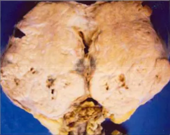
female, mongrel dog was presented to the clinics with a growing mass around anus and defecation problems related to the mass and euthanasia was applied upon the owner’s request and also taking into consideration the age of the animal and the size of the mass. The mass spreading into subcutis of left hind leg out from the ventro-lateral of anus, was of 20x15x10 cm size and 1110 g weight. It was covered with nodules varying from size of a chickpea to a walnut (Figure 1). The cut section of tumoral mass was yellowish in color and containing cystic structures filled with a yellowish-red fluid. Oval or round shaped neoplastic cells with hyperchromatic nuclei, eosinophilic cytoplasm, showing solid distribution were observed microscopically (Figure 2). In some areas, these cells formed rosettes by surrounding a pink colored homogenous secrete-like material (Figure 3). These structures were accompanied by a thin stroma consisting of capillaries and connective tissue. These cells were observed to react positively with PCNA in ABC-P staining and gain dark brown color (Figure 4). It was
Sevil Atalay Vural - Rıfkı Hazıroğlu - Zafer Özyıldız - Şule Yurdagül Özsoy - Yusuf Sinan Şirin 202
Figure 3. Rozette formations of the neoplastic cells HE, x400 Figure 1. Cross section of the anal sac carcinoma
Figure 4. PCNA positive neoplastic cells, ABC-P, x 250 Figure 2. Neoplastic cells arranged solid sheets HE, x100
diagnosed as anal sac carcinoma obtained the clinical and pathomorphological findings. Anal sac carcinomas are reported frequently in dogs and rarely in cats (4, 5). In dogs, race predisposition is not mentioned however, unilateral occurrence in females and old animals is reported to be often (81%). Its increased incidence in castrated animals is also mentioned (4). Detection of the tumor in a 12-year old, female, mongrel dog in this case has supported the present opinion. Meuten et al. (4) has grouped anal sac adenocarcinomas into three types as solid, tubular, and rosette. Additionally this, Ogawa at al. has reported forth type as a papillary (6). As for this case, both solid and rosette types were observed. For that reason, it is thought to be more appropriate to grouped the tumor as mixed type. It is recorded that the tumor metastated %90 to the regional lymph nodes and %40 to the internal organs (3,8). Having observed no metastasis in the case was thought to be due to rapid development in
two months time and euthanasia applied. Low number of PCNA positive cells lead to the idea that neoplastic cells have already passed the proliferation phase.
As a conclusion, the tumor was diagnosed as anal sac carcinoma in the light of clinical and pathomorphological findings. It was considered as substantial by means of being the first case reported in dogs, investigation of PCNA positivity in neoplastic cells and being informative regarding clinicians in our country
References
1. Esplın DG, Wılson SR, Hullınger GA (2003): Squamous
cell carcinoma of the anal sac in five dogs. Vet. Pathol, 40,
332-334.
2. Gröne A, Weckmann MT, Blomme EAG, Capen CC, Rosol TJ (1998): Dependence of humoral hypercalcemia
of malignancy on parathyroid hormone-related protein expression in the canine anal sac apocrine gland
Ankara Üniv Vet Fak Derg, 52, 2005 203
adenocarcinoma (CAC-8) nude mouse model. Vet Pathol,
35, 344-351.
3. Hoelzler MG, Bellah JR, Donofro MC (2001):
Omentalization of cystic sublumbar lymph node metastases for long-term palliation of tenesmus and dysuria in a dog with anal sac adenocarcinoma. JAVMA, 219, 1729-1731.
4. Meuten DJ (2002): Tumors of the Skin and Soft Tissues. 74-78; 672-679. In: MH Goldschmidt, MJ Hendrick, Tumors of the Endocrine Glands, ed. CC. CAPEN (Ed), Tumors in Domestic Animals, 4th ed. Iowa State Press, Iowa.
5. Nielsen SW, Moulton JE (1990): Tumors of the Skin and
Soft Tissues. 73-75. In: LT Pulley, AA Stannard (Ed),
Tumors in Domestic Animals 3rd ed. University of California Press, Berkely.
6. Ogawa K, Tateyama S, Nosaka D (1991): Usefullnes of
paradoxical con a staining for diagnosis of papillary adenocarcinoma of the apocrine anal sac gland in the dog.
J. Vet. Med. Sci., 53, 931-932.
7. Rosol TJ, Capen CC, Danks JA, Suva LJ, Steinmeyer CL, Hayman J, Ebelıng PR, Martın TJ (1990):
Identification of parathyroid hormone- related protein i canine apocrine adenocarcinoma of the anal sac. Vet Pat.,
27, 89-95.
8. Williams LE, Gliatto JM, Dodge RK, Johnson JL, Gamblin RM, Thamm DG, Lana Se, Szymkowski M, Moore AS (2003): Carcinoma of the apocrine glands of
the anal sac in dogs: 113 cases (1985-1995). JAVMA, 15,
825-831.
Geliş tarihi : 30.12.2004 / Kabul tarihi: 19.01.2005
Corresponding adress: Assoc. Prof. Sevil Atalay Vural Ankara University faculty of Veterinary Medicine Department of Pathology, 06110 Diskapi, Ankara