173 Ankara Üniversitesi Tıp Fakültesi Mecmuası 2009, 62(4) DAHİLİ BİLİMLER / MEDICAL SCIENCES
Olgu Sunumu / Case Report
Başvuru tarihi: 28.01.2010 • Kabul tarihi: 08.06.2010 İletişim
Dr. Gonca Sandal
Zekai Tahir Burak Kadın Sağlığı Eğitim ve Araştırma Hastanesi Yenidoğan Kliniği
GSM : 0 505 487 10 27 E-Posta Adresi : [email protected]
Greig cephalopolysyndactyly is a rare multiple congenital anomaly characterized by clinical triad of polysyndactyly, macrocephalia and hypertelorism. In general, although it is characterized with autosomal dominant inheritance, there is also autosomal recessive inheritance pattern. Here, we aimed to discuss an infant who was born from multiple gestations arising from assisted reproduc-tion techniques and suspected to be Greig syndrome with no familial history. Best of our knowl-edge this is the first case related with assisted reproduction technique.
Key Words : Greig Cephalopolysyndactyly, Assisted Reproduction Technique
Greig sefalopolisindaktili sendromu klinik olarak polisindaktili, makrosefali ve hipertelorizm tri-adı ile karakterize nadir multipl konjenital bir anomalidir. Genelde otozomal dominant kalıtımla karakterize olmasına rağmen, otozomal resesif geçiş paterni de vardır. Biz burada aile öyküsü ol-maksızın Greig sendromu olduğundan şüphelenilen, yardımcı üreme tekniği ile olan çoğul gebe-likten doğan bir yenidoğanı tartışmayı amaçladık. Yardımcı üreme tekniği ile ilişkili bizim bildiği-miz ilk vakadır.
Anahtar Sözcükler: Greig Sefalopolisindaktili, Yardımcı Üreme Tekniği
Zekai Tahir Burak Kadın Sağlığı Eğitim ve Araştırma Hastanesi Yenidoğan Kliniği
A Greig Syndrome Case Diagnosed In One Of The Singleton Of
Twin Preterm Who Were Obtained By Assisted Reproduction
Yardımcı Üreme Tekniği İle Olan Preterm İkiz Eşinde Tanımlanmış Bir Greig Sendromu Olgusu
Gonca Sandal, Nurdan Uraş, Ömer Erdeve, Şerife Suna Oğuz, Uğur Dilmen
The Greig cephalopolysyndactyly syn-drome (GCPS) is a pleiotropic, multi-ple congenital anomaly syndrome. It is rare, but precise estimates of incidence are difficult to determine, as ascertain-ment is erratic. The primary findings include hypertelorism, macrocephaly with frontal bossing, and polysyn-dactyly. The polydactyly is most commonly preaxial of the feet and postaxial in the hands, with variable cutaneous syndactyly, but the limb findings vary significantly (1, 2). Oth-er low frequency findings include cen-tral nervous system (CNS) anomalies, hernias, and cognitive impairment it was first described by Greig (1926) in an affected mother and daughter (1). Mutations in the GLI3 gene located on chromosome 7p3 are responsible (3). In addition to mutations, translo-cations that interrupt the gene, micro-deletions, and cytogenetically detect-able deletions have been described (3). In literature, several reports have been published describing variable expres-sion of this syndrome (4-6). However,
association with assisted reproduction technique was not reported. Here, we aimed to discuss an infant who was born from multiple gestations arising from assisted reproduction techniques and suspected to be Greig syndrome with no familial history.
Case Report
One of the twins who were born from first pregnancy of the 25-year old mother at 28th gestational week with birth weight of 1180 gram was hospitalized to neonatal intensive care unit due to dyspnea. The pregnancy was result of assisted reproduction technique. Considering physical examination findings, birth weight was 1180 gram (50p), height was 42 cm (90p) and head circumference was 32 cm(>90p). General status was intermediate, face had dysmorphic appearance, forehead was wide and hypertelorism was pres-ent (Fig 1). In cardiovascular examina-tion, 2/6 systolic murmur was present.
174 A Greig Syndrome Case Diagnosed In One Of The Singleton Of Twin Preterm Who Were Obtained By Assisted Reproduction Ankara Üniversitesi Tıp Fakültesi Mecmuası 2009, 62(4)
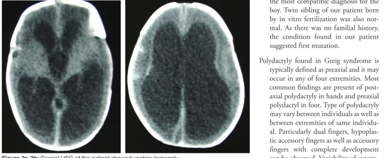
Partial subcutaneous syndactyly was present at bilateral hand and foot in-dex fingers and there was one acces-sory finger with normal development at hands and feet (Fig 2). Findings of other system examinations were normal. In echocardiographic exami-nation, patent ductus arteriosus was found. In cranial USG, width of right lateral ventricle was 10 mm and width of left lateral ventricle was 12 mm that ventriculomegaly was observed.
Tri-ventricular hydrocephalus was found in cranial CT. Chromosome analysis showed a normal 46, XY karyotype. Further genetic investigations have not been performed due to techni-cal difficulties. We performed cranial USG and head circumference mea-surements weekly. At postnatal 21th day cranial ultrasonografi revealed that hydrocephalus was increased and intraparanchymal hemorrhage was developed. As head circumference
increased 2.3 cm/week, he was dis-charged for shunt operation at another center at postnatal 47th day. We did not detect any clinical convulsion and rapid increase in head circumference at the follow up period. The patient is on follow up with pediatric neurology and neurologic surgery units.
Discussion
Greig cephalopolysyndactyly is a rare multiple congenital anomaly char-acterized by clinical triad of polysyn-dactyly, macrocephalia and hyper-telorism (2). Incidence is estimated as 1-9/1.000.000. As molecular diagnosis methods are not widely used, it is dif-ficult to definitely estimate incidence. At least 75 % of cases with clinical di-agnosis of cephalopolysyndactyly had GL13 gen mutation. GL13 mutation has a wide spectrum. In general, al-though it is characterized with auto-somal dominant inheritance, there is also autosomal recessive inheritance pattern (2, 3). Many families with Greig polysyndactyly syndrome were reported. Risk of non-GCPS individu-als within involved families to have child with GCPS is less than 1% per each conception. In our case, there was no other individual with GCPS in the family (5, 7). When we search the London Dysmorphology Data-base using his multiple signs, the Greig cephalopolysyndactyly syndrome was the most compatible diagnosis for the boy. Twin sibling of our patient born by in vitro fertilization was also nor-mal. As there was no familial history, the condition found in our patient suggested first mutation.
Polydactyly found in Greig syndrome is typically defined as preaxial and it may occur in any of four extremities. Most common findings are present of post-axial polydactyly in hands and prepost-axial polydactyl in foot. Type of polydactyly may vary between individuals as well as between extremities of same individu-al. Particularly dual fingers, hypoplas-tic accessory fingers as well as accessory fingers with complete development can be observed. Variability of
Figure 2a-2b: Cranial USG of the patient showed ventriculomegaly
175
Gonca Sandal, Nurdan Uraş, Ömer Erdeve, Şerife Suna Oğuz, Uğur Dilmen
Journal Of Ankara University Faculty of Medicine 2009, 62(4)
ous syndactyly is also high. Large part of patients with cutaneous syndactyly has partial cutaneous syndactyly in several fingers(2,4,6). However, there are also cases with complete cutaneous syndactyly in all fingers. Our case had partial cutaneous syndactyly in bilat-eral foot and hand thumb and index fingers and polydactyly.
In this syndrome, cranio-facial mani-festations may also vary. Most cases have hypertelorism with or without telechantus. In our case, hypertelorism was present without telechantus. Mac-rocephalus not related with central nervous system is found in many cases. In our case, hydrocephalus associated by triventricular hydrocephalus and secondary macrocephalus were pres-ent. Although rare, craniocynostosis
mental retardation, corpus callosum agenesis, umbilical and diaphragmatic hernia may also be present in Greig syndrome. Our case manifested none of above mentioned findings.
Teebi hypertelorism syndrome, Carpender syndrome and Gorlin syndrome are found among differential diagnosis. In Teebi syndrome, plolydactyly is typi-cally not preaxial. Carpenter syndrome is characterized by polysyndactly, cra-niocynostosis and mental retardation. Gorlin syndrome (nevoid basal cell carcinoma syndrome) is characterized by macrocephalus and particularly hypertelorism and polydactyly. More-over, mutations were found in genes related with PTCH1 and GLI-SHH pathways (6,8,9).
Factors related to infertility of both part-ners, quality of sperm and oocyte cell, age of couple and tozin exposure in-creased secondary to advanced age are among factors causing genetic dis-orders in assisted reproduction tech-nique pregnancies. Quality of sperm and oocyte cells may particularly increase incidence of chromosome anomaly diagnosed during antenatal period as well as congenital malfor-mations found in live births (10-13). Nevertheless, our case led to inquire relationship between assisted repro-duction techniques and chromosomal disorders, although there is no defini-tive relationship between assisted re-production techniques and chromo-somal disorders .
REFERENCES
1. Biesecker LG. The Greig cephalopolysyndac-tyly syndrome. Orphanet J Rare Dis 2008; 24:10.
2. Balk K, Biesecker LG.The Clinical Atlas Of Greig Cephalopolysyndactly Syndrome. Am J Med Genet A 2008; 146: 548-57. 3. Biesecker LG.What you can learn from one
gene : GL13. J Med Genet 2006; 43: 465-69.
4. Winter RM, Huson SM. Greig Cephalopoly-syndactly Syndrome: A possible mouse ho-mologue (xt- extratoes). Am j Med. Genet 1998; 31:93-8
5. Marafie MJ, Temtamy SA, Rajaram U, al-Awadi SA, el-Badramany MH, Farag TI. Greig cephalopolysyndactyly syndrome with dysgenesis of the corpus callosum in a Bedouin family. Am j Med Genet 1996; 66: 261-64
6. Wang CH, Tsai FJ, Shi YR. Greig Cephalo-polysyndactly Syndrome in a family. Acta Paediatr Taiwan 2006; 447: 97-9.
7. Kunze J, Kaufmann HJ (1985). Greig cepha-lopolysyndactyly syndrome. Report of a spo-radic case. Helv Paediatr Acta 1985; 40:489-95.
8. Williams PG, Hersh JH, Yen FF, Barch MJ, Kleinert HE, Kunz J, Kalff-Suske M. Greig Cephalopolysyndactly Syndrome : Altered phenotype of a microdeletion syndrome due to presence of a cytogenetic abnormality. Clin Genet 1997; 52:436-41
9. Biesecker LG. Polydactyly : How many disor-ders and how many genes ? Am J Med Genet 2002; 112: 279-283.
10. Clementi E, Palka C, Lezzi L, Stuppia L, Franchi P, Tiboni G M. Prevalence of chro-mosomal abnormalities in 2078 infertile couples referred for ART. Human Reprod 2005; 20:437-42.
11. Buckett WM, Tan SL. Congenital abnor-malities in children born after assisted re-productive techniques: how much is associ-ated with the presence of infertility and how much with its treatment? Fertil Steril. 2005 Nov;84(5):1318-9
12. Simpson JL. Registration of congenital anomalies in ART populations: pitfalls. Hum Reprod. 1996 Dec;11 Suppl 4:81-8. 13. Nancy S. Green Risks of Birth Defects and
Other Adverse Outcomes Associated With Assisted Reproductive Technology Pediatrics Vol. 114 No. 1 July 2004, pp. 256-259.