Thoracoscopic Cystostomy of Pulmonary Hydatid Cyst in
Children
Çocuklarda Akciğer Kist Hidatiğinde Torakoskopik Kistostomi
Ufuk Ateș
1, Ergun Ergün
1, Gülnur Göllü
1, Aydın Yağmurlu
1, A. Murat Çakmak
1,
Hüseyin Dindar
1, Meltem Bingöl-Koloğlu
11 Ankara University Medical Faculty, Department of Pediatric Surgery,
Ankara, Turkey Amaç: Ekinokok enfeksiyonu dünyada; özellikle de Akdeniz ülkelerinde önemli bir sağlık problemi olmaya
devam etmektedir.İnsanlardaki ekinokok enfeksiyonlarında akciğer, ikinci en sık etkilenen organdır. Bu enfek-siyon çocuklarda da görülebilir ve tedavi bazen zor olabilir. Çocuklarda küçük akciğer kist hidatikleri medikal tedaviye olumlu cevap verir. Büyük ve komplike kistlerde ise yaklașım cerrahidir. Çocuklardaki akciğer kist hidatiklerinde günümüzde uygulanan cerrahi tedavi torakotomi ile kistostomi ve kapitonaj gibi parankim ko-ruyucu cerrahi ișlemlerdir fakat torakoskopik cerrahi deneyimi çocuklarda sınırlıdır. Bu çalıșmanın amacı, ço-cuklarda akciğer kist hidatiklerinde torakoskopik cerrahi deneyiminin sunulmasıdır.
Hastalar ve yöntem: 2008 ve 2016 yılları arasında akciğer kist hidatiği nedeniyle torakoskopik kistotomi
ya-pılmıș olan çocukların tıbbi kayıtları gözden geçirildi. Medikal tedaviye karșın semptomatik olan ve kist çapı 6 cm’nin üzerinde olan hastalara cerrahi tedavi önerildi. Hastalarda kistostomi, germinatif membranın çıkarıl-ması ve hava kaçağının önlenmesi gibi parankim koruyucu cerrahi uygulandı. Akciğer kapasitesini kötüleș-tirme riski nedeniyle kapitonaj tercih edilmedi.
Bulgular: Çalıșmaya alınan 15 çocukta akciğer kist hidatiği nedeniyle 16 torakoskopik ișlem gerçekleștirildi.
Bir hastada bilateral komplike hidatik kist mevcuttu. Üç hastada (%20) hava kaçağının güvenli bir șekilde kont-rol edilememesi nedeniyle mini torakotomiye geçildi. On iki çocukta ișlem torakoskopik olarak tamamlandı. Torakoskopik olarak tamamlanan üç olguda (%25) uzamıș hava kaçağı (1 haftanın üzerinde) gerçeklești ve bu hastalardan birinde bronkopulmoner fistülü kontrol altına alabilmek için torakotomi yapıldı. Olgulardan bi-rine karaciğer kist hidatiği nedeniyle laparoskopik kistostomi ve kapitonaj uygulandı. Ortalama 60 aylık ta-kipte rekürrens saptanmadı ve hastaların takiplerinde problem izlenmedi.
Sonuç: Çocuklarda görülen komplike olmayan kist hidatik olgularında torakoskopik kistostomi önerilse de
komplike kist hidatiklerde torakoskopik wedge rezeksiyon önerilmektedir. Bronșial hava kaçaklarının kontro-lünde zorlanılırsa mini torakotomiye geçilebilir.
Anahtar Sözcükler: Çocuk, Hidatik Kist, Torakoskopi
Aim: Echinococcosis is still an important health problem throughout the world, particularly in the
Mediterranean area. In humans, the lungs are the second most commonly affected sites. The disease may affect children and its treatment may be challenging. In children, small hydatid cysts of the lungs respond favorably to the medical treatment. Surgery is the standard option for the treatment of large and complicated cysts. In current practice, thoracotomy and parenchyme-saving procedures such as cystostomy and capitonnage remain the standard surgical approach for pulmonary hydatid cysts in children. However, surgical experience with thoracoscopy is limited. The aim is to present the experience in thoracoscopic management of pulmonary hydatid cysts in children.
Patients and Methods: Medical records of children who underwent thoracoscopic cystostomy of pulmonary
hydatid cysts between 2008- 2016 were reviewed. Surgical treatment was recommended for patients who remained symptomatic on medical treatment and when the cyst size was larger than 6 cm in diameter. Parenchyme-saving surgery was preferred which included cystostomy, removal of germinative membrane and control of air leaks. Capitonnage was not preferred because of the risk of deterioration in lung capacity.
Results: Fifteen patients underwent 16 thoracoscopy procedures for pulmonary hydatid cysts. One of the patients
had bilateral complicated pulmonary hydatid cysts. There were conversions to mini-thoracotomy in 3 (20%) procedures because the air leaks could not be controlled safely. The procedure was completed thoracoscopically in 12 patients. In thoracoscopically completed cases, prolonged air leak (over a week) occurred in 3 (25%) patients and one of them underwent thoracotomy to control bronchopulmonary fistula. One of the patients underwent laparoscopic cystostomy and capitonnage for associated liver hydatid cyst. There were no recurrences during 60 months of mean follow-up time and no problem was observed during the follow-up period.
Conclusion: Thoracoscopic management is recommended in children with uncomplicated hydatid cyst. In
complicated hydatid cysts, however, thoracoscopic wedge resection is recommended. If there is difficulty in controlling bronchial openings, mini-thoracotomy could be considered.
Key Words: Children, Hydatid Cyst, Thoracoscopy
Received: Dec 19, 2016 Accepted: June 01, 2017 Corresponding Author:
Gülnur GÖLLÜ, Assist Prof, E-mail: drggollu@yahoo.com GSM: +90 505 502 56 97 Tel: +90 312 595 84 35 Fax: +90 312 595 58 23
Department of Pediatric Surgery Ankara University Medical Faculty 06100, Dikimevi, Ankara, TURKEY
Hydatid Disease (HD) is a parasitic disease, which is caused by Echinococcus granulosus (1,2). The infection is common in areas in which humans and carnivores are in contact consistently (3). It is endemic in Mediterranean Region, Middle East, South America, Australia, India, and the Balkans (4). HD is also a concerning problem in terms of public health for Turkey due to its incidence of 20/100,000, especially in eastern and southeastern parts of Turkey (3,5). Hydatid Disease (HD) most frequently
involves the parenchyma of the liver (55-75%); however some of the parasites escape through the microvascular barrier and reach the lung (15-40%) (1,6). There is no specific symptom for pulmonary hydatid disease (PHD). Symptoms are usually due to the size and complications of the cysts (6). The treatment should be scheduled after performing laboratory and radiologic tests and differentiating the diagnosis properly. Surgical treatment has remained the prevailing option in the management of HD although medical treatment and combination of medical and surgical treatment choices are also other options (6,7).
Thoracotomy and sternotomy are the classical surgical procedures but in last decade, minimal invasive surgery; video assisted thoracoscopic surgery (VATS) to speak specifically has become a
considerable alternative for these patients (4).
In this study, we aimed to present the retrospective data of surgical procedures and perioperative complications of PHD patients who have undergone thoracoscopic surgical procedures.
Patient and Methods
The study was performed in adherence to the Declaration of Helsinki. Charts of 15 patients who underwent thoracoscopic surgery due to PHD between 2008-2016 were retrospectively investigated. Surgery was recommended if the cyst size was larger than six cm and if the patients stayed symptomatic after medical treatment. Children who could not tolerate the operation, had hydatid cysts in more than two organs and whose parents did not allow the operation were considered as inoperable. As our routine medical treatment, all of the patients received Albendazole. All patients were administered parenteral Ceftriaxone during induction of anesthesia. All 15 patients were performed thoracoscopic surgery after selective intubation. Selective intubation was performed in case of bronchial opening of the cyst, to prevent contralateral lung contamination. Right selective intubation was performed by
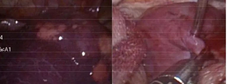
insertion of endotracheal tube into the right bronchus and left selective intubation was performed by a Fogarty catheter insertion to right bronchus with an insufflated balloon. Children were positioned lateral decubitus position and made sure that the cyst side was up. CO2 insufflation was performed with 6-8 mmHg pressure. A 4 mm, 30-degree lens camera (Karl Storz Endoscopy, Tuttlingen, Germany) was used. Working ports (3-5mm) were inserted to thorax. A sponge with iodine was placed surrounding the cyst to prevent intrathoracic infection. The different stages of the procedure were the same as in open surgery: cystostomy; removal of germinative membrane; control of air leaks. When excising the lung hydatid cyst; after the cystostomy, germinative membrane was removed. After cystostomy, germinative membrane was totally removed by an endobag (Figure 1 and Figure 2). Then the air leaks were controlled by filling the thoracic cavity with sterile saline solution after deflating the fogarty. Air leaks were controlled with intracorporeal 3/0 or 4/0 poliester sutures. Chest tube was inserted after the control of thoracic cavity.
Patient demographics, surgical procedure, perioperative complications, chest tube removal time, hospital stay and recurrence rates were evaluated.
Figure 2: Germinative membrane removal
Results
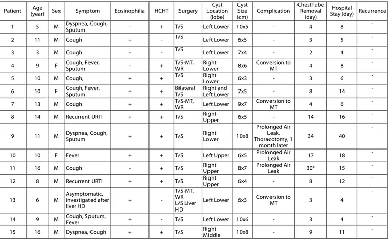
Between 2008 and 2016, fifteen patients with PHD have undergone 16 thoracoscopic procedures. Patients’ demographic data, symptoms, blood tests, radiologic tests, surgical
procedures and outcomes were analyzed (Table 1). Eighty percent of the patients (n=12) were male and 20% of them (n=3) were female. The median age was 10 (3-16) years. Fourteen children were symptomatic and one of them was asymptomatic. Out of these 15 patients, 67% of them
(n=10) had cough, 33% of patients (n=5) had sputum, 27% of patients (n=4) had fever and 20% of patients (n=3) had dyspnea. Seventy-three percent of the patients (n=11) had eosinophilia and 73% of them (n=11) had positive hemagglutination test for echinococcosis.
Table 1: Patients’ demographic data, symptoms, blood tests, radiologic tests, surgical procedures and outcomes
Patient (year) Age Sex Symptom Eosinophilia HCHT Surgery Location Cyst (lobe) Cyst Size (cm) Complication ChestTube Removal (day) Hospital
Stay (day) Recurrence
1 5 M Dyspnea, Cough, Sputum - + T/S Left Lower 10x5 - 4 8 -
2 11 M Cough + - T/S Left Lower 6x5 - 3 5 -
3 3 M Cough - - T/S Left Lower 7x4 - 2 4 -
4 9 F Cough, Fever, Sputum - + T/S-MT, WR Right Lower 8x6 Conversion to MT 4 8 -
5 10 M Cough, + + T/S Right Lower 6x3 - 3 6 -
6 10 F Cough, Fever, Sputum + + Bilateral T/S Right and Left Lower 7x5 - 8 14 -
7 13 M Cough + + T/S-MT, WR Left Lower 9x7 Conversion to MT 4 6 -
8 14 M Recurrent URTI + + T/S Right Upper 6x5 - 14 16 -
9 11 M Dyspnea, Cough, Sputum + + T/S Right Lower 10x8
Prolonged Air Leak, Thoracotomy, 1 month later 34 40 -
10 10 F Fever + + T/S Left Upper 6x5 Prolonged Air Leak 17 18 -
11 16 M Cough - + T/S Right Upper 8x7 Prolonged Air Leak 30* 15 -
12 8 M Recurrent URTI + + T/S Right Upper 6x4 - 8 12 -
13 6 M Asymptomatic, investigated after
liver HD + -
T/S-MT, WR L/S Liver HD
Left Lower 6x3 Conversion to MT 3 4
-
14 9 M Cough, Sputum, Fever + - T/S Left Lower 10x6 - 3 4 -
15 16 M Dyspnea, Cough + + T/S Right Middle 10x8 - 9 11 -
According to the results of the study, seven patients (47%) had left lower lobe, four patients (27%) had right lower lobe, three patients (20%) had right upper lobe, one patient (7%) had left upper lobe and one patient (7%) had right middle lobe cysts. A ten-year-old girl had bilateral lower lobe cysts in these patients (Table 2).
Table 2: Anatomic location of the cyst
Right upper 20% (n=3)
Right lower* 27% (n=4)
Right middle 7% (n=1)
Left lower* 47% (n=7)
Left upper 7% (n=1)
*One patient with bilateral lower lobe PHD.
There were conversions to mini-thoracotomy in 3 (20%) patients because the air leaks could not be controlled safely. The procedure was completed thoracoscopically in 12 patients. In thoracoscopically completed cases, prolonged air leak (over a week) occurred in 3 (25%) patients and one of them underwent thoracotomy to control bronchopulmonary fistula. Out of these three patients, two of them had suffered from bronchial rupture preoperatively. One of the patients underwent laparoscopic cystostomy and capitonnage for associated liver hydatid cyst. The chest tube was removed after an average duration of four days (2-34) and average duration of hospital stay of the patients was eight days (4-40). One patient, who suffered from prolonged air leak, was discharged with a Heimlich valve and the valve was removed one month later uneventfully. There were no recurrences during 60 months of mean follow-up time and all patients are doing well.
Discussion
We found that HD is still an important health problem especially in rural areas of Turkey. In our center, thoracoscopy can be performed in even complicated PHD in children.
Human beings serve as intermediate hosts. The infection is acquired by ingesting food or soil contaminated with eggs excreted from the canines (5). Due to the life cycle of parasite, the medical history of patient has a crucial importance on the diagnosis. In the literature, it has been reported that
only 10–19% of patients are asymptomatic. Most common symptoms are cough, chest pain, fever, hemoptysis, sputum production (5,6,8). The same pattern can be seen in our patients too. The most common symptom was cough (67%) and only one of our patients was asymptomatic (7%). Sputum production, dyspnea and fever were other common symptoms.
Although there is not a certain data in the literature about age, sex and disease incidence, it has been observed that the incidence seems to increase by age and male patients are more common than females (1,3). In our study, 63% of the patients were older than nine years while our patients’ mean age was 10 years and 80 % of our patients were male (n=12).
Indirect hemagglutination test for echinococcosis and eosinophil count are some valuable tests to support radiological and clinical diagnosis (9,10). Out of 15 patients of in our study, 11 of them had positive hemagglutination test (73%) and 11of them had eosinophilia(73%) in blood count. When radiologic findings are not certain, these tests can guide the surgeon to prepare for the operation. Chest X-Ray and Computed Tomography
(CT) are the radiologic tools to diagnose exact size and location of these cysts in the thoracic cavity. All of the patients in this study were performed a chest CT before the surgery. PHD usually appears in lower lobes and in right lung according to the literature (3,7,8). Not likely the literature, in our study, the majority of patients had cyst in left lower lobe. But the PHD can be presented in any location of any lobe, as it was in our study (7). More commonly, PHD
presents as a solitary/unilateral cyst but bilateral cysts and associated liver cysts can be found (5). In these 15 patients, majority of cysts were solitary, one was associated with a liver cyst and one of them presented with bilateral PHD, which was concordant with the literature.
The standard treatment procedure for PHD is patient – specific and a combination of medical and surgical protocols (3,9,10). The treatment decision is made depending on cyst size and complications but commonly surgery is needed when the cyst is bigger than five cm in diameter (1). Albendazole is the first medical treatment choice because of its better cyst penetration among other antihelmintic agents and is used widely in HD even if there is no concencus in antihelmintic usage in children (3,9). In our routine, we used albendazole in all patients who was diagnosed with HD in inoperable patients or before the surgery too. PHD rarely regresses by spontaneous
evacuation into the bronchus. Intrabronchial rupture, anaphylactic reaction, rupture into the pleural cavity with hydropneumothorax, rupture into mediastinum with sudden occlusion of bronchus or trachea, infection, lung abscess, atelectasis, bronchiectasis and dissemination of the disease are rare and possibly fatal complications (7,11). In addition, due to the smaller hemithorax volume of children, the risk of parenchyma and airway compression is higher (8). The main aim of surgical intervention in
pediatric patients is to keep the maximum amount of healthy lung tissue while achieving total removal of all parasitic material (3-5,7).
The traditional therapy for PHD is surgical, as it is for abdominal hydatid cysts (10). Various surgical procedures have been described in the literature such as excision of cyst by enucleation, excision of pericyst, cystostomy, capittonage, pulmonary wedge resection, segmentectomy, and
lobectomy (1,6,7). In classical approach, all of these procedures were carried out by lateral thoracotomy or median sternotomy (6). But recently, as it has been in all of the surgical interventions, minimal invasive techniques started gain their places in PHD (7).
Various minimally invasive techniques have been used in the treatment of PHD. Aspiration of the cyst by needle in combination with chemotherapy before and after the aspiration, which is generally not recommended due to the risk of anaphylaxis and the danger in spreading the disease by spilling cyst contents (7).
Another feasible minimal invasive surgery method is VATS. Video assisted thoracoscopic surgery (VATS and mini-thoracotomies can be used in selected patients with relatively small solitary cysts (11). In the literature, minimal-access surgery is observed as a safe, effective and viable option for the management of selected patients with hydatid cysts. It also comes along with less pain, good cosmesis, rapid recovery, earlier chest tube removal and short hospital stay compared to thoracotomy (4,11).
Long-term results have not been declared yet in the literature, follow up would be necessary to detect recurrences. In one study, it has been stated that there were no difference in recurrence rates between thoracotomy and VATS group in adults; while we also did not observe any recurrences in any of our patients (4). In our 15 patients, the most compulsive part of the surgery was performing cystostomy without
lung tissue resection and controlling the air leaks. Lung sparing surgery should be recommended especially in pediatric patients. We are aware that there may be risks as recurrence, prolonged air leak and residual disease of which we encountered only the air leak after the surgery. Even though the cyst may not open to a main bronchus, after dissecting the region and removing the germinative membrane, air leak usually occurs. In three patients, it was not possible to repair the leak thoracoscopically and mini thoracotomy and wedge resection was performed (20%). In another three, air leak was controlled intraoperatively, but prolonged air leak (over a week) occurred and one of them underwent thoracotomy to control bronchopulmonary fistula. Even though it has been stated that bigger
cysts can be associated with higher morbidity and mortality rates (8). We did not find any association of these parameters with cyst size in our patients.
Capitonnage is a controversy in PHD surgery and it is stated that capitonnage is associated with postoperative atelectasis (3). In children, we believe the non-capitonnage method is better serve preserving healthy lung tissue so we routinely perform non-capitonnage surgery. We have not been encountered any postoperative complications or recurrences associated with this method in mean 60 months of follow up period. Surgery for HD is associated with
morbidity of 0 to 13% and mortality
of 0 to 5% (5). Thoracoscopic surgery is a feasible method for HD surgery and it is at least equal to thoracotomy with regard to complications (4). In an adult study, the authors found that the patients who underwent thoracoscopic surgery had shorter hospital stay, shorter chest tube removal and faster return to daily activities (12). They found the chest tube removal time was 5 days in thoracoscopy group and 8.8 in thoracotomy group. Even though we do not have data on thoracotomy cases; in thoracoscopies the mean chest tube removal time was 4 days mean. Besides, cystostomy without resecting any healthy lung tissue is the optional treatment choice in pediatric patients.
There are some limitations in our study. First of all, it is a retrospective study and does not have a control group of patients that outcomes can be compared. Another limitation is our relatively small number of patients. But considering only a small number of patients who acquired HD in pediatric age and were diagnosed before adulthood, the number can be accepted (1,3,5).
Conclusion
Thoracoscopic management is recommended in children with uncomplicated hydatid cyst. In complicated hydatid cysts, thoracoscopic wedge resection, if possible and conversion to mini-thoracotomy when there is difficulty in controlling bronchial openings is recommended.
REFERENCES
1. Cevik M, Boleken ME, Kurkcuoglu IC, et al. Pulmonary hydatid disease is difficult recognized in children. Pediatric Surgery International 2014;30:737-741.
2. Arroud M, Afifi MA, El Ghazi K, et al. Lung hydatic cysts in children: comparison study between giant and non-giant cysts. Pediatric Surgery International 2009;25:37-40.
3. Ozyurtkan MO, Balci AE. Surgical treatment of intrathoracic hydatid disease: a 5-year experience in an endemic region. Surgery today 2010;40:31-37.
4. Alpay L, Lacin T, Ocakcioglu I, et al. Is video-assisted thoracoscopic surgery adequate in treatment of pulmonary hydatidosis? The Annals of Thoracic Surgery 2015;100:258-262.
5. Arinc S, Kosif A, Ertugrul M, et al. Evaluation of pulmonary hydatid cyst cases. International Journal of Surgery 2009;7:192-195.
6. Turk F, Yuncu G, Karabulut N, et al. A single-center large-volume experience in the surgical management of hydatid disease of the lung with and without extrapulmonary involvement. World Journal of Surgery 2013;37:2306-2312. 7. Parelkar SV, Gupta RK, Shah H, et al.
Experience with video-assisted thoracoscopic removal of pulmonary hydatid cysts in children. Journal of Pediatric Surgery 2009;44:836-841. 8. Kanat F, Turk E, Aribas OK. Comparison
of pulmonary hydatid cysts in children and adults. ANZ Journal of Surgery 2004;74:885-889.
9. Aydogdu B, Sander S, Demirali O, et al. Treatment of spontaneous rupture of lung hydatid cysts into a bronchus in children. Journal of Pediatric Surgery 2015;50:1481-1483.
10. Kurkcuoglu I, Eroglu A, Karaoglanoglu N, et al. Surgical approach of pulmonary hydatidosis in childhood. International Journal of Clinical Practice 2005;59:168-172.
11. Tullu MS, Lahiri KR, Kumar S, et al. Minimal access therapy in pediatric pulmonary hydatid cysts. Pediatric pulmonology 2005;40:92-95.
12. Mehta KD, Gundappa R, Contractor R, et al. Comparative evaluation of thoracoscopy versus thoracotomy in the management of lung hydatid disease. World Journal of Surgery 2010;34:1828-1831


