Introduction
Free radicals, which are very reactive molecules, change the structures of the other molecules by their oxidation reactions. Normally quite a number of free radicals are formed in the body by aerobic metabolism. When this oxidant compounds level is above that necessary for the antioxidant mechanism, tissue injury
occurs. Free radicals can effect any component of the cell. However, in particular, polyunsaturated fatty acids (PUFA), proteins, DNA and carbohydrates are more susceptible to free radicals (1).
The reaction of thiobarbituric acid (TBA) with free radical-triggered lipid peroxides leads to the formation of MDA, by whose measurement, damage caused by free
Effects of Phorbol Myristate Acetate on the Liver:
a Biochemical and Ultrastructural Study
M. Gülhal BOZKIR
Department of Anatomy, Çukurova University, Faculty of Medicine, 01330 Balcal›-Adana - TURKEY
Ufuk Ö. METE
Department of Histology-Embryology, Çukurova University, Faculty of Medicine, 01330 Balcal›-Adana - TURKEY
Özkan O⁄UZ
Department of Anatomy, Çukurova University, Faculty of Medicine, 01330 Balcal›-Adana - TURKEY
Received: 03.08.2000
Abstract: The effects of free radicals in the liver tissue of 20 mice were studied. The mice, separated into acute and chronic groups, were injected with phorbol myristate acetate (PMA) to trigger the release of free radicals.
For evaluation of the effects of free radicals, malandialdehyde (MDA) measurement and electron microscopic examination were performed.
The results showed that the level of lipid peroxide in the chronic PMA group was significantly higher than it was in the acute PMA group (P<0.005).
In the electron microscopic examination of liver biopsy materials of the acute group, a disruption of the inner mitochondrial membranes, an increase in lysosomes and a slight dilatation of the granular endoplasmic reticulum were observed. However, these structural changes were more prominent in the chronic group. It is concluded that injection of PMA results in the formation of free radicals and liver degeneration.
Key Words: free radicals, phorbol myristate acetate, malandialdehyde, liver, ultrastructure
Phorbol Myristate Acetate’in Karaci¤er Üzerine Etkileri: Biyokimyasal ve Ultrastrüktürel Bir Çal›flma
Özet: 20 Farenin karaci¤er dokusunda serbest radikallerin etkileri çal›fl›ld›. Akut ve kronik olmak üzere iki gruba ayr›lan farelere serbest radikallerin sal›n›m›n› bafllatmak için Phorbol Myristate acetate (PMA) enjekte edildi.
Serbest radikallerin etkilerini de¤erlendirmek için; malandialdehyde (MDA) ölçümleri ve elektron mikroskobik çal›flma uyguland›. Çal›flmam›zda, kronik PMA grubundaki lipid peroksit düzeyinin, akut PMA grubundakinden belirgin olarak yüksek oldu¤u gözlendi (P<0,005).
Akut grubun karaci¤er biyopsi materyallerinin elektron mikroskobik incelemesinde iç mitokondrial membranlarda parçalanma, lizozomlarda artma ve granüler endoplazmik retikulum sisternalar›nda hafif geniflleme görüldü. Di¤er taraftan, bu yap›sal de¤ifliklikler kronik grupta daha belirgindi. PMA enjeksiyonunun serbest radikal oluflumuna ve karaci¤erde rejenerasyona yol açt›¤› sonucuna var›ld›.
radicals can be assessed (2). It has been reported that PMA causes significant damage to the tissue by increasing the formation of free radicals (3-7). The liver is expected to be affected by the free radicals due to its high metabolism. Actually, it is also reported that free radicals cause fatty degeneration, organelle dysfunction, increase in collagen formation, necrosis and cells death, particularly in the cells of the periportal area of the liver (8-11).
The aim of this study was to examine the biochemical and ultrastructural effects of free radicals, triggered by PMA, on the hepatocytes in mice.
Materials and Methods
Twenty Balb/c male mice, weighing 18-30 g and 2.5-3 months old were used. The animals were divided into four groups, with five in each group. The first and second groups were the control groups. While physiological saline was injected into the mice of the first group only once, the second group received 25 injections of physiological saline every other day for 50 days. While PMA was injected into the mice of the third group only once , the fourth group received 25 injections of PMA every other day during 50 days. A solution of 0.1 mg/ml PMA in dimethyl sulfoxide was prepared, and 1 ml was injected intraperitoneally. Biopsies were taken from the liver tissue in the control and experimental groups under urethane anesthesia (5% urethane solution, 0.2 ml/10g) and the biopsy materials were immediately put into fixative solution. Biopsies were taken from the first and third groups 30 min after the injection and from the second and fourth groups 30 min after the last injection. Samples for lipid peroxidation measurement (250 mg) and for electron microscopic examination (1mm3) were taken.
We measured lipid peroxides in animal tissues by thiobarbituric acid (TBA) reaction, by a technique developed by Ohkawa et al. in 1979 (2). The tissue homogenates were washed with 0.9% NaCl, and 9ml of 1.15% KCI was added to 1g of wet tissue. Homogenization was done by a Teflon Potter homogenizer in ice. To 0.1 ml of the 10% of the homogenate, 0.2ml of 8.1% sodium dodecyl sulfate (SDS), with NaOH 1.5 ml of 20% of acetic acid solution (pH 3.5), and 1.5 ml of 0.8% of aqueous solution of TBA were added. The mixture was finally made up to 4.0 ml
with distilled water, and heated at 95ºC for 60 min. After cooling with tap water, 1.0ml of distilled water and 5.0 ml of a mixture of n-butanol and pyridine (15:1, v/v) were added. The mixture was shaken vigorously. After centrifugation at 4000 rpm for 10 min, the absorption of the organic layer (upper layer) was measured at 532 nm. Tetramethoxypropane (TMP) was used as an external standard. The level of lipid peroxides is expressed in terms of n mol MDA/g wet weight. For assessment of level difference of lipid peroxides in the groups Student’s t-test was utilized.
Tissues for electron microscopic examination were immediately placed in 5% glutaraldehyde buffered at pH 7.4 with Millonig phosphate buffer (12) for four hours. The tissue pieces were subsequently fixed in 1% osmic acid for two hours. The samples were then dehydrated in graded ethanol, embedded in araldite, and examined with a Zeiss EM 900 electron microscope.
Results
Lipid Peroxidation Results
Malondialdehyte (MDA), which is a lipid peroxidation product that shows the free radical damage, was 148.0 ± 16.4 nmol MDA/g wet tissue (average ± SD) in the acute control group livers, and 164.1 ± 27.2 nmol MDA/g wet tissue in the acute PMA group. There was no significant difference between the groups according to Student’s t-test (P>0.1). However, the MDA levels in the livers of the chronic PMA group mice (238.9 ± 41.6 nmol MDA/g wet tissue) were significantly higher than those in the livers of the chronic control group (12.5 ± 33.6 nmol MDA/wet tissue) (p<0.005). Lipid peroxide levels of the chronic PMA group were also significanty higher than those of the acute PMA group (p<0.005).
Electron Microscopic Results
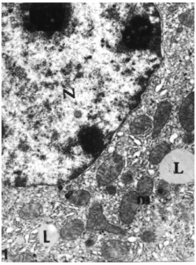
Control group: It was observed that the hepatocytes, bile canaliculus and perisunisoidal spaces exhbited normal structures (Figs. 1,2).
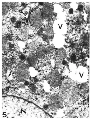
Acute group: There were prominent degenerative changes in the cytoplasm of hepatocytes in many sections. Slight dilatation of perinuclear cisternae and granular endoplasmic reticulum cisternae (Fig. 3), partial or total disruption of the inner mitochondrial membranes, increase in lysosomes, vacuolization and membranous whorl-like structures were observed. Lipid droplets were Effects of Phorbol Myristate Acetate on the Liver: a Biochemical and Ultrastructural Study
Figure 1. Control group. Normal appearance of nuclei (N), well-developed endoplasmic reticulum, pleomorphic mitochondria (m) and lipid droplets (L) are seen. X 12,250.
Figure 2. Control group. There were numerous mitochondria (m) with electron dense matrices and well-developed endoplasmic reticulum (ER). Nuclei (N), lipid droplets (L). X 7,700.
Figure 3. Acute group. There were dilated perinuclear cisternae (thin arrows) and endoplasmic reticulum cisternae (thick arrows) in the cytoplasm of hepatocyte. Nuclei (N), mitochondria (m). X 7,700.
Figure 4. Acute group. Irregular outlined nuclei (N) and slight mitochondrial degeneration (arrows) are seen. Sinusoid (S), bile canaliculi (Bc). X 7,700.
increased in comparison to the control group. Glycogen particles were arranged as small clumps throughout the cytoplasm (Figs. 4,5).
Chronic group: In this group, there were more severely degenerative changes than in the acute group; these changes were prominent in the organelles of hepatocytes, particularly in the mitochondria.
Most of the hepatocytes exhibited dilated perinuclear cisternae and granular endoplasmic reticulum cisternae forming small vacuoles throughout the cytoplasm (Fig. 6). Mitochondria were enlarged, their inner membranes were disrupted and their matrices were lytic. The distribution of the lipid droplets and glycogen particles was similar to that of the acute group (Figs. 7,8).
Discussion
The electron microscopic and biochemical findings in this study revealed that the various degrees of degenerative change occurring in the liver are a result of lipid peroxide derived from PMA. An increase in lysosomal fragility, changes in the microsomal enyzmes, cell death and collagen formation caused by free radicals in various organs have been reported previously (10). In our study,
the lipid peroxide level, which is an important sign of free radical damage, was found to be higher in the chronic group than in both control and acute groups. However, the lipid peroxide amount in the acute group was not significantly higher than that in the control group. This was related to the low free radical formation in the acute group. Moreover, we observed that the cell destruction in the acute group with less lipid peroxide was significantly smaller than that in the chronic group with more lipid peroxide.
In most of the sections, the mitochondria were the most affected organelles. There were severe degenerative changes such as disruption of inner membranes of mitochondria and formation of membranous whorl-like structures. The formation of these structures might be related to the dissolution of the inner mitochondrial membrane, which is particularly rich in PUFA. Sokol et al. suggested that an oxidant injury to the mitochondria of hepatocytes may be one of the initiating factors in hepatocellular damage (13).
Dilatation of endoplasmic reticulum cisternae and increase in lipid droplets were other prominent structural alterations observed in this study. The degeneration may be caused by peroxidative decomposition of membrane Effects of Phorbol Myristate Acetate on the Liver: a Biochemical and Ultrastructural Study
Figure 5. Acute group. Mitochondrial degeneration and vacuolization (V) are seen. Nucleus (N), membranous structures (arrows). X 7,700.
Figure 6. Chronic group. Dilated perinuclear cisterna (arrows) and granular endoplasmic reticulum cisternae (GER) are seen in the cytoplasm of the hepatocyte. X 12,250.
phospholipids of the cell organelles due to free radical attacks. In previous experiments, in which the effects of free radicals were triggered in mice livers by various methods, electron microscopic results disclosed the fragmentation of endoplasmic reticulum, degeneration of mitochondria, dilatation of the Golgi apparatus, disruption of plasma membranes, formation of large vacuoles and lipid droplets, degenerative changes in the cells of periportal areas and cell death (8-10, 13-16).
In conclusion, after injecting PMA into mice, lipid peroxide formed as an important sign of free radicals. Consequently, liver degeneration occurred moderately in the acute group and severely in the chronic group. We believe that the formation of free radicals increases after an injection of PMA, and this in turn causes cellular degeneration. This assumption is also supported by the high level of lipid peroxide measured in the chronic group.
Figure 7. Chronic group. Disruption of the mitochondrial membranes and cristae (arrows) are present. Nuclei (N). X. 7,700.
Figure 8. Chronic group. Irregular outlined nucleus (N), degenerated mitochondria (m) and dilated granular endoplasmic reticulum cisternae (arrows) are seen. Lipid (L). X 7,700.
References
1. Reilly, P.M., Schilder, H.J. and Bulkley, G.B. Pharmacologic approach to tissue injury mediated by free radicals and other reactive oxygen metabolites. Am J Surg. 1991. 161: 488-503. 2. Ohkawa, H., Ohishi, N. and Yagi, K. Assay for lipid peroxides in
animal tissues by thiobarbituric acid reaction. Anal Biochem. 1979. 95: 351-358.
3. Duyster, J., Schwende, H., Fitzke, E., Hidaka, H. and Dieter, P. Different roles of protein kinase C-beta and alfa in arachidonic acid cascade, superoxide formation and phosphoinositide hydrolysis. Biochem J. 1993. 292 (Pt 1): 203-207.
4. Gabrielian, E.S., Akopov, S.E., Grigorian, M.R. and Toumasian, K.S. Cerebrovascular injuries induced by activation of platelets and leukocytes in vivo and their correction by neurotropin Jpn J Pharmacol. 1992 60: 51-54.
5. Johnson, R.J., Couser, W.G., Chi, E.Y., Adler, S. and Klebanoff, S.J. New mechanism for glomerular injury. J Clin Invest. 1987. 79: 1379-1387.
6. Levy, R., Schlaeffer, F., Keynan, A., Nagauker, O., Yaari, A. and Sikuler, E. Increased neutrophil function induced by bile ligation in a rat model. Hepatology. 1993. 17: 908-914.
Effects of Phorbol Myristate Acetate on the Liver: a Biochemical and Ultrastructural Study
7. Pick, E. and Keisari, Y. Superoxide anion and hydrogen peroxide production by chemically elicited peritoneal macrophages-induction by multiple nonphagocytic stimuli. Cell Immunol. 1981. 59: 301-318.
8. French, S.W., Wong, K., Jui, L., Albano, E., Hagbjork, A.L., and Sundberg, M.I. Effect of ethanol on cytochrome P450 2E1 (CYP2E1), lipid peroxidation, and serum protein adduct formation in relation to liver pathology pathogenesis. Exp Mol Pathol. 1993. 58: 61-75.
9. Lieber, C.S., Interaction of alcohol with other drugs and nutrients. Implication for the therapy of alcoholic liver disease. Drugs 40 Suppl. 1990. 3: 23-44.
10. Stal, P. and Hultcrantz, R. Iron increases ethanol toxicity in rat liver. J Hepatol. 1993. 17: 108-115.
11. Towner, R.A., Reinke, L.A., Jansen, E.G. and Yamashiro, S. Enhancement of carbon tetrachloride-induced liver injury by a single dose of ethanol: proton magnetic resonance imaging (MRI) Studies in vivo. Biochim Biophys Acta. 1991. 15, 1096:222-230.
12. Millonig, G. Advantages of a phosphate buffer for O5O4 solutions and fixation. J Appl Physics. 1961. 32, 1637.
13. Sokol, R.J., Devereaux, M.W., Traber, M.G. and Shikes, R.H. Copper toxicity and lipid peroxidation in isolated rat hepatocytes: effect of vitamin E. Pediatr Res. 1989. 25: 55-62.
14. Hirai, K., Ikeda, K. and Wang, G.Y. Paraquat damage of rat liver mitochondria by superoxide production depends on extramitochondrial NADH. Toxicology. 1992. 72: 1-16. 15. Högberg, J., Moldeus, P., Arborgh, B., O'Brien, P.J. and Orrenius,
S. The consequences of lipid peroxidation in isolated hepatocytes. Eur J Biochem. 1975. 59: 457-462.
16. Shorma, B.K., Bacon, B.R., Britton, R.S., Park, C.H., Magiera, C.J., O'Neill, R., Dalton, N., Smanik, P. and Speroff, T. Prevention of hepatocyte injury and lipid peroxidation by iron chelators and alpha-tocopherol in isolated iron-loaded rat hepatocytes. Hepatology. 1990. 12: 31-39.