COBALT AND ASCORBIC ACID TREATMENT: EFFECTS ON TISSUE NITRIC OXIDE LEVELS IN
STREPTOZOTOCIN-DIABETIC RAT
ÖZLEM YILDIRIM
Ankara University, Science Faculty, Department of Biology, 06100 Tandogan/ANKARA
e-mail:[email protected]
(Received March 15, 2010; Accepted June 4, 2010)) ABSTRACT
The aim of present study was to determine the effect of cobalt (II) chloride (CoCl2) and CoCl2
plus ascorbic acid (AA) on liver and kidney nitric oxide level at different stage of diabetes. Three days after streptozotocin (STZ) injection, rats were divided into six groups: control, diabetic, control and diabetic treated with cobalt chloride, control and diabetics treated with cobalt chloride plus ascorbic acid. 0.5 mM CoCl2 or 0.5 mM CoCl2 plus 1 g/l ascorbic acid
were added to their drinking water daily. At the end of the second, fourth and sixth week of the treatment all animals were sacrified by decapitation. Liver and kidney tissues were removed for the determination of nitric oxide (NO) levels. Cobalt treatment effectively decreased the nitric oxide levels both in liver and kidney tissues of diabetic rats. Thus, the effect of oral administration of cobalt treatment may prove effective in improving the impaired nitrite status during the early stage of diabetes and ascorbic acid supplementation at this dose (1g/l) potentiates the effectiveness of cobalt action.
KEYWORDS: Nitric oxide; diabetes; cobalt; ascorbic acid; liver and
kidney
INTRODUCTION
Diabetes affects a variety of metabolic processes and characterized by alterations in tissue trace element and antioxidant concentrations which causes oxidative damages. Hyperglycemia is associated with excessive free radical generationand oxidant stress and reduction in the antioxidant status.
In recent studies, in vitro and in vivo insulinlike activity of several elements were showed by researchers (Abrams and Murrer, 1993; Brichard and Henquin, 1995; Novelli and Rodrigues, 1988; Özçelikay et al., 1996; Shisheva, Gefel and Shechter, 1992). Saker and co-workers (1998) were determined the potential role of enhanced tissue glucose uptake vs. decreased systemic glucose production in mediating the glycemia-lowering effect of cobalt chloride (CoCl2) in diabetic rats. In principle, the
glycemia-lowering effect of CoCl2 could be decreased systemic glucose production,
increased tissue glucose uptake, or a combination of the two mechanism. It has been previously reported that addition of 2 mM Co(II) in drinking water reduced the glycemia of streptozotocin-induced diabetic rats (Ybarra, Behrooz, Gabriel, Köseoğlu and Ismail-Beigi, 1997). Its action has been mainly ascribed to increased GLUT1 expression (Ybrra et al., 1997) and inhibition of gluconeogenesis (Saker at al., 1998) in STZ-diabetic rats. On the other hand, hydrophilic vitamin, ascorbic acid is used as a scavenger of reactive oxygen species. In aqueous solutions, ascorbate also scavenges reactive nitrogen oxide species efficiently. A sufficient supply of antioxidants either from the diet or from supplements might help to prevent or delay the occurrence of oxidative burden (Behrens and Madere, 1991; Siman and Eriksson, 1997). Cobalt alone or with a combination of ascorbate has been previously demonstrated to decrease lipid peroxidation in STZ-diabetic rats in various organs such as the liver, kidney (Yıldırım and Büyükbingöl, 2002, 2003a), heart and aorta (Yıldırım and Büyükbingöl, 2003b).
NO is a free radical gas that is synthesized from L-arginine by three different isoforms of NO synthase (NOS). NO plays an important role in homeostatic vasodilatation and regulation of blood flow, but excess release induces tissue disorders because of increased oxidative stress, especially caused by the production of peroxynitrite. It has been reported that NO is responsible for various vascular complications such as coronary arteriosclerosis and diabetes (Ignarro, Buga, Wood, Byrns and Chaudhuri, 1987; Ischiropoulos, Zhu and Beckman, 1992; Moncada and Higgs, 1993; Satoh, Fujimoto, Haruna, Arakawa, Horike, Komai, Sasaki, Tsujioka, Makino and Kashihara, 2005). NO is extremely unstable and easily oxidized, it is difficult to measure NO levels in vivo. Recently, evaluations of the stable NO end products nitrite (NO2−) and nitrate (NO3−), collectively
NOx, in biologic fluid, based on the Griess method, have been widely conducted to estimate NO production.
The objective of this study was to determine the effects of cobalt chloride and cobalt chloride plus ascorbate on nitric oxide levels which accompanies with diabetes. The levels of nitric oxide were measured in liver and kidney tissues of both control and diabetic rats (2 to 6 weeks).
MATERIALS AND METHODS
Animals
Diabetes was induced in male Sprague Dawley rats weighing 200-220 g, by a single intraperitoneal injection of streptozotocin (STZ, 45 mg/kg) prepared in 0.1M citrate buffer (pH 4.5). Control animals were injected with the buffer (0.1 ml/100 g) only. Diabetic state was confirmed by detecting the presence of hyperglycemia in blood drops obtained following tail snipping at 48 and 72 h after STZ injection, using Reflolux (Boehringer Mannheim, East Sussex, UK) glucometer and test strips. The minimal blood glucose value accepted for a diabetic rat was 13.9 mM. 2, 4 and 6 weeks of diabetes were studied. All animals were housed in air/humidity-controlled room, with 12 h dark/light cycle, and allowed food and water (control animals only) ad libitum throughout the experiment.
Animals were randomly assigned to 6 groups for each week: (1) C, control group (n=7), (2) D, diabetic group (n=9), (3) CCo, control group with cobalt treatment (n=7), (4) DCo, diabetic group with cobalt treatment (n=9), (5) CCoAA, control group with cobalt plus ascorbic acid treatment (n=7) and (6) DCoAA, diabetic group with cobalt plus ascorbic acid treatment (n=9). 0.5 mM cobalt (II) chloride were added to the drinking water of the CCo group, on the other hand, 0.5 mM cobalt (II) chloride and 1 g/l ascorbic acid were added to the CCoAA and DCoAA’s drinking water every day.
Tissue Sample Preparation
2, 4 and 6 weeks after STZ injection, animals were sacrified by decapitation without anaesthesia. Liver and kidney tissues of each rat were immediatelly removed, cleaned of gross adventitial tissue, blotted dry and placed in aliminum foil and stored at –80oC and assayed within 3 weeks. Tissue
samples were homogenized in 3 volumes of % 1.15 KCl at 4oC for 30 sec
(2x15 sec with 1 min cooling interval) at 3000 x g using a Teflon glass pestle with Heidolph homogenizer and centrifuged at 20,000 x g for 15 min. and supernatant was kept at -80oC for determination of NO levels.
Experimental Procedure
Nitric oxide, as an unstable molecule, that reacts with oxygen and biological molecules to form several end products (e.g.nitrite, nitrate and S-nitrosothiols) that can be determined by different methods (Braman and Hendrix, 1989; Mesaros, 1999). The most preferred method is based on Griess reaction which can be easily applied in the laboratory. The tissue sample homogenized as mentioned above (0.5 ml) was incubated with nitrate reductase (EC 1.6.6.2) from Aspergillus sp. (50 mU/100µl of sample) with NADPH (final concentration, 80 µmol/l) diluted in 20 mmol/l Tris buffer (pH 7.6) for 30 min at room temperature for nitrate reduction (Guevara, Iwanejko, Dembinska-Kiec, Pankiewicz, Wanat, Anna, Golabek, Bartus, Malczewska-Malec, and Szczudlik, 1998). After the reduction, 5% (w/v) ZnSO4 was added for deproteinization. Then this mixture was
cetrifuged at 5000 x g for 10 min. The samples nitrite levels were measured by Griess reaction (Green, Wagner, Glogowski, Skipper, Wishnok and Tannenbaum, 1982). The absorbance at 540 nm was measured. Nitric oxide levels were calculated from the standard curve constructed using known nitrite concentrations. Tissue nitrite levels were expressed as µmol g-1 wet
weight.
All experiments were performed using at least 7 animals in each group and the results were expressed as mean ± standard deviation (SD). Data were analyzed using Duncan’s multiple range test. A p value of < 0.05 was considered significant.
RESULTS
Induction of diabetes by STZ was confirmed by the presence of glucosuria within 24 hr. The general biochemical parameters and body weights in control and diabetic groups are shown in Table 1. As expected, diabetic animals were hyperglycemic (minimal blood glucose level was 13.9 mM) and their body weights were decreased, while nondiabetics were
normoglycemic and high body weight. Data also indicated that the plasma cholesterol and triglyceride levels were elevated in diabetics.
General features of the experimental model
CONTROL DIABETIC 2 WEEKS Glucose (mM) 6.8 ± 0.5 23.5 ± 3.9∗ Cholesterol (mM) 1.7 ± 0.3 3.8 ± 0.4∗ Triglycerides (mM) 1.1 ± 0.2 8.4 ± 1.3∗ Body weight (g) 249 ± 6 220 ± 8∗ Fluid intake (ml) 44 ± 5 170 ± 10∗ 4 WEEKS Glucose (mM) 7.3 ± 0.6 25.8 ± 5.9∗ Cholesterol (mM) 1.8 ± 0.5 4.3 ± 0.3∗ Triglycerides (mM) 1.2 ± 0.5 9.1± 2.7∗ Body weight (g) 273 ± 7 212 ± 5∗ Fluid intake (ml) 48 ± 4 160 ± 8∗ 6 WEEKS Glucose (mM) 7.8 ± 0.6 27.3 ± 3.3∗ Cholesterol (mM) 1.7 ± 0.3 4.2 ± 0.7∗ Triglycerides (mM) 1.3 ± 0.1 8.9 ± 2.1∗ Body weight (g) 288 ± 12 232± 13∗ Fluid intake (ml) 52 ± 5 168 ± 10∗
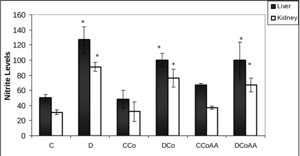
All values are means ± SD. Each group consisted of eight animals. Significance of difference was assessed by Duncan’s test. p values are shown as ∗p < 0.05 vs. control groups. At the end of second week, nitrite levels were determined in liver and kidney tissues of all groups (Fig 1). In diabetic rat liver and kidney, we determined 254% and 293% increase in nitrite levels, respectively. Increased levels of nitrite were lowered 21% by CoCl2 and CoCl2 plus AA
treatment.
Figure 1. Nitrite levels (µmol g-1 wet weight) of liver and kidney tissues at
the end of second week. Control (C), diabetic (D), cobalt treated control (Co), cobalt treated diabetic (DCo), cobalt plus ascorbic acid treated control (CCoAA) and cobalt plus ascorbic acid treated diabetic (DCoAA) groups. (Results are expressed as means ± SD (n=7 for C, Co and CoAA; n=9 for D, DCo and DCoAA groups). p values are shown as * p < 0.05 versus control groups. p < 0.05 versus diabetic groups)
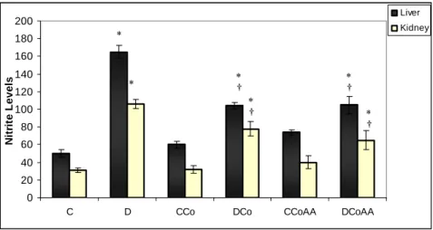
At the end of 4th week we determined significant decrease (p<0.05) in
diabetic liver and kidney nitrite levels compairing to controls (Fig 2). In diabetic liver which is treated by CoCl2, we determined 47% decrease in
0 20 40 60 80 100 120 140 160
C D CCo DCo CCoAA DCoAA
N it ri te L evel s Liver Kidney * * * * * *
nitrite levels compairing to control group. CoCl2 plus AA treatment also
made a significant reduction (p<0.05) in diabetic liver nitrite levels (56%). We also demonstrated a significant decreases (p<0.05) in diabetic kidney nitrite levels, with the treatment of CoCl2 and CoCl2 plus AA (Fig 2).
24% and 48% reduction were investigated respectively compairing with the control group.
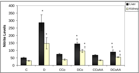
At the end of 6th week we determined liver and kidney nitrite levels (± SD) in control, diabetic, treated controls and treated diabetic groups (Fig 3). The nitrite levels of the CoCl2 and CoCl2 plus AA treated diabetic rats
exibited a statistically significant recovery in both tissues. CoCl2 with a
combination of AA treated diabetic rats, exibited 75% recovery for liver and 59% for kidney approaching to control levels. However, nitrite levels increased progressively in diabetic group and significantly higher as compared to controls at all studied times.
Figure 2. Nitrite levels (µmol g-1 wet weight) of liver and kidney tissues at
the end of fourth week. Control (C), diabetic (D), cobalt treated control (Co), cobalt treated diabetic (DCo), cobalt plus ascorbic acid treated control (CCoAA) and cobalt plus ascorbic acid treated diabetic (DCoAA) groups (Results are expressed as means ± SD 0 20 40 60 80 100 120 140 160 180 200
C D CCo DCo CCoAA DCoAA
N itr it e L eve ls Liver Kidney * * ∗† ∗ † ∗ † ∗ †
(n=7 for C, Co and CoAA; n=9 for D, DCo and DCoAA groups). p values are shown as * p < 0.05 versus control groups. p < 0.05 versus diabetic groups).
Figure 3. Nitrite levels (µmol g-1 wet weight) of liver and kidney tissues at
the end of sixth week. Control (C), diabetic (D), cobalt treated control (Co), cobalt treated diabetic (DCo), cobalt plus ascorbic acid treated control (CCoAA) and cobalt plus ascorbic acid treated diabetic (DCoAA) groups (Results are expressed as means ± SD (n=7 for C, Co and CoAA; n=9 for D, DCo and DCoAA groups). p values are shown as * p < 0.05 versus control groups. p < 0.05 versus diabetic groups). DISCUSSION
The biological importance of nitric oxide has been reported by several investigators (Ignarro, et al., 1987; Ischiropoulos, Zhu and Beckman, 1992; Moncada and Higgs, 1993; Satoh, Fujimoto, Haruna, et al., 2005). Excess or deficient NO has been related to several pathological conditions. Corbett et al. indicated that, NO formation may play a role in the destruction of the pancreatic β-cells during the development of diabetes (Corbett, Mikhael, Shimizu, Frederick, Misco, McDaniel and Kanagawa, 1993). Under diabetic conditions, free radicals such as O2− and NO are produced as a result of the
0 50 100 150 200 250 300 350 400
C D CCo DCo CCoAA DCoAA
N it rit e Le v e ls Liver Kidney * * ∗ † ∗ † ∗† ∗ †
induction of the glycation reaction in β-cells that have been affected by diabetic oxidative stress. NO interacts with O2− to form the highly reactive
hydroxyl radical that leads to reactive oxidative damage (Beckman, Beckman, Chen, Marshall and Freeman, 1990). It is also well established that the increase in glucose blood levels in diabetes mellitus increase the cytosolic levels of NADPH oxidase resulting in an increase in nitric oxide production (Grisham, Jourd-Heuil and Wink, 1999).
In our recent study, we have demonstrated increased nitrite levels of diabetic and treated groups (p < 0.05) compared to control groups. In addition, NO targets intracellular antioxidative enzymes, resulting the loss of their function. At the end of 2nd, 4th and 6 th weeks treatment with cobalt
chloride and cobalt chloride plus ascorbic acid, nitrite–nitrate levels were significantly decreased compared to diabetic group (p < 0.05) (Fig. 1, 2 and 3). Our previous studies indicate that the free radical scavenging property of CoCl2 and CoCl2 plus AA suggests that CoCl2 might protect against diabetic
oxidative stress (10-12). Low molecular weight compounds potentially capable to remove excess pathogenic NO due to NO overproduction. It has been reported that NO binding is followed by oxidation of Co(II) to Co(III) with the formation of a Co-NO complex formally containing Co(III) (Rochelle, Morana, Kruszyna, Russell, Wilcox, Smith, 1995; Kruszyna, Megyar, Rochelle, Russell, Wilcox and Smith, 1998).
In present study, we have shown that there is a decrease in the level of nitric oxide by the treatment with cobalt alone and vitamin C plus cobalt in liver and kidney tissues during the progression of diabetes. Our study was done at different stages of diabetes to determine whether there is a significant difference in cobalt chloride and cobalt chloride plus ascorbic acid treatment on nitrite levels. The nitrite content in control group is remained unchanged throughout the 6th week diabetic group the nitrite
levels were increased up to 6th week comparing with control groups. Kidney
nitrite level was decreased at 2nd week because of advanced glycation end
products which have oxidizing potential can quench nitric oxide as reported in Bucala and his colleagues research (Bucala, Tracey and Cerami, 1991). At 4th and 6 th weeks renal nitrite level was increased because of some
disturbances in amino acid metabolism in diabetes may contribute to increased intrarenal NO production. Increased glucose concentration in diabetes induces elevation in intracellular levels of diacylglycerol activates protein kinase C, which has been shown to activate nitric oxide synthases.
Studies on patients with diabetes mellitus, a disorder associated with hyperglycemia and accompanying complications, have revealed oxidative stress loads. Superoxide, hydrogen peroxide, hydroxyl radical and singlet oxygen which are named as reactive oxygen species have been implicated to play important roles in diabetes (Halliwell, 1997; Baynes and Thorne, 1999; Opara, 2002). NO may protect against reactive oxygen species from the tissue (Wink, Vodovotz, Grisham, Degraff, Cook, Pacelli, Krishna and Mitchell, 1999). It has been also indicated that, nitric oxide is an important mediator, governing a range of physiological functions in animals, from controlling smooth muscle tone in the cardiovascular, gastrointestinal, respiratory and genitourinary systems, to neurotransmission and a role in immune function and inflamation (Singh and Evans, 1997).
Diabetes mellitus is associated with increased lipid peroxidation, which may contribute to long-term tissue damage and increased oxidative stress (Yıldırım and Büyükbingöl, 2002; 2003a; 2003b). Our data have shown that administration of the cobalt and cobalt plus ascorbic acid to diabetic rats decreased the tissue levels of nitrite significantly, indicating that it may be useful for correcting oxidative stress.
ÖZET: Bu çalışmanın amacı kobalt II klorür (CoCl2) ve kobalt II klorür ile birlikte verilen
askorbik asitin (AA) diyabetin çeşitli haftalarında karaciğer ve böbrek nitrik oksit düzeylerine olan etkisini belirlemektir. Streptozotozin (STZ) uygulamasından üç gün sonra sıçanlar altı gruba ayrılmışlardır: kontrol, diyabetik, kontrol ve diyabetiklerin kobalt klorür ile tedavili grupları, kontrol ve diyabetiklerin kobalt klorür ve askorbik asit ile tedavili grupları. 0.5 mM CoCl2 veya 0.5 mM CoCl2 artı 1 g/l askorbik asit, sıçanların günlük içme suyuna katılarak
verilmiştir. Çalışmanın ikinci, dördüncü ve altıncı haftaları sonunda sıçanlar anestezi altında öldürülmüşlerdir. Karaciğer ve böbrek dokuları nitrik oksit (NO) düzeyleri belirlenmek amacı ile çıkarılmıştır. Kobalt tedavisinin hem karaciğer hem de böbrek dokusunda nitrik oksit düzeylerini etkili bir şekilde azalttığı bulunmuştur. Bu durum, oral yolla kobalt alımının diyabetin erken döneminde bozulan nitrit düzeylerini normale döndürmede etkili olduğunu ve askorbik asit ilavesinin de belirtilen dozda (1g/l) kobalt etkisine katkıda bulunduğunu göstermektedir.
REFERENCES
Abrams MJ, Murrer BA (1993). Metal compounds in therapy and diagnosis. Scien. 261: 725- 730.
Baynes JW, Thorne S (1999). Role of oxidative stress diabetic complications: a new perspective on an old paradigm. Diabetes 48: 1-9. Beckman JS, Beckman TW, Chen J, Marshall PA, Freeman BA (1990). Apparent hydroxyl radical production by peroxiynitrite: implications for endothelial injury from nitric oxide and superoxide. Proc. Nat. Acad. Scien. USA, 87: 1620–1624.
Behrens WA, Madere R (1991). Vitamin C and vitamin E status in the spontaneously diabetic BB rat before the onset of diabetes. Metabol. 40(1): 72-76.
Braman RS, Hendrix SA (1989). Nanogram nitrite and nitrate determination in environmental and biological materials by vanadium (III) reduction with chemiluminescence detection. Anal. Chem. 61: 2715-2718.
Brichard SM, Henquin JC (1995). The role of vanadium in the management of diabetes. TiPS 16: 265-270.
Bucala R, Tracey KJ, Cerami A (1991). Advanced glycosylation products quench nitric oxide and mediate defective endothelium-dependent vasodilation in experimental diabetes. J. Clin. Invest. 87: 432-438.
Corbett JA, Mikhael A, Shimizu J, Frederick K, Misco TP, McDaniel ML, Kanagawa O (1993). Nitric oxide production in islets from nonobese diabetic mice: aminoguanidine-sensitive and resistant stages in the immunological diabetic process. Proc. Nat. Acad. Scien. USA 90: 8992-8995.
Green LC, Wagner DA, Glogowski J, Skipper PL, Wishnok JS, Tannenbaum SR (1982). Analysis of nitrate, nitrite, and [15N] nitrate in
biological fluids. Anal.Bioch. 126: 131-138.
Grisham MB, Jourd-Heuil D, Wink DA (1999). Nitric oxide. I. Physiological chemistry of nitric oxide and its metabolites: implications in inflammation. Am. J. Physiol. 276: G315–21.
Guevara I, Iwanejko J, Dembinska-Kiec A, Pankiewicz J, Wanat A, Anna P, Golabek I, Bartus S, Malczewska-Malec M, Szczudlik A (1998).Determination of nitrite/nitrate in human biological material by the simple Griess reaction. Clin. Chim. Acta 274: 177-188.
Halliwell B (1997). Antioxidant and human disease: a general introduction. Nut. Rev. 55(1): S44-S52.
Ignarro LJ, Buga GM, Wood KS, Byrns RE, Chaudhuri G (1987). Endothelium-derived relaxing factor produced and released from artery and vein is nitric oxide. Proc. Nat. Acad. Scien.USA 84 (24): 9265–9269. Ischiropoulos H, Zhu L, Beckman JS (1992). Peroxynitrite formation from macrophage-derived nitric oxide. Arch. Bioch. Biophy. 298 (2): 446–451. Kruszyna H, Megyar JS, Rochelle LG, Russell MA, Wilcox DE, Smith RP (1998). Spectroscopic studies of nitric oxide (NO) interactions with cobalamins: reaction of NO with superoxocobalamin(III) likely accounts for cobalamin reversal of the biological effects of NO. J. Pharmacol. Exper. Therap. 285: 665–671.
Mesaros S (1999). Determination of nitric oxide saturated solution by amperometry on modified microelectrode. Meth. Enzymol. 301: 160-168. Moncada S, Higgs A (1993). The L-arginine-nitric oxide pathway. New England J. Med. 329 (27): 2002–2012.
Novelli ELB, Rodrigues NL (1988). Effect of nickel chloride on streptozotocin-induced diabetes in rats. Can. J. Physiol. Pharmacol. 66: 663-665.
Opara EC (2002). Oxidative stress, micronutrients, diabetes mellitus and its complications. J. Royal Soc. Prom. Health 122: 28-34.
Özçelikay TA, Becker DJ, Ongemba LN, Pottier A-M, Henquin J-C. Brichard S M. (1996). Improvement of glucose and lipid metabolism in diabetic rats treated with molybdate. Am. J. Physiol. 270: E344-E352. Rochelle LG, Morana SJ, Kruszyna H, Russell MA, Wilcox DE, Smith RP (1995). Interactions between hydroxocobalamin and nitric oxide (NO): evidence for a redox reaction between NO and reduced cobalamin and reversible NO binding to oxidized cobalamin. J. Pharmacol. Exp. Ther. 275: 48–52.
Saker F, Ybarra J, Leahy P, Hanson RW, Kalhan SC Ismail-Beigi F (1998). Glycemia-lowering effect of cobalt chloride in the diabetic rat: role of decreased gluconeogenesis. Am. J. Physiol. 274: E984-E991.
Satoh M, Fujimoto S, Haruna Y, Arakawa S, Horike H, Komai N, Sasaki T, Tsujioka K, Makino H, Kashihara N (2005). NAD(P)H oxidase and uncoupled nitric oxide synthase are major sources of glomerular superoxide in rats with experimental diabetic nephropathy. Am. J. Physiol. Renal Physiol. 288: F1144–F1152.
Shisheva A, Gefel D, Shechter Y (1992). Insulinlike effects of zinc in vitro and in vivo. Diabetes, 41: 982-988.
Siman CM, Eriksson UJ (1997). Vitamin C supplementation of the maternal diet reduces the rate of malformation in the offspring of diabetic rats. Diabetologia 40(12): 1416-1424.
Singh S, Evans TW (1997). Nitric oxide, the biological mediator of the decade: fact of fiction?. Eur. Res. J 10: 699-707.
Ybarra J, Behrooz A, Gabriel A, Köseoğlu MH, Ismail-Beigi F (1997). Glycemia-lowering effect of cobalt chloride in the diabetic rat: increased GLUT1 mRNA expression. Mol. Cell. Endocrinol. 133: 151-160.
Yıldırım Ö, Büyükbingöl Z (2002). Effects of supplementation with a combination of cobalt and ascorbic acid on antioxidant enzymes and lipid peroxidation levels in streptozotocin diabetic rat liver. Biol. Trace Elem. Res. 90: 143–154.
Yıldırım Ö, Büyükbingöl, Z (2003a). In vivo effects of vitamin C with cobalt on oxidative stress in experimental diabetic rat kidney, Diabetes Nut. Metab. 6: 208–213.
Yıldırım Ö, Büyükbingöl, Z. (2003b). Effect of cobalt on the oxidative status in heart and aorta of streptozotocin-induced diabetic rats. Cell Biochem. Funct. 21: 27–33.
Wink DA, Vodovotz Y, Grisham MB, Degraff W, Cook JC, Pacelli R, Krishna MC, Mitchell JB (1999). Antioxidant effects of nitric oxide. Method. Enzymol. 301: 413-424.