101 Case Report / Olgu Sunumu
Turkish Journal of Thoracic and Cardiovascular Surgery 2021;29(1):101-104
http://dx.doi.org/doi: 10.5606/tgkdc.dergisi.2021.20988
Transcatheter closure of the aortopulmonary window in a three-month-old infant
with a symmetric membranous ventricular septal defect occluder device
Üç aylık bebekte simetrik membranöz ventriküler septal defekt oklüder cihazı ile transkateter aortopulmoner pencere kapatılması
Ayşe Yıldırım1, Abdullah Erdem2, Aysu Türkmen Karaağaç3
ÖZ
Aortopulmoner pencere olgularının birçoğu cerrahi olarak kapatılsa da, uygun hastalarda perkütan kapatma da tercih edilebilmektedir. Pulmoner ve aort kapaklarından, koroner arterden ve pulmoner arter bifurkasyonundan uzak olan, yeterli septal kenarlara sahip defektlerin perkütan kapatma için uygun olduğu düşünülmektedir. Üç aylık, 4 kg ağırlığında bir erkek bebek, emzirme sırasında çabuk yorulma, kilo alamama, kalp üfürümü ve solunum sıkıntısı şikâyetleri ile çocuk kardiyoloji bölümümüze sevk edildi. Ekokardiyografide geniş aortopulmoner pencere (5.3 mm) ve sol kalp boşluklarında genişleme izlendi. Geniş aortopulmoner pencere, simetrik membranöz ventriküler septal defekt oklüder cihaz kullanılarak kapatıldı. Kapatma işlemi antegrad yolla arteriyovenöz lup oluşturmadan gerçekleştirildi. Sonuç olarak, seçilmiş bebeklerde, geniş aortopulmoner pencereyi kapatmak için simetrik membranöz ventriküler septal defekt oklüder cihazı kullanımı, cerrahiye güvenilir ve etkili bir alternatif olarak görünmektedir.
Anah tar söz cük ler: Aortopulmoner pencere, kalp yetmezliği, yenidoğan, transkateter kapatma.
ABSTRACT
Although most of aortopulmonary window cases are closed surgically, percutaneous closure can be also used in suitable patients. Defects which are far from the pulmonary and aortic valves, coronary artery, and pulmonary artery bifurcation, with adequate septal rims are considered suitable for percutaneous closure. A three-month-old male infant weighing 4 kg was referred to our pediatric cardiology department with the complaints of fatigue while breastfeeding, difficulty in weight gain, heart murmur, and respiratory distress. A large aortopulmonary window (5.3 mm) and left heart chamber dilatation were detected on echocardiography. The large aortopulmonary window was closed using a symmetric membranous ventricular septal defect occluder device. The closure procedure was performed via the antegrade route without forming an arteriovenous loop. In conclusion, the use of a symmetric membranous ventricular septal defect device for closure of large aortopulmonary window seems to be a safe and effective alternative to surgery in selected infants.
Keywords: Aortopulmonary window, heart failure, infant, transcatheter closure.
Institution where the research was done:
University of Health Sciences, Kartal Koşuyolu Training and Research Hospital, Istanbul, Turkey
Author Affiliations:
1Department of Pediatric Cardiology, University of Health Sciences, Kartal Koşuyolu Training and Research Hospital, Istanbul, Turkey 2Department of Pediatric Cardiology, Medipol University Medical Faculty, Istanbul, Turkey
3Department of Pediatric, University of Health Sciences Kartal Koşuyolu Research and Training Hospital, Istanbul, Turkey
Received: October 20, 2020 Accepted: December 01, 2020 Published online: January 13, 2021
Correspondence: Abdullah Erdem, MD. Medipol Üniversitesi Tıp Fakültesi Pediatrik Kardiyoloji Bilim Dalı, 34214 Bağcılar, İstanbul, Türkiye.
Tel: +90 212 - 487 39 93 e-mail: [email protected]
©2021 All right reserved by the Turkish Society of Cardiovascular Surgery.
This is an open access article under the terms of the Creative Commons Attribution-NonCommercial License, which permits use, distribution and reproduction in any medium, provided the original work is properly cited and is not used for commercial purposes (http://creativecommons.org/licenses/by-nc/4.0/).
Yıldırım A, Erdem A, Türkmen Karaağaç A. Transcatheter closure of the aortopulmonary window in a three-month-old infant with a symmetric membranous ventricular septal defect occluder device. Turk Gogus Kalp Dama 2021;29(1):101-104
Cite this article as: According to the Society of Thoracic Surgeons Congenital Heart Surgery Nomenclature and Database Project, there are four types of aortopulmonary window (APW): type I is a proximal defect with
very small inferior aortopulmonary septum above the semilunar valves; type II consists of distal defects located in the upper portion of the ascending aorta without a superior septum; type III defects are the
102
Turk Gogus Kalp Dama 2021;29(1):101-104
large ones which extend from the semilunar valves to the pulmonary artery bifurcation; and type IV is the intermediate type which has a central defect
with adequate superior and inferior rims.[1] The
intermediate APW is the most suitable type for percutaneous closure (PC). Early closure is required to avoid cardiac failure or irreversible pulmonary vascular disease in large APWs. In the literature, several successful studies have been reported since 1995 using different devices.[2-10]
In this article, we report an APW closure with a symmetric membranous ventricular septal defect (VSD) occluder device without forming an arteriovenous loop.
CASE REPORT
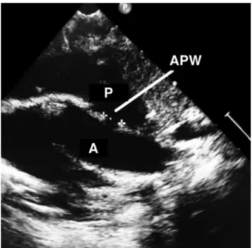
A three-month-old male infant weighing 4 kg was referred to our pediatric cardiology department with the complaints of fatigue while breastfeeding, difficulty in weight gain, heart murmur, and respiratory distress. On admission, the patient was tachypneic with a hyperdynamic precordium and bounding pulses. The chest X-ray showed cardiomegaly with prominent pulmonary vascular fields. Transthoracic echocardiography revealed an enlarged left ventricle and left atrium, pulmonary arterial dilatation, moderate mitral valve regurgitation, and predominant left-to-right shunt of intermediate type APW (5.3 mm). There were
no associated cardiac abnormalities. The defect had adequate superior and inferior rims with a reasonable distance from the ostium of the left coronary artery, both semilunar valves, and the origin of the right pulmonary artery. Therefore, it was considered to be suitable for PC (Figure 1). A written informed consent was obtained from each parent.
The procedure was performed under general anesthesia. The 4Fr and 5Fr sheaths were located into the right femoral artery and right femoral vein, respectively. Unfractionated heparin (100 U/kg) was administered intravenously. The mean aorta and pulmonary artery pressures were 48 and 33 mmHg, respectively. An aortic root angiography was performed using a pigtail catheter, which revealed a 5.1-mm APW located 8-mm away from the left coronary artery. This diameter was compatible with that measured by transthoracic echocardiography. The defect was crossed via the antegrade route from the pulmonary artery to the aorta using a 5Fr Judkins right coronary catheter (Medtronic Inc., Minneapolis, MN, USA) with an angled tip of 0.035-inch hydrophilic guidewire. The wire was advanced through the descending aorta and exchanged by a 0.035-inch stiff wire. Over this wire, a 6Fr CeraTM membranous VSD occluder delivery system (Lifetech Scientific Medical Co. Ltd., Shenzhen, China) was advanced through the femoral vein and APW into the descending aorta. A 7-mm symmetric membranous
VSD occluder (CeraTM; Lifetech Scientific Medical
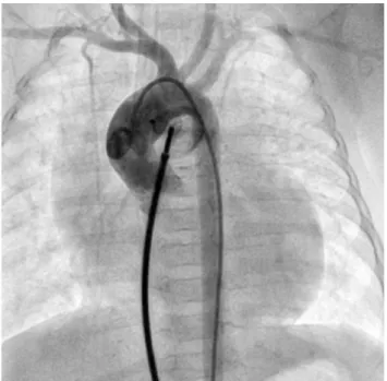
Co. Ltd., Shenzhen, China) device was advanced to descending aorta through the 6Fr delivery system. After the first disc was opened in the aorta and the entire system was withdrawn to the pulmonary side of the defect, the second disc was opened under fluoroscopic and transthoracic echocardiographic guidance. As the control aortography and echocardiography showed no protrusion into the aortic and pulmonary sides, no residual shunt and no compression on the left coronary artery ostium, the device was released and the defect was occluded successfully (Figure 2, Video 1). The total procedural time was 40 min and the total fluoroscopy time was 3.5 min. On control echocardiography on the following day, there was no residual shunt (Video 2). The patient received antibiotherapy before and after the procedure. Acetylsalicylic acid was advised at a dose of 3 mg/kg/day for three months, and the infant was discharged on Day 3 of the procedure. At one-month follow-up, the infant gained 600 g and the dilatation in the left heart cavities regressed. The patient has been under follow-up for one year without any complications.
Figure 1. An echocardiographic view showing aorta and
pulmonary artery and centrally located aortopulmonary window.
103 Yıldırım et al.
Transcatheter closure of aortopulmonary window
DISCUSSION
As the large APW carries a high risk for cardiac failure and irreversible pulmonary vascular disease, surgical or transcatheter closure is required at early
ages. Although the conventional treatment is surgery, successful PC procedures have been reported in the literature, since 1995. The potential risks of the PC of a large APW in low-weight infants include damage to the semilunar valves by the device discs and the possibility of device protrusion into the arterial lumen and risk of coronary compression.[3,4] Selection of the device is important in infants to avoid these complications. There are only a few reports in the literature about the PC of large APW in small infants.[3-6,9,10] Trehan
et al.[4] reported three symptomatic infants with large
defects (8 to 10 mm), and two were intermediate-type windows, closed by duct and muscular VSD occluders, while the third was the window with a mild inferior rim deficiency, closed by a perimembranous VSD occluder. The authors recommended this device, owing to its relatively flat profile (a waist diameter of 1.5 mm), and asymmetric discs suitable for closure of an APW with some deficiency. Kosmač et al.[9] used a 5-mm perimembranous Amplatzer™ VSD occluder device (St. Jude Medical Inc., MN, US) for the intermediate-type APW of a 3,600 g newborn. They considered that the tunnel-shaped anatomy of the patient’s defect facilitated the positioning of the device. In another case report, Sabnis et al.[5] reported the closure of a 4.4-mm defect with adequate rims using an 8-6 Amplatzer™ duct occluder (ADO; St. Jude Medical Inc., MN, USA) in a 2,600-g newborn. Although there are a few successful closure reports in the literature, duct occluders tend to protrude toward the main pulmonary artery, thus carrying a risk of obstructing the vessel and coronary artery.[3,4,9] Atrial septal defect occluders with very large rims, on the other hand, may obstruct the semilunar valves or left coronary ostium, due to
Video 1. An angiographic appearance showing the device before
and after detachment.
Video 2. An echocardiographic view showing the correct position
of device with no residual shunt or protrusion into vessels.
Figure 2. Control aortography before device releasing. The
device position is correct, and no residual shunt and protrusion into vessels are seen.
104
Turk Gogus Kalp Dama 2021;29(1):101-104
their bulky device structure; therefore, these devices seem to be inappropriate for patients with a low weight and large APW.[3,4]
The proximal and distal type small APW can be closed by the ADO™ II additional sizes. This device seems to be suitable for the PC of small defects (<4 mm) in close proximity to pulmonary bifurcation, aortic valve and/or coronary ostium, thanks to its smaller disc size, which minimizes the risk of obstruction of vessel lumen, semilunar valve, and left coronary artery.[7,8] Alternatively, Odemis et al.[10] used a 7-mm
Nit-Occlud® PDA-R device (BVM Medical Ltd.,
Leicestershire, UK) and successfully closed the APW, type I defect of 5 mm, in a three-month-old patient. They offered this device owing to its lower profile, requiring smaller sheet sizes, and the completely flat retention disk. Despite limited experiences, its small skirt at the distal side and very flat design may provide the advantage of reducing the risk of compression on the coronary arteries.
The type, size, and location of the defect, suitability of the device to be selected, and the body weight of the infant are critical factors in the selection of an appropriate device. The ideal device for PC of APW should be double-disc with a short waist and short skirt rim, thereby, reducing the risk of protrusion into the pulmonary artery and compression of coronary artery and requiring a small sheath. Therefore, in the present case, we preferred a 7-mm symmetric membranous VSD occluder device (waist length: 3 mm, right disc: 11 mm and left disc: 11 mm) through its short waist and two equal and short retention discs (only 2 mm for each side) and the device was fitted to the defect. After the procedure, we observed no residual shunt or complications such as protrusion into the pulmonary vessels or compression onto the coronary arteries. In addition, requirement of a small 6Fr sheath prevented the damage to the small vessels of the infant.
During the procedure, we passed directly from the antegrade route using a soft hydrophilic guidewire and a 5Fr right Judkins catheter from pulmonary artery to ascending aorta. This technique facilitated the procedure and the procedure completed without using a snare in a short time. In our technique, we used only 4Fr sheath in the arterial side and we used the arterial way to show defect before and after releasing the device. Of note, the interventional cardiologist should know the properties of the materials at shelf and choose the most appropriate one according to each individual case. In addition, a flexible strategy should be followed according to the characteristics of the case and the easiest and simplest way should be preferred, particularly in infants.
In conclusion, it is challenging to choose the appropriate device and, to date, there is no consensus on the choice of an optimal device for aortopulmonary window closure. The intermediate type, as in our patient, consists of a central defect with adequate superior and inferior rims, which makes it most suitable for percutaneous closure. We recommend the symmetric membranous ventricular septal defect occluder and antegrade passing technique as a safe and effective alternative to surgery for percutaneous closure of aortopulmonary window in infants with large defects soon after the diagnosis.
Declaration of conflicting interests
The authors declared no conflicts of interest with respect to the authorship and/or publication of this article.
Funding
The authors received no financial support for the research and/or authorship of this article.
REFERENCES
1. Jacobs JP, Quintessenza JA, Gaynor JW, Burke RP, Mavroudis C. Congenital Heart Surgery Nomenclature and Database Project: aortopulmonary window. Ann Thorac Surg 2000;69(4 Suppl):S44-9.
2. Stamato T, Benson LN, Smallhorn JF, Freedom RM. Transcatheter closure of an aortopulmonary window with a modified double umbrella occluder system. Cathet Cardiovasc Diagn 1995;35:165-7.
3. Campos-Quintero A, García-Montes JA, Zabal-Cerdeira C, Cervantes-Salazar JL, Calderón-Colmenero J, Sandoval JP. Transcatheter device closure of aortopulmonary window. Is there a need for an alternative strategy to surgery? Rev Esp Cardiol (Engl Ed) 2019;72:349-51.
4. Trehan V, Nigam A, Tyagi S. Percutaneous closure of nonrestrictive aortopulmonary window in three infants. Catheter Cardiovasc Interv 2008;71:405-11.
5. Sabnis GR, Shah HC, Lanjewar CP, Malik S, Kerkar PG. Transcatheter closure of large aortopulmonary window in a neonate. Ann Pediatr Cardiol 2018;11:228-30.
6. Pillekamp F, Hannes T, Koch D, Brockmeier K, Sreeram N. Transcatheter closure of symptomatic aortopulmonary window in an infant. Images Paediatr Cardiol 2008;10:11-7. 7. Uçar T, Karagözlü S, Ramoğlu MG, Tutar E. Transcatheter closure of aortopulmonary window with Amplatzer duct occluder II: additional size. Cardiol Young 2020;30:424-6. 8. Fiszer R, Zbroński K, Szkutnik M. Percutaneous closure of
an aortopulmonary window using Amplatzer Duct Occluder II: Additional Sizes: the first reported case. Cardiol Young 2017;27:812-5.
9. Kosmač B, Eicken A, Kühn A, Heinrich M, Ewert P. Percutaneous device closure of an aortopulmonary window in a small infant. Int J Cardiol 2013;168:e102-3.
10. Odemis E, Guvenc O, Saygi M, Demir IH. Closure of aortopulmonary window using Nit-Occlud® PDA-R device in a 3-month-old infant. Pediatr Int 2016;58:754-6.