Treatment of osteonecrosis of the femoral head
with free vascularized fibular grafting:
results of 7.6-year follow-up
Correspondence: Hakan Çift, MD. İstanbul Medipol Üniversitesi Tıp Fakültesi,
Ortopedi ve Travmatoloji Anabilim Dalı, İstanbul, Turkey. Tel: +90 212 – 440 10 00 e-mail: [email protected]
Submitted: July 23, 2015 Accepted: January 14, 2016
©2016 Turkish Association of Orthopaedics and Traumatology
Available online at www.aott.org.tr doi: 10.3944/AOTT.2015.15.0328 QR (Quick Response) Code
doi: 10.3944/AOTT.2015.15.0328
Mehmet Bekir ÜNAL1, Eren CANSÜ2, Fatih PARMAKSIZOĞLU3, Hakan ÇİFT4, Serkan GÜRCAN5
1Göztepe Medicalpark Hospital, Department of Orthopedics and Traumatology, İstanbul, Turkey 2Marmara University Faculty of Medicine, Department of Orthopedics and Traumatology, İstanbul, Turkey 3Yeni Yüzyıl University Faculty of Medicine, Department of Orthopedics and Traumatology, İstanbul, Turkey 4İstanbul Medipol University Faculty of Medicine, Department of Orthopedics and Traumatology, İstanbul, Turkey
5Private Medicana Hospital, Department of Orthopedics and Traumatology, İstanbul, Turkey
Osteonecrosis of the femoral head is a disabling disease that frequently affects adults aged 20–50 years old.[1]
Re-gardless of etiology, the disease often progresses to femo-ral head collapse and hip joint arthritis if left untreated.
[2] Treatment options for the disease mainly depend
upon the stage and age of the patient. According to many studies, the etiology of the disease is not a determinant for a particular modality of treatment.[3,4] Older age,
se-Objective: The aim of this study was to determine long-term follow-up results of patients with femo-ral head osteonecrosis who were treated with free vascularized fibular grafting (FVFG).
Methods: The results of 28 hips of 21 patients (16 male, 5 female) who underwent FVFG for treat-ment of osteonecrosis of the femoral head were retrospectively reviewed. Mean age of patients at time of surgery was 30.7 years (range: 15–53 years). Mean follow-up duration was 7.6 years (range: 5–9.2 years).
Results: During follow-up, 1 patient died because of leukemia, and 1 patient was lost. The remaining 26 hips of 19 patients were evaluated. According to Ficat classification, at time of surgery, 17 hips were grade II, and 9 hips were grade III, 3 hips underwent total hip arthroplasty. Postoperative Harris Hip Score (HHS) in grade II disease was excellent in 12 patients, good in 3 patients, and fair in 1 patient. In grade III disease, 1 patient was excellent, 5 patients were good, and 1 patient was fair. There was a sig-nificant increase in HHS scores from preoperatively to postoperatively (61±9.7 vs 84±17.8, p<0.001). Conclusion: FVFG yields extremely good results, particularly in pre-collapse stages of the disease in young patients. The operation time does not markedly increase if the surgical team is knowledgeable of the procedure and the residual fibular defect of the donor site does not impair functions of daily living. Keywords: Free vascularized fibular grafting; osteonecrosis of the femoral head.
verely collapsed femoral head, and arthritic patients are accepted as candidates for arthroplasty. Treatment op-tions before collapse of the femoral head are intended to achieve regeneration of the subchondral necrotic bone to prevent collapse and preserve sphericity of the femoral head. Core decompression with or without cancellous bone grafting, non-vascularized strut grafts, tantalum rods, electrode insertions, different type of osteotomies, and free or muscle pedicled viable bone grafts have been presented for this purpose.[5–7] Among these techniques,
core decompression with vascularized fibular grafting technique removes all necrotic bone and fills the remain-ing cavity with vascularized bone graft and spongious graft that contains both osteoinductive and osteocon-ductive properties. While the procedure is not new, most of the reports in the literature are from a few pioneer-ing clinics utilizpioneer-ing the free vascularized fibular graft-ing (FVFG) technique. In this study, we presented our experience in treatment with FVFG in pre- and post-collapse stages of the disease, reporting medium-term (mean: 7.6 years) follow-up results.
Patients and methods
In this study, 28 hips of 21 patients (16 male, 5 female) who underwent FVFG for treatment of osteonecrosis of the femoral head between 2005 and 2009 were ret-rospectively reviewed. Mean age of the patients at time of surgery was 30.7 years (range: 15–53 years). Seven patients who had bilateral hip involvement were oper-ated on in 5- to 8-month intervals. The etiologies were collum femoris fracture in 2 hips (7.1%), steroid use in 18 hips (64.3%), and idiopathic in 8 hips (28.5%). The osteonecrosis of patients was diagnosed and staged according to Ficat classification by evaluation of plain roentgenogram and magnetic resonance imaging (MRI) findings. Mean follow-up duration of patients was 7.6 years (range: 5–9.2 years). Due to less than 10-year follow-up, conversion to total hip arthroplasty (THA) was accepted as failure of the procedure. Radiologic progress of treatment was followed with plain roent-genograms taken in 3-month intervals for the first year and once yearly thereafter. Radiographs of patients were evaluated for a trabecular bone appearance at the tip of the vascularized fibular graft, indicating new bone for-mation. Preservation of spherical femoral head and loss of subchondral cystic lesions and crescent sign were also accepted as part of the healing of the lesion. Loss of sphericity and decrease in the joint space indicated pro-gression of the disease. Functional outcomes of survived hips were evaluated using the Harris Hip Score (HHS) system. Scores greater than 90 points were accepted as
excellent, scores between 80 and 89 points were good, scores between 70 and 79 points were fair, and scores less than 70 points were poor. The original technique described by Urbaniak and Aldridge et al. was used in all patients.[8] All patients were operated under general
anesthesia in the lateral decubitus position by the same surgical team, who were experienced in microsurgery. To reduce operation time, preparation of the hip and harvesting of fibular graft were performed concurrently with 2 teams (Figure 1). Preoperatively, the extent and spatial localization of the lesion was studied on mag-netic resonance images. Intraoperative C-arm fluoro-scope was used for anteroposterior and frog-leg lateral views (Figure 2). Prior to placement of the fibular graft, bone graft mixture composed of cancellous autografts harvested from the greater trochanter and 5–10 cc de-mineralized bone matrix (DBM) allograft (Osteoplant Activagen Injectable Paste OGS-ACI5, Bioteck S.p.A.,
Fig. 1. Completely scrubbed lower extremity; sterile tourniquet
used. [Color figures can be viewed in the online issue, which is available at www.aott.org.tr]
Fig. 2. Debridement of osteonecrosis field and evaluation of cavity
formed by contrast agent. [Color figures can be viewed in the online issue, which is available at www.aott.org.tr]
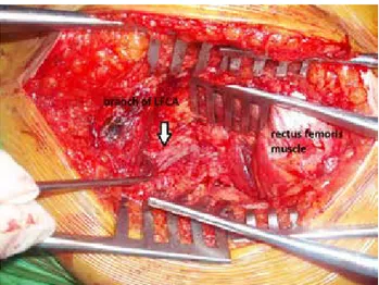
Arcugnano, Italy) was used in all patients. The ascend-ing branch of the lateral femoral circumflex artery and accompanying veins were used as recipient vessels (Fig-ure 3).
Patients were treated with low-molecular weight heparin (20 mg enoxaparin subcutaneous/day) for 3 weeks postoperatively and intravenous antibiotic pro-phylaxis during hospitalization. All patients were kept in absolute bed rest for 5 days and mobilized with crutch-es without weight bearing on the affected side on the sixth day. Weight bearing was prohibited until the third month, after which partial weight bearing (20–25 kg) was initiated using a single Canadian-type crutch for the affected side for 45 days and gradually increased until full weight bearing was gained in the sixth month. Pa-tients were informed about thumb flexion contractures and were encouraged to stretch to extension.
Categorical variables were presented as a percent-age in a number of cases, with continuous variables as mean±standard deviation. Normal distribution was tested with skewness and kurtosis. A paired t-test was used to compare the changes in each patient’s pre- and postoperative HHS scores. A p-value of <0.05 was con-sidered significant for all tests. SPSS software (version 11.0, SPSS Inc., Chicago, IL, USA) was used.
Fig. 3. View of lateral femoral circumflex artery in fatty tissue over
the vastus intermedius muscle. [Color figures can be viewed in the online issue, which is available at www.aott.org.tr]
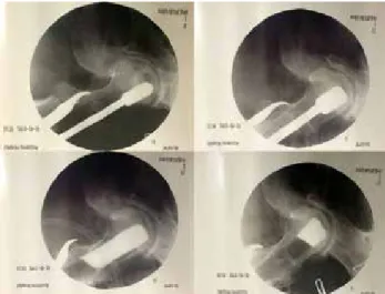
Fig. 4. Roentgenogram of both hips of patient which were operated at stage II at sixth postoperative year. [Color
figures can be viewed in the online issue, which is available at www.aott.org.tr]
Fig. 5. Roentgenogram of both hips of patient which were operated at stage III at seventh and eighth postoperative
Results
During follow-up, 1 patient died because of leukemia, and 1 patient was lost. The remaining 26 hips of 19 pa-tients were evaluated. According to Ficat classification, 17 hips were grade II, and 9 hips were grade III at time of surgery. At final follow-up, the disease had progressed and eventually had been treated with THA in 3 hips. No complications related to surgery–such as infection, deep venous thrombosis, femoral neck fracture, subtrochan-teric fracture, peroneal nerve palsy, or severe flexion con-tracture of the great toe–were seen. Postoperative HHS scores in grade II disease were excellent in 12 patients, good in 3 patients, and fair in 1 patient. In grade III dis-ease, 1 patient was excellent, 5 patients were good, and 1 patient was fair. There was a significant increase in HHS scores from preoperatively to postoperatively (61±9.7 vs 84±17.8, p<0.001). Radiographic trabecular bone for-mation at the tip of the fibular graft was detected in all patients with stage II and stage III disease. There was no sign of collapse or joint narrowing in patients who had HHS greater than 90 points (Figure 4). In 6 pa-tients, the progress of collapse and joint narrowing was observed on plain X-rays (Figure 5). The degree of col-lapse and joint narrowing were directly correlated with HHS score.
Discussion
If the osteonecrotic lesion at the femoral head is large and involves the lateral pillar, the progress to collapse is inevitable; the time of progress to collapse is usually less than 3 years, according to the study of Ohzono, which observed the natural progress of 115 untreated hips.[9]
Many treatment modalities have been proposed for this disease. Osteotomies of the proximal femur, including the femoral neck and intertrochanteric region, have been recommended to replace the osteonecrotic lesion area under the stress of body weight with an unaffected vi-able healthy portion of the femoral head. Rotation, var-us, flexion, extension and medializing-type osteotomies have been proposed for this purpose.[2] The rotational
osteotomy described by Sugioka is a very demanding procedure, with regards to both the planning required and the need to preserve the branches of the medial femoral circumflex artery during the procedure.[5]
Oste-otomies also distort the anatomy and biomechanics of the hip joint. In a study of 115 osteotomies performed by Schneider et. al., all osteotomies were associated with a high incidence of complications and a low survival rate, and the benefits provided were only temporary.[10] Core
decompression alone may be accepted, similar to the first generation of intralesional surgical interventions. Arlet
and Ficat proposed the cause of osteonecrosis as the in-crease in intraosseous hypertension, intramedullary ve-nous stasis, and edema due to distortion of blood supply to the femoral head.[11] It was believed that the procedure
would diminish intraosseous pressure and allow restora-tion of blood flow in the hypoxic femoral head. Accord-ing to the results of a large series, core decompression may be effective in small lesions and early stages (stage I) of the disease, but without mechanical and osteogenetic support of bone grafts, the treatment will eventually fail in large lesions and advanced stages of the disease.
[6] According to a meta-analysis by Castro et al., further
surgical interventions were necessary in 16%, 37%, and 71% of cases after core decompression of osteonecrosis at Steinberg stages I, II, and III, respectively.[12]
Combin-ing core decompression with porous tantalum implants was proposed as structural support to the subchondral bone required for prevention of collapse, without leading to donor-site morbidity of the fibular graft. According to a meta-analysis of 6 randomized controlled trials includ-ing 256 cases, this treatment option is recommended for young patients with early-stage small lesions. Large le-sions and advanced stages of the disease were associated with poor results.[7] Vascularized iliac bone grafting was
also combined with tantalum rods to achieve better re-sults in large lesions and advanced stages of the disease.
[13] The FVFG technique described by Urbaniak and
Al-dridge contains unique steps for treatment of osteone-crosis of the femoral head. Adequate curettage of lesions shortens the healing period by removing all dead bone that would require additional time to replace with new bone formation if the lesion were to heal without treat-ment. Control scans of a contrast-filled cavity during the procedure ensure complete removal of necrotic bone.[8]
Filling the remaining cavity with cancellous autologous bone grafts with osteoinductive and osteoconductive properties provides the scaffold for new bone formation. We believe that grafting the cavity properly is important for new bone formation. In the present study, specially designed instruments were used for implantation of grafts into the cavity (Figure 6). DBM allograft was also used as a bone graft extender and mixed with autologous cancellous grafts in all lesions. Adding DBM allografts extends the amount of grafts to properly fill the cavity. In large lesions particularly, the cancellous graft har-vested from a trochanteric region may be insufficient for the amount, and emptying the trochanteric region may cause iatrogenic fracture. Furthermore, it was shown that DBM allograft extenders enhanced the consolida-tion of spinal fusions.[14] Therefore, high survival rates in
large lesions may be related to the use of DBM allografts in our patients. Following cancellous graft placement,
in-troducing a strut graft is the theoretical approach to me-chanically support an undermined subchondral cortex of the femoral head to prevent collapse. Nonvascularized strut cortical bone, autografts, and allografts have been used for this purpose.[15] Besides mechanical support,
the hypothesis governing the vascularization of a strut graft is to provide continuous osteoinductive support to impacted cancellous grafts for ossification. As described in the original technique, folding the periosteum at the tip of the fibula exposes the cambium layer, allowing it to benefit from the osteogenic effect (Figure 7).[16]
Sup-porting this theory, vascularized bone grafts yield better results than nonvascularized grafts.[17,18] Several authors
reported very consistent and satisfactory results with this procedure.[19–21] Brunelli reported good-to-excellent
results (average: 78%) with more than 5-year follow-up.
[19] Yoo et al. reported a 91% success rate in a follow-up
interval of 3–10 years.[20] Urbaniak et al. showed an 83%
survival rate of the fibular graft in 646 procedures after follow-up of up to 17 years.[21] They also reported the
results of a prospective study of 103 consecutive hips treated with FVFG for symptomatic osteonecrosis of the femoral head.[1] Age, etiology, size, location, and stage of
the osteonecrotic lesion have all been proposed as factors influencing the outcome of vascularized fibular grafting for osteonecrosis of the femoral head. However, upon review of the literature, we found conflicting results re-garding the effect of these factors on clinical outcomes. Higher survival rates in younger patients are a common finding of most reports.[22,23] While some authors report
the use of steroids as a poor prognostic factor, others associated idiopathic and alcohol-related osteonecrosis of the femoral head with worse prognoses.[23,24] On the
other hand, some reports present no relationship be-tween outcomes and etiology.[1] The extent of
osteone-crotic lesions may be a determining factor for prognosis of FVFG treatment. According to Kerboull, the necrotic
angle, which shows the extent of the lesion, is calculated by adding the areas of osteonecrosis on the anteropos-terior and frog lateral views.[25] Lesions with an angle
larger than 200° frequently experience poor results with femoral head-preserving procedures.[23,26] Contrarily,
ac-cording to a long-term (mean: 14.4 years) survival analy-sis of 65 hips, Eward et al. found no correlation between the size of lesion and conversion to THA. However, all patients in that study were at the pre-collapse stage.[24] In
the present study, patients were not classified according to lesion size.
Radiographic stage of the disease is another point of interest for predicting the prognosis of treatment. According to many reports, the probability of THA increases with advanced Steinberg stages, whereas it is 0–10% in stage II, 12–23% in stage III, 17–30% in stage IV, and 27–60% in stage V.[20,27] Eward found no
differ-ence between Ficat stage I and stage II regarding conver-sion to THA.[24] Marciniak also found no relationship
Fig. 6. (a) Straight reamers in increasing size provide a tunnel size matching with fibula. Ball-tipped
reamers (b) and reverse cutters (c) are used inside the lesion. Graft impactor composed of
a drill with large helix angle inside the metal tube with distal openings. The grafts are first placed in the tube and then the drill is advanced through. (d) Clockwise spinning of the drill
impacts the grafts inside the cavity. Removal of the tube leaves a space to inset the fibular graft. [Color figures can be viewed in the online issue, which is available at www.aott.org.tr]
(a) (b) (c) (d)
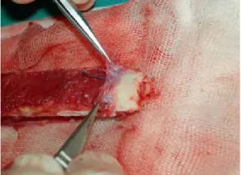
Fig. 7. Periosteum stripped and folded at the end of the fibula and
view of the cambium layer. [Color figures can be viewed in the online issue, which is available at www.aott.org.tr]
between initial radiographic stage and final clinical out-come or overall rate of graft survival.[28] In the present
series, 3 THAs (10.7%) were applied to 2 bilaterally in-volved patients, both hips of 1 patient at 5.5 years and 1 side of another patient at the sixth year. Both hips of the bilateral THA patient were stage III at time of primary surgery. The other patient’s hip was stage II at time of surgery.
The patency of all microvascular anastomoses in the postoperative period is the subject of debate, particularly if no monitor-like skin paddle is present. To eliminate this doubt, some authors recommended to inset the fib-ular graft with a skin paddle.[29] Although skin paddle
shows the patency of anastomoses, it does not provide information regarding revascularization inside the femo-ral head. According to our experience, localization of the perforator of the skin paddle is not consistent and may lift and kink the pedicle of the graft when sutured to the skin. Bone scintigraphy using Technetium-99m methy-lene diphosphonate and single-photon emission com-puted tomography are effective methods in assessment of bone blood flow in the postoperative period. How-ever, these 2 tests are usually performed within the first week following surgery (preferably within 72 hours) to rule out thrombus formation.[30] According to Berggren
et al., a positive bone scan a week or longer after surgery does not show patency of the vessels or bone viability, as a result of new bone formation at the surface of the graft due to creeping substitution. Thus, it has been recom-mended to perform bone scans at the week of surgery to avoid false-positive uptake.[31,32] Dynamic
contrast-enhanced MRI is another method used to evaluate the degree of bone marrow perfusion of the fibular graft fol-lowing injection of gadolinium contrast medium.[33] To
the best of our knowledge, there are no studies in the existing literature using these tests for long-term follow-up.
In the present study, patients were followed with standing anteroposterior roentgenograms and evaluated for joint space narrowing and shape of the femoral head. We believe that these 2 criteria are sufficient to evalu-ate the radiologic outcomes of the FVFG technique. Furthermore, we believe that proper surgical technique is another important factor for success. Specifically, pre-venting the pedicle of the graft from being stranded in-side the femoral tunnel and adjustment of pedicle length so as not to be kinked or stretched are the technical key points of the procedure.[34] Vascularized fibular grafting
provides the most consistently successful results of any joint-preserving method, including core decompression, conventional bone grafting, and osteotomy.[35] We believe
that FVFG yields extremely good results, particularly in pre-collapse stages of the disease in young patients. In this study, 15 of 17 (88%) Ficat stage II hips achieved ex-cellent or good results according to HHS. The outcomes of less-invasive techniques such as core decompression are unpredictable, and published results are not superior to FVFG.[36] Vascularization of fibular strut graft does
not result in a marked increase in operation time if the surgical team is familiar with the procedure. The residual fibular defect of the donor site does not impair the func-tions of daily living. As mentioned above, theoretically there are many factors present which may interfere with the expected results of FVFG, but we found that there was no clear evidence of contraindications for FVFG of avascular necrosis of the femoral head, except in cases of advanced stages of the disease and patients of advanced age.
Conflics of Interest: No conflicts declared. References
1. Urbaniak JR, Coogan PG, Gunneson EB, Nunley JA. Treatment of osteonecrosis of the femoral head with free vascularized fibular grafting. A long-term follow-up study of one hundred and three hips. J Bone Joint Surg Am 1995;77:681–94.
2. Scher MA, Jakim I. Intertrochanteric osteotomy and au-togenous bone-grafting for avascular necrosis of the femo-ral head. J Bone Joint Surg Am 1993;75:1119–33. 3. Zalavras CG, Lieberman JR. Osteonecrosis of the femoral
head: evaluation and treatment. J Am Acad Orthop Surg 2014;22:455–64.
4. Moya-Angeler J, Gianakos AL, Villa JC, Ni A, Lane JM. Current concepts on osteonecrosis of the femoral head. World J Orthop 2015;6:590–601.
5. Sugioka Y. Transtrochanteric anterior rotational osteoto-my of the femoral head in the treatment of osteonecrosis affecting the hip: a new osteotomy operation. Clin Orthop Relat Res 1978;130:191–201.
6. Smith SW, Fehring TK, Griffin WL, Beaver WB. Core decompression of the osteonecrotic femoral head. J Bone Joint Surg Am 1995;77:674–80.
7. Zhang Y, Li L, Shi ZJ, Wang J, Li ZH. Porous tantalum rod implant is an effective and safe choice for early-stage femoral head necrosis: a meta-analysis of clinical trials. Eur J Orthop Surg Traumatol 2013;23:211–7.
8. Aldridge JM 3rd, Berend KR, Gunneson EE, Urbaniak JR. Free vascularized fibular grafting for the treatment of post-collapse osteonecrosis of the femoral head. Surgical tech-nique. J Bone Joint Surg Am 2004;86-A Suppl 1:87–101. 9. Ohzono K, Saito M, Takaoka K, Ono K, Saito S, Nishina
avas-cular necrosis of the femoral head. J Bone Joint Surg Br 1991;73:68–72.
10. Schneider W, Aigner N, Pinggera O, Knahr K. Intertro-chanteric osteotomy for avascular necrosis of the head of the femur. Survival probability of two different methods. J Bone Joint Surg Br 2002;84:817–24.
11. Schroer WC. Current concepts on the pathogenesis of os-teonecrosis of the femoral head. Orthop Rev 1994;23:487– 97.
12. Castro FP Jr, Barrack RL. Core decompression and con-servative treatment for avascular necrosis of the femoral head: a meta-analysis. Am J Orthop (Belle Mead NJ) 2000;29:187–94.
13. Zhao D, Zhang Y, Wang W, Liu Y, Li Z, Wang B, et al. Tantalum rod implantation and vascularized iliac graft-ing for osteonecrosis of the femoral head. Orthopedics 2013;36:789–95.
14. Lee KJ, Roper JG, Wang JC. Demineralized bone matrix and spinal arthrodesis. Spine J 2005;5(6 Suppl):217–23. 15. Keizer SB, Kock NB, Dijkstra PD, Taminiau AH,
Ne-lissen RG. Treatment of avascular necrosis of the hip by a non-vascularised cortical graft. J Bone Joint Surg Br 2006;88:460–6.
16. Augustin G, Antabak A, Davila S. The periosteum. Part 1: Anatomy, histology and molecular biology. Injury 2007;38:1115–30.
17. Kim SY, Kim YG, Kim PT, Ihn JC, Cho BC, Koo KH. Vascularized compared with nonvascularized fibular grafts for large osteonecrotic lesions of the femoral head. J Bone Joint Surg Am 2005;87:2012–8.
18. Tetik C, Başar H, Bezer M, Erol B, Ağir I, Esemenli T. Comparison of early results of vascularized and non-vascularized fibular grafting in the treatment of osteone-crosis of the femoral head. Acta Orthop Traumatol Turc 2011;45:326–34.
19. Brunelli G, Brunelli G. Free microvascular fibular transfer for idiopathic femoral head necrosis: long-term follow-up. J Reconstr Microsurg 1991;7:285–95.
20. Yoo MC, Kim KI, Hahn CS, Parvizi J. Long-term follow-up of vascularized fibular grafting for femoral head necro-sis. Clin Orthop Relat Res 2008;466:1133–40.
21. Korompilias AV, Lykissas MG, Beris AE, Urbaniak JR, Soucacos PN. Vascularised fibular graft in the manage-ment of femoral head osteonecrosis: twenty years later. J Bone Joint Surg Br 2009;91:287–93.
22. Berend KR, Gunneson EE, Urbaniak JR. Free vascularized fibular grafting for the treatment of postcollapse osteone-crosis of the femoral head. J Bone Joint Surg Am 2003;85-A:987–93.
23. Kawate K, Yajima H, Sugimoto K, Ono H, Ohmura T, Kobata Y, et al. Indications for free vascularized fibular grafting for the treatment of osteonecrosis of the femoral
head. BMC Musculoskelet Disord 2007;8:78.
24. Eward WC, Rineer CA, Urbaniak JR, Richard MJ, Ruch DS. The vascularized fibular graft in precollapse osteone-crosis: is long-term hip preservation possible? Clin Orthop Relat Res 2012;470:2819–26.
25. Kerboul M, Thomine J, Postel M, Merle d’Aubigné R. The conservative surgical treatment of idiopathic aseptic necro-sis of the femoral head. J Bone Joint Surg Br 1974;56:291– 6.
26. Mont MA, Hungerford DS. Non-traumatic avascu-lar necrosis of the femoral head. J Bone Joint Surg Am 1995;77:459–74.
27. Chen CC, Lin CL, Chen WC, Shih HN, Ueng SW, Lee MS. Vascularized iliac bone-grafting for osteonecrosis with segmental collapse of the femoral head. J Bone Joint Surg Am 2009;91:2390–4.
28. Marciniak D, Furey C, Shaffer JW. Osteonecrosis of the femoral head. A study of 101 hips treated with vascular-ized fibular grafting. J Bone Joint Surg Am 2005;87:742– 7.
29. Cho BC, Kim SY, Lee JH, Ramasastry SS, Weinzweig N, Baik BS. Treatment of osteonecrosis of the femoral head with free vascularized fibular transfer. Ann Plast Surg 1998;40:586–93.
30. Schuepbach J, Dassonville O, Poissonnet G, Demard F. Early postoperative bone scintigraphy in the evaluation of microvascular bone grafts in head and neck reconstruction. Head Face Med 2007;3:20.
31. Berggren A, Weiland AJ, Ostrup LT. Bone scintigraphy in evaluating the viability of composite bone grafts revascu-larized by microvascular anastomoses, conventional autog-enous bone grafts, and free non-revascularized periosteal grafts. J Bone Joint Surg Am 1982;64:799–809.
32. Droll KP, Prasad V, Ciorau A, Gray BG, McKee MD. The use of postoperative bone scintigraphy to predict graft re-tention. Can J Surg 2007;50:261–5.
33. Bey E, Paranque A, Pharaboz C, Cariou JL. Postoperative monitoring of free fibular grafts by dynamic magnetic reso-nance imaging. Preliminary results in three cases of man-dibular reconstruction. [Article in French] Ann Chir Plast Esthet 2001;46:10–7. [Abstract]
34. Tan CH, Sathappan SS, Chew YC, Ong HS. Technical challenges of advanced hip osteonecrosis managed using a vascularised fibular graft. Singapore Med J 2007;48:299– 303.
35. Mont MA, Jones LC, Hungerford DS. Nontraumatic os-teonecrosis of the femoral head: ten years later. J Bone Joint Surg Am 2006;88:1117–32.
36. Scully SP, Aaron RK, Urbaniak JR. Survival analysis of hips treated with core decompression or vascularized fibu-lar grafting because of avascufibu-lar necrosis. J Bone Joint Surg Am 1998.