164
Journal of Neurological Sciences [Turkish] 31:(1)# 39; 164-174, 2014
http://www.jns.dergisi.org/text.php3?id=757
Research Article
The Expression of MMP-11 In Benign Meningiomas
Erdogan AYAN1, Süheyla Bozkurt UYAR2, Ajlan ÇERÇI3, Murat ALTAŞ4, Fatihhan BÖLÜKBAŞI5, İlhan ELMACI5, Aydın SAV6
1Department of Neurosurgery, Gazi Osman Pasa University Faculty of Medicine, Tokat, Turkey 2Department of Pathology, Marmara University Faculty of Medicine, İstanbul, Turkey
3Department of Neurosurgery, Goztepe Education and Research Hospital, İstanbul, Turkey 4Department of Neurosurgery, Tayfur Ata Sokmen Medical Faculty, Mustafa Kemal University, Hatay, Turkey 5Department of Neurosurgery, Medipol University Faculty of
Medicine, İstanbul, Turkey 6Department of Pathology, Acıbadem University Faculty of Medicine, İstanbul, Turkey
Summary
The WHO categorises tumours based on their histological properties; however, this classification falls short of understanding their biological behaviours. Recently, there has been an increase in the number of studies on extracellular matrix components with the aim of understanding the biological behaviours of tumours. MMP-11 is a metalloproteinase from the MMP family that has a different expression pattern, mechanism of action, and substrate range compared with the other members of this family.
Of the 55 benign meningioma cases examined, 29 were transitional, 16 were meningothelial, and 8 were fibroblastic in nature. In the meningotheliomatous cases, the MMP-11 expression score was high (75%), whereas it was low in the fibrous cases (87.5%). The Ki-67 levels in the cases with high MMP-11 expression scores were significantly higher than those of cases with low MMP-11 expression scores. Although there was no statistically significant correlation between recurrence and the MMP-11 expression score, 5 out of 7 cases with recurrence were found to have high MMP-11 expression scores.
According to our study, MMP-11 is expressed in Grade I meningiomas. The level of MMP-11 expression appears to be high in the meningotheliomatous tumours and low in the fibrous subtypes. The tumours with a high level of MMP-11 expression also demonstrated high Ki-67 values.
Key words: Meningioma, MMP11, Extracellular Matrix
Benign Meningiomalarda MMP-11 Ekspresyonu Özet
WHO sınıflaması tümörlerin histolojik özellikleri değerlendirilerek yapılan bir sınıflama olup, onların biyolojik davranışlarının anlaşılmasında yetersiz kalmaktadır. Son zamanlarda tümörlerin biyolojik davranışlarını gösteren ekstrasellüler matriks komponentleri ile ilgili çalışmaların sayısı artmaktadır. MMP-11; MMP ailesi üyesi olan bir metalloproteinaz olup diğer aile üyelerinden etki mekanizması,subtrat yelpazesi,ve ekspresyon paterni bakımından farklılık gösterir. Çalışmamızda; 55 benign meningioma olgusu incelendi.Bunların 29' u transisyonel,16' sı meningotelyamatöz,8' i fibroblastik alttipte idi. Meningotelyamatöz olanlarda MMP-11 ekspresyon katsayısının yüksek(%75), fibroblastik olanlarda düşük (%87.5) olduğu tespit edildi. Ki-67 düzeyi MMP-11 ekspresyon katsayısı yüksek olgularda anlamlı derecede yüksek iken diğerlerinde düşük bulundu. Rekurrens gösteren olgularda
165
istatistiksel olarak anlamlı sonuç çıkmasa da, rekurrens gösteren 7 olgunun 5'inde MMP-11 ekpresyon katsayısı yüksekti. Bu çalımaya göre Grade I meningiomalarda MMP-11 eksprese edilmektedir. MMP-11 ekpresyonu meningotelyamatöz tümörlerde daha yüksek iken fibroblastiklerde düşüktür. MMP-11 ekspresyonu yüksek tümörlerde Ki-67 ekspresyonu da yüksektir.
Anahtar Kelimeler: Meningioma, MMP11, Extracellular Matrix INTRODUCTION
Meningiomas are the most common tumours of the CNS (central nervous system), comprising 34% of all primary CNS tumours(5). The female to male ratio is 2:1(4). Although it has not been definitively proven, these tumours are thought to arise from arachnoid cap cells. These cells are located in the outer layer and villi of the arachnoid membrane. They share several similarities with meningioma cells(18). Meningiomas have been classified into various histologic subtypes, the most common of which are meningothelial, transitional, and fibrous forms(10). The meningotheliomatous subtype is comprised of lobules separated by thin collagen septae, whereas the fibrous subtype presents with fusiform tumour cells generally embedded in a dense collagen stroma. The transitional subtype falls between these two forms(19). The WHO (World Health Organization) recognises three histological tumour grades. The grading system reflects histological characteristics; however, it falls short of demonstrating the biological behaviour of tumours(1). Benign meningiomas show recurrence in 7-20% of cases despite total resection(17). Some of those cases may
show extracranial metastasis or multiple recurrences(14). The WHO classification, which is particularly inadequate for the identification of benign tumours with recurrence despite the revisions performed in 2007, has underscored the importance of markers such as Ki-67 and PR (progesterone receptor), although these markers are not included in the related criteria(6). Therefore, studies have focused on the molecular differences of the meningioma subtypes, the migration of
neoplastic cells, and angiogenesis and have revealed the critical roles of serine, cysteine, and matrix metalloproteases (MMPs) in cell migration and angiogenesis(19). These markers have been investigated to understand the behaviour patterns of meningiomas.
The extracellular matrix (ECM) is a dynamic structure that determines tissue functions. This complex organisation plays a critical role in the pathogenesis of many diseases and a multitude of physiological events. MMPs are proteases within the structure of the extracellular matrix. MMP-11, or stromelysin-3, is a member of the zinc-dependent endopeptidase family known as matrix metalloproteinases, which are involved in extracellular matrix degradation(12). The MMP-11 gene was first identified in invasive breast carcinomas(3). Unlike other MMPs, it is secreted in an active form. This protein takes part in tissue remodelling and tumour progression(12). It has been shown to be expressed in invasive malignant epithelial tumours of the breast, colon, lung, head and neck, skin, and bladder. It is also expressed in some colonic adenomas and laryngeal dysplastic lesions, which have the potential to become malignant. Therefore, it is hypothesised that MMP-11 might be an invasion marker(15).
In this study, we evaluated the three most common subtypes of benign meningiomas. We measured the MMP-11 expression levels in these tumours and investigated any relationship between MMP-11 expression and the Ki-67 proliferation index, clinical and pathologic characteristics, histologic subtypes, tumour localisation, and survey.
166 MATERIAL AND METHODS
Fifty-three meningioma cases from the Goztepe Teaching and Research Hospital that received surgery and were determined to be benign were included in this study. The pathological materials and clinical records of the cases were acquired. After obtaining sections stained with hematoxylin and eosin from specimens embedded in paraffin from the Pathology Laboratory of the Institute of Neurological Sciences at Marmara University, the meningioma subtypes were determined based on the WHO classification criteria. Immunohistochemical staining was performed using MMP-11 and Ki-67 antibodies on specimens taken from the block representing the tumour.
Immunohistochemistry
Immunohistochemistry was performed in the Pathology Laboratory of the Institute of Neurological Sciences at Marmara University. Sections (4 µm thick) were placed on 3-aminopropyl ethylene-covered slides and deparaffinised at 60 ºC for 1 h. The slides were dewaxed in xylene and dehydrated in 96% alcohol. Then, the sections were immersed in 10 mmol/L citrate buffer (pH 6.0) and dried in a microwave for 20 min for antigen retrieval. The slides were cooled to room temperature and rinsed in phosphate-buffered saline (PBS). The endogenous peroxidase activity was blocked by pretreatment with 3% H2O2/distilled water
for 20 min at room temperature. After a thorough washing with PBS, blocking solution (Lab Vision Co., Fremont, CA, 94539) was applied to inhibit nonspecific antibody binding. The sections were incubated with primary antibodies against mouse MMP-11 (TaKaRa Bio Inc, Shiga, Japan, clone ON1-1, 8 µg/ml concentration) and rabbit Ki-67 (monoclonal, NeoMarkers Inc, Fremont, USA, clone SP6, ready to use) for 1 h at RT. After a 10-min rinse in PBS, biotinylated goat anti-polyvalent immunoglobulin (Lab Vision Co.,
Fremont, CA, 94539) and a streptavidin-biotin peroxidase complex were applied at room temperature for 20 min each. 3,3'Diaminobenzidine (DAB) chromogen was used to visualise the antigen-antibody binding. The slides were counterstained with hematoxylin, dehydrated, cleared in xylene, and coverslipped with Entellan. Immunohistochemical Analysis Ki-67
The immunohistochemical hot spots (significantly stained areas) were determined under light microscopy at 100x magnification by scanning the entire tumour field. The neoplastic cells in these hot spots were counted under 1000x magnification. The staining index was calculated as the percentage of the neoplastic cells with nuclear staining relative to the total number of neoplastic cells over the same area.
MMP-11
The immunostaining for MMP-11 was evaluated according to both the staining intensity and the frequency of tumour cells. Ten representative fields were evaluated. The staining intensity was scored as 0 for no staining, weak staining (1), medium staining (2) and strong staining (3). The frequency of MMP-11 immunostaining was also scored as 0 when no staining of the tumour cells was observed, as 1 when less than 1-10% of the tumour cells were stained, as 2 when 10-50% of the tumour cells were stained, as 3 when 50-75% of the tumour cells were stained, and as 4 when more than 75% of the tumour cells were stained. The total immunohistochemical score was determined as the sum of the frequency and intensity scores for tumour cells. The results obtained from these two evaluations were multiplied by each other to calculate a single immunohistochemical score. The median value of the acquired results was set as the cut-off value. The cases with a score below 6, which was the cut-off value, were categorised as having a low
167 expression score, while the ones with a score above 6 were categorised as having a high expression score (Table 1).
Statistical Analysis
In this study, any relationship between the meningioma subtype, WHO grade, Ki-67 expression, recurrence parameters, degree of MMP-11 staining or percentage of immunopositive areas and the expression score was investigated.
The statistical analyses were performed using the SPSS (Statistical Package for Social Sciences) 10.0 program for Windows. While analysing the study data, descriptive statistics (mean and standard deviation) and the Kruskal-Wallis test (for the comparison of more than 2 groups for non-normally distributed parameters) were employed. The comparison of normally distributed parameters between two groups was carried out using Student's t-test, whereas the comparison of non-normally distributed parameters between two groups was performed using the Mann-Whitney U-test. The quantitative data were compared using a chi-square test and Fisher's exact chi-square test. The results were evaluated within a 95% confidence interval. p<0.05 was considered statistically significant.
RESULTS
Patients and Follow-up
The present study included 53 patients: 37 females and 16 males. The average age at diagnosis was 54.9±14.10 y. Clinical follow-up data for a period of up to 72 months were obtained for 52 patients. The mean follow-up time was 37.53 months (range: 3-72 months). During this period, 3 patients died and 10 recurrences were detected in 7 patients. All of these meningiomas were Grade 1. The
histological diagnosis was defined according to the new WHO classification: 29 tumours were transitional, 16 were meningotheliomatous, and 8 were fibrous(Table 1).
Immunosuppression of MMP-11 and Ki-67 in Meningiomas
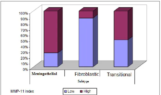
Among our cases, 45.5% had low MMP-11 immunohistochemical scores, while 54.4% had high scores. A review of the cases with low scores revealed that 7 cases were fibrous, 4 cases were meningotheliomatous, and 14 cases were transitional. In the cases with a high immunohistochemical score, 1 case was fibrous, 12 cases were meningotheliomatous, and 15 cases were transitional. The MMP-11 score was observed to be high in 12.5% of the fibrous subtypes, in 51.7% of the transitional subtypes, and in 75.0% of meningotheliomatous subtypes (Graphic 1). Five (71.4%) of the 7 recurrence cases and 25 (52.1%) of the 48 cases without recurrence had high MMP-11 expression scores. The mean Ki-67 proliferation index in the cases with low MMP-11 expression scores was 1%, whereas it was 3.25% in cases with high MMP-11 expression scores. There was a statistically significant relationship between the MMP-11 expression score and the tumour subtypes (p<0.05). The MMP-11 expression score was high (75%) in meningotheliomatous cases and low (87.5%) in fibrous cases. The Ki-67 levels in cases with high MMP-11 expression scores were significantly higher than the Ki-67 levels in cases with low MMP-11 expression scores (p>0.05) (Graphic 2). There was no statistically significant difference between the MMP-11 expression scores and recurrence (p>0.05) (Table 2).
168
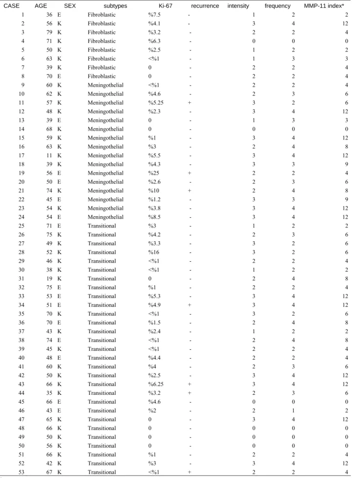
Table 1: Demographic information, number of recurrent cases showing MMP-11 staining, the degree of enhancement, and the MMP-1 index
* MMP-11 intensity score X MMP-11 frequency score
CASE AGE SEX subtypes Ki-67 recurrence intensity frequency MMP-11 index*
1 36 E Fibroblastic %7.5 - 1 2 2 2 56 K Fibroblastic %4.1 - 3 4 12 3 79 K Fibroblastic %3.2 - 2 2 4 4 71 K Fibroblastic %6.3 - 0 0 0 5 50 K Fibroblastic %2.5 - 1 2 2 6 63 K Fibroblastic <%1 - 1 3 3 7 39 K Fibroblastic 0 - 2 2 4 8 70 E Fibroblastic 0 - 2 2 4 9 60 K Meningothelial <%1 - 2 2 4 10 62 K Meningothelial %4.6 - 2 3 6 11 57 K Meningothelial %5.25 + 3 2 6 12 48 K Meningothelial %2.3 - 3 4 12 13 39 E Meningothelial 0 - 1 3 3 14 68 K Meningothelial 0 - 0 0 0 15 59 K Meningothelial %1 - 3 4 12 16 63 K Meningothelial %3 - 2 4 8 17 11 K Meningothelial %5.5 - 3 4 12 18 39 K Meningothelial %4.3 - 3 3 9 19 56 E Meningothelial %25 + 2 2 4 20 50 E Meningothelial %2.6 - 2 3 6 21 74 K Meningothelial %10 + 2 4 8 22 45 E Meningothelial %1.2 - 3 3 9 23 54 K Meningothelial %3.8 - 3 4 12 24 54 E Meningothelial %8.5 - 3 4 12 25 71 E Transitional %3 - 1 2 2 26 75 K Transitional %4.2 - 2 3 6 27 49 K Transitional %3.3 - 3 2 6 28 52 K Transitional %16 - 3 2 6 29 46 K Transitional <%1 - 2 2 4 30 38 K Transitional <%1 - 1 2 2 31 19 K Transitional 0 - 2 4 8 32 75 E Transitional %1 - 2 2 4 33 53 E Transitional %5.3 - 3 4 12 34 51 E Transitional %4.9 + 3 4 12 35 70 K Transitional <%1 - 3 2 6 36 70 E Transitional %1.5 - 2 4 8 37 43 K Transitional %2.4 - 1 2 2 38 74 E Transitional <%1 - 2 4 8 39 45 K Transitional <%1 - 2 2 4 40 48 E Transitional %4.4 - 2 2 4 41 60 K Transitional %4 - 2 3 6 42 50 K Transitional %2.5 - 3 4 12 43 66 K Transitional %6.25 + 3 4 12 44 35 K Transitional %3.2 + 2 3 6 45 66 E Transitional %4.6 - 0 0 0 46 43 E Transitional %2 - 2 1 2 47 65 K Transitional 0 - 3 4 12 48 66 K Transitional 0 - 0 0 0 49 50 K Transitional 0 - 0 0 0 50 56 K Transitional 0 - 0 0 0 51 66 K Transitional %1 - 2 2 4 52 42 K Transitional %3 - 3 4 12 53 67 K Transitional <%1 + 2 2 4
169
Table 2: Percentage of Ki-67 and MMP-11 Expression by Sub-Type and Recurrent Asset Coefficient Evaluation
Expression of MMP-11 Coefficient (Index)
Low
(mean±SD)
High (mean±SD)
p
Percentage of Ki-67 (Median) 2,75±5,06
(1) 3,77±3,31 (3,25) Z:-2,403 p:0,016* n (%) n (%) Meningothelial 4 (% 25,0) 12 (% 75,0) Fibroblastic 7 (% 87,5) 1 (% 12,5) Subtype transitional 14 (% 48,3) 15 (% 51,7) χ2:8,392 p:0,015* yes 2 (% 28,6) 5 (% 71,4) Recurre nce no 23 (% 47,9) 25 (% 52,1) Fχ2 p:0,436
Z: Mann-Whitney U test χ2: Chi-square Test Fχ2:Fisher's Exact Test
* p <0.05
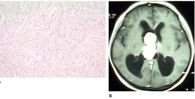
Figure 1: 1a, Strong MMP-11 expression in a grade 1 psammomatous meningioma (MMP11x200); 1b, contrast-enhanced computed tomography of the axial section of a left frontal lobe meningioma.
170
Figure 2: 2a, Strong MMP-11 expression in a grade 1 meningothelial meningioma (MMP-11, x 200); 2b, an MRI contrast-enhanced axial T1-section of a meningioma located in the left temporal lobe.
Figure 3: 3a, Moderate MMP-11 expression in a grade 1 transitional meningioma (MMP-11, x 200); 3b, an axial cross-section contrast-enhanced T1 MRI of an intraventricular meningioma.
171 DISCUSSION
Benign meningiomas may exhibit recurrence, give rise to extracranial metastasis, or present with multicentric characteristics. The most important factors involved in recurrence are tumour histology and the extent of surgical
resection. In addition, the extracellular matrix ultrastructure of the tumour is believed to play a significant role. Tumours with similar morphologies may exhibit different ECM components. In particular, MMPs, members of the metalloproteinase family, play important Graphic 2: Ki-67 Percentage Distribution by the Expression Level of MMP-11
172 roles in many physiological and pathological activities and are known to induce tumour growth and facilitate metastasis(7).
This study shows that benign meningiomas express MMP-11 and that this expression is particularly high in cases with recurrence. In 7 cases of recurrence, 5 had a high expression index. Furthermore, the MMP-11 index and Ki-67 levels showed a significant correlation. While the mean Ki-67 value in cases with a low MMP-11 index was 1%, the Ki-67 value was 3.25% in cases with a high MMP-11 index. This observation may suggest that MMP-11 can be a marker of benign meningiomas with an aggressive course. Unsurprisingly, MMP-11 is noted as a potential marker for pre-invasive lesions in the relevant literature (20, 13, and 21). MMP-11 is hypothesised to play a role in tumour progression via pathways that are different from those of other metalloproteinases. It has also been reported that MMP-11 is not involved in the degradation of major ECM components and has no ability to bind to ECM proteins, such as laminin, fibronectin, and elastin(12). Unlike other MMPs, MMP-11 is activated in the cytoplasm and excreted in the activated form. Kim et al. noted that MMP-11 has a significant role in tissue remodelling, adding that the common ground of embryonic development, wound healing, tumour invasion, and dermatofibroma pathogenesis is extensive tissue remodelling(9).
Boulay et al. found that MMP-11 caused no neoangiogenesis or proliferation of malignant cells in tumours; however, they showed that MMP-11 caused tumour progression by reducing malignant epithelial cell death(4). Various studies have shown that MMP-11 is expressed at a higher rate in the B-cells of the immune system, which includes substrates such as
ILGFBP1, α-2-macroglobulin, and
casein(2,8,11). In the study by Perret et al., MMP-11 was mentioned as a
metalloproteinase that is expressed in atypical meningiomas and is regarded as a potential marker of tumour invasion(15,16). In our opinion, there may be a relationship between the distinctive effects of MMP-11 and its expression in benign lesions, or the invasion may cause the benign tumour to take an aggressive course, which might have different mechanisms than those in malignant tumours. As mentioned by Boulay and Kim(4,9), the problem in benign lesions with an aggressive course may be the decreased apoptosis of tumour cells. Therefore, we believe that MMP-11 expression in benign lesions could also be a marker for histopathologically benign tumours exhibiting an aggressive clinical course.
In our study, MMP-11 was observed to have a particularly high expression rate in the meningotheliomatous subtypes. In the relevant literature, MMP-11 has been noted to be of mesenchymal origin and is expressed by fibroblasts adjacent to the epithelial or tumour cells(12,4). Perret used this opinion to explain the fibrous nature of the only MMP-11-positive case in his series and associated it with the mesenchymal origin of MMP-11(15). Naturally, we do not have a completely different opinion; however, because our study exhibited a high rate of expression in cases with a meningotheliomatous subtype and given that 11 of the 13 cases in Perret's study(15) were a meningotheliomatous subtype, we believe that MMP-11 and the meningotheliomatous subtype may be associated. In fact, these results suggest that there are significant ultrastructural differences between the two main meningioma subtypes and that the mechanism of action and substrates of MMP-11 have yet to be determined, unlike those of other MMPs. In this study, similar to the results reported by Perret, we observed that MMP-11 was expressed in both tumour and stromal cells(15). Unlike our study, Wolf reported that MMP-11 was expressed in fibroblasts adjacent to the neoplastic cells(21).
173 The fibroblastic and meningotheliomatous subtypes have different matrix components, which suggests that these meningioma subtypes may originate from different cell types. This difference may help us to understand the developmental mechanisms and clinical courses of those tumours.
Furthermore, we evaluated MMP-11 expression by establishing a staining index based on the degree of staining and the percentage of stain-positive areas. We believe that this index may prove to be beneficial in the comparison of immunohistochemical studies performed in different centres at different times.
In conclusion, there are significant ultrastructural differences between the two main subtypes of meningiomas. The morphological classification is not sufficient for predicting the clinical course of meningiomas. Although MMP-11 is an indispensable proteinase in tumour growth, its mechanism of action and substrates are still not clear. However, it is obvious that they are different from those of other MMPs. Benign meningiomas express MMP-11, and this expression rate is higher in cases with the meningotheliomatous subtype, recurrence, or an elevated proliferative index.
Correspondence to: Erdogan Ayan
E-mail: [email protected]
Received by: 07 May 2013 Revised by: 17 February 2014 Accepted: 07 March 2014
The Online Journal of Neurological Sciences (Turkish) 1984-2014
This e-journal is run by Ege University Faculty of Medicine,
Dept. of Neurological Surgery, Bornova, Izmir-35100TR
as part of the Ege Neurological Surgery World Wide Web service.
Comments and feedback: E-mail: [email protected] URL: http://www.jns.dergisi.org
Journal of Neurological Sciences (Turkish) Abbr: J. Neurol. Sci.[Turk]
ISSNe 1302-1664
REFERENCES
1. Babu S, Uppin SG, Uppin MS, Panigrahi MK, Saradhi V, Bhattacharjee S, Sahu BP,Purohit AK, Challa S. Meningiomas: correlation of Ki67 with histological grade.Neurol India. 2011 Mar-Apr;59(2):204-7.
2. Bar-Or A, Nuttall RK, Duddy M, Alter A, Kim HJ, Ifergan I, Pennington CJ, Bourgoin P, Edwards DR, Yong VW. Analyses of all matrix metalloproteinase members in leukocytes emphasize monocytes as major inflammatory mediators in multiple sclerosis. Brain. 2003 Dec;126(Pt 12):2738-49.
3. Basset P, Bellocq JP, Wolf C, Stoll I, Hutin P, Limacher JM, Podhajcer OL, Chenard MP, Rio MC, Chambon P. A novel metalloproteinase gene specifically expressed in stromal cells of breast carcinomas. Nature. 1990 Dec 20-27;348(6303):699-704.
4. Boulay A, Masson R, Chenard MP, El Fahime M, Cassard L, Bellocq JP, Sautès-Fridman C, Basset P, Rio MC. High cancer cell death in syngeneic tumors developed in host mice deficient for the stromelysin-3 matrix metalloproteinase. Cancer Res. 2001 Mar 1;61(5):2189-93.
5. CBTRUS. Statistical Report: Primary Brain Tumors in the United States, 2004-2006. Hinsdale, IL: Central Brain Tumor Registry of the United States, 2010
6. Commins DL, Atkinson RD, Burnett ME.
Review of meningioma histopathology.Neurosurg Focus. 2007;23(4):E3.
7. Das A, Tan WL, Smith DR. Expression of extracellular matrix markers in benign meningiomas. Neuropathology. 2003 Dec;23(4):275-81.
8. del Mar Barbacid M, Fernández-Resa P, Buesa JM, Márquez G, Aracil M, Quesadaand AR, Mira E. Expression and purification of human stromelysin 1 and 3 from baculovirus-infected insect cells. Protein Expr Purif. 1998 Jul;13(2):243-50.
174
9. Kim HJ, Lee JY, Kim SH, Seo YJ, Lee JH, Park JK, Kim MH, Cinn YW, Cho KH, Yoon TY. Stromelysin-3 expression in the differential diagnosis of dermatofibroma and dermatofibrosarcoma protuberans: comparison with factor XIIIa and CD34. Br J Dermatol. 2007 Aug;157(2):319-24.
10. Louis DN, Ohgaki H, Wiestler OD, Cavenee WK, Burger PC, Jouvet A, Scheithauer BW, Kleihues P. The 2007 WHO classification of tumours of the central nervous system. Acta Neuropathol. 2007 Aug;114(2):97-109. Erratum in: Acta Neuropathol. 2007 Nov;114(5):547.
11. Mañes S, Mira E, Barbacid MM, Ciprés A, Fernández-Resa P, Buesa JM, Mérida I, Aracil M, Márquez G, Martínez-A C. Identification of insulin-like growth factor-binding protein-1 as a potential physiological substrate for human stromelysin-3. J Biol Chem. 1997 Oct 10;272(41):25706-12.
12. Matziari M, Dive V, Yiotakis A. Matrix metalloproteinase 11 (MMP-11; stromelysin-3) and synthetic inhibitors. Med Res Rev. 2007 Jul;27(4):528-52. Review.
13. Munck-Wikland E, Heselmeyer K, Lindholm J, Kuylenstierna R, Auer G, Engel G.Stromelysin-3 mRNA expression in dysplasias and invasive epithelial cancer of the larynx. Int J Oncol. 1998 Apr;12(4):859-64.
14. Nakasu S, Fukami T, Jito J, Nozaki K. Recurrence and regrowth of benign meningiomas. Brain Tumor Pathol. 2009;26(2):69-72.
15. Perret AG, Duthel R, Fotso MJ, Brunon J, Mosnier JF. Stromelysin-3 is expressed by aggressive meningiomas. Cancer. 2002 Feb 1;94(3):765-72.
16. Perry A, Gutmann DH, Reifenberger G. Molecular pathogenesis of meningiomas. J Neurooncol. 2004 Nov;70(2):183-202.
17. Perry A, Scheithauer BW, Stafford SL, Lohse CM, Wollan PC. "Malignancy" in meningiomas: a clinicopathologic study of 116 patients, with grading implications. Cancer. 1999 May 1;85(9):2046-56
18. Riemenschneider MJ, Perry A, Reifenberger G. Histological classification and molecular genetics of meningiomas. Lancet Neurol. 2006 Dec;5(12):1045-54. Review. Erratum in: Lancet Neurol. 2007 Feb;6(2):105.
19. Rooprai HK, van Meter TE, Robinson SD, King A, Rucklidge GJ, Pilkington GJ.Expression of MMP-2 and -9 in short-term cultures of meningioma: influence of histological subtype. Int J Mol Med. 2003 Dec;12(6):977-81. 20. Urbanski SJ, Edwards DR, Hershfield N,
Huchcroft SA, Shaffer E, Sutherland L, Kossakowska AE. Expression pattern of metalloproteinases and their inhibitors changes with the progression of human sporadic colorectal neoplasia. Diagn Mol Pathol. 1993 Jun;2(2):81-9.
21. Wolf C, Rouyer N, Lutz Y, Adida C, Loriot M, Bellocq JP, Chambon P, Basset P. Stromelysin 3 belongs to a subgroup of proteinases expressed in breast carcinoma fibroblastic cells
and possibly implicated in tumor progression. Proc Natl Acad Sci USA,1993 Mar 1;90(5):1843-7