The All-Seeing Needle Instead of the Veress Needle
in Pediatric Urologic Laparoscopy
Mesrur Selcuk Silay, MD,1Abdulkadir Tepeler, MD,1Ahmet Ali Sancaktutar, MD,2Huseyin Kilincaslan, MD,3 Bulent Altay, MD,4Mehmet Remzi Erdem, MD,1Namik Kemal Hatipoglu, MD,2Muzaffer Akcay, MD,1
Tolga Akman, MD,1 and Abdullah Armagan, MD1
Abstract
Purpose: To investigate the feasibility of the all-seeing needle for safe entry and creation of pneumoperitoneum in pediatric urologic laparoscopy.
Patients and Methods: A total of 14 children underwent various transperitoneal urologic laparoscopic proce-dures. The all-seeing needle, which is 4.85F in diameter, was used for safe entry into the abdominal cavity at the site of the umblicus in all cases. The microoptic was integrated with the light system and connected via a zoom ocular enabling direct visualization of the layers between the skin and the peritoneal cavity. Once the intra-peritoneal access was obtained, CO2pneumoperitoneum was created from one port of the three-way connector
attached to the proximal part of the needle. Then the laparoscopic trocars were placed under vision of the microoptical system.
Results: Mean age of the children was 4.5 – 2.9 years. In all children, the all-seeing needle was safely introduced into the abdominal cavity under direct vision. Then, CO2pneumoperitoneum was succesfully performed. The
mean time for optical puncture was calculated as 1.1 – 0.8 minutes. No complication was encountered during the introduction of the needle, creation of the pneumoperitoneum, and placement of the trocars.
Conclusions: The all-seeing needle appears to be beneficial in safe entry and for creating pneumoperitoneum in laparoscopic pediatric urology cases. It eliminates the disadvantages of the Veress needle, which is blunt in-sertion, and may possibly prevent complications.
Introduction
T
he technique for creating pneumoperitoneumand the first trocar insertion is a critical step during lapa-roscopic procedures. The two main and worldwide ac-cepted approaches for intra-abdominal entry are the open insertion of the trocar with Hasson technique and blind insertion with the Veress needle.1,2 Both techniques have unique advantages, depending on the age and character-istics of the patients, in terms of safety, but neither can completely prevent iatrogenic injuries. Nevertheless, the debate has still continued for the best available option for this first step of laparoscopic procedures, even during the last decade.3–7The all-seeing needle has recently been introduced for con-firming renal access during percutaneous nephrolithotomy.8It has been stated that this instrument increases the safety for creation of the percutaneous renal tract and may decrease the risk of tract-related complications, such as bleeding and bowel
injury. This very thin (1.6 mm) instrument, however, has not been applied in laparoscopy yet.
In this study, we aimed to investigate the feasibility and safety of the all-seeing needle (microoptical system) for safe entry and creation of pneumoperitoneum in pediatric uro-logic laparoscopy. The main advantages of this technique are performance under direct vision and its smaller size. It elim-inates the most important disadvantage of the Veress needle, which is the blind insertion.
Patients and Methods
The all-seeing needle is a novel instrument consisting of three main pieces (Fig. 1A). The first one is the 4.85F (1.6 mm, 16 gauge) needle that has an inner stylet. The diameter of the inner stylet is 3.9F (1.3 mm). The second piece of this system is the Y-shaped three-way connector. The third piece is the highly flexible 2.7F (0.9 mm) microfiber optics with resolution of 10,000 pixels (PolyDiagnost, Pfaffenhofen, Germany).
1Department of Urology, Faculty of Medicine, Bezmialem Vakif University, Istanbul, Turkey. 2
Department of Urology, Faculty of Medicine, Dicle University, Diyarbakir, Turkey.
3Department of Pediatric Surgery, Faculty of Medicine, Bezmialem Vakif University, Istanbul, Turkey. 4Department of Urology, Faculty of Medicine, Medipol University, Istanbul, Turkey.
ª Mary Ann Liebert, Inc. Pp. ---–---DOI: 10.1089/end.2013.0054
Those three pieces are connected with each other in the fol-lowing order:
1. The inner stylet of the 16-gauge needle is removed. 2. Three-way connector is attached to the proximal end of
the needle.
3. Microfiber optic is inserted inside the middle port of the three-way connector.
4. The length of the optical system is adjusted with the aid of an adapter that is attached on the working sheath. After completion of these steps, the all-seeing needle or the microoptical system is ready for use (Fig. 1B). The microoptic is integrated with the light system and connected via a zoom ocular. Those parts (ocular, camera, light cables) are located outside the sterile area and are stabilized with a special arm, which helps the surgeon for comfortable use. The pressure pump system for temporary irrigation is attached to another port of the three-way connector and is controlled by the sur-geon with a foot pedal. Water dissection gives the possibility of better visualization whenever the vision is blurred. Phy-siologic saline (0.9% NaCl) is used as the irrigant. Once in-traperitoneal access is obtained, CO2 pneumoperitoneum is
created via the other port of the connector with high-flow insufflation under direct vision.
Operative technique
The all-seeing needle was used instead of a Veress needle for various treansperitoneal laparoscopic urologica proce-dures in children (Figs. 2A, 2B). All proceproce-dures were per-formed under general anesthesia. The prone position and the 60-degree left or right lateral decubitus positions were used according to the type and the site of the procedure. In all cases, the puncture was performed at the edges of the umblicus, and the skin was retracted by two Langenbeck retractors. In-troduction of the needle was visualized from the video monitor in real time, making it possible to identify the
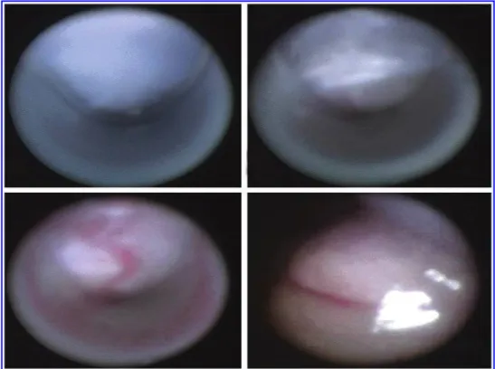
sub-cutaneous tissue, fascial layer, the peritoneum, and the ab-dominal cavity (Figs. 3A–D).
After successful entrance of the all-seeing needle and the creation of penumoperitoneum, the trocars were inserted under direct vision of the optical system. The location of the trocars is decided according to the type and the site of the procedure.
Patient demographics including age, sex, and type of op-eration were recorded. The duration of the optical puncture was also documented for each patient. In addition, operative time and complications were noted.
Results
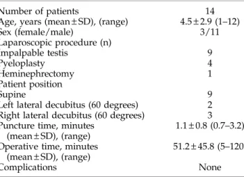
A total of 14 children underwent various transperitoneal laparoscopic procedures between November 2011 and De-cember 2012 (Table 1). The mean age of the study population was 4.5 – 2.9 years. The majority of the children were placed in the supine position (64.2%) for laparoscopic intra-abdominal testis exploration, whereas the 60-degree lateral decubitus position was chosen for the rest of the patients. In all cases, the all-seeing needle was safely introduced into the abdominal cavity. CO2pneumoperitoneum was successfully performed
under direct vision in all children. The mean time for optical puncture was calculated as 1.1 – 0.8 minutes. No complication was encountered during the introduction of the needle, cre-ation of the pneumoperitoneum, and placement of the trocars. Discussion
Regardless of the recent technical advances in minimally invasive surgery and the development of safer instruments, the best option for laparoscopic access still remains a hot spot in the literature. Many severe complications, including death, have been reported related to Veress needle insertion in sev-eral series.5–7The occurence of those complications has been
attributed to the blind needle/trocar insertion. Although an open technique was suggested to be safer than the Veress FIG. 1. (A)Three main pieces of
microoptical system: Left to right, fiberoptic, 16F needle, three-way connector. (B) Microoptical system ready for use.
needle insertion in terms of bowel and major vascular injuries, the meta-analyses reveal that there is no evidence that the open technique is superior to other available techniques.9,10
As a consequence, optical access trocars were developed and met with entusiasm.11,12 The main purpose of the in-vention of the optical trocars was the visualization of the layers between the skin and the peritoneal cavity to reduce the risk of complications. It has been suggested that those trocars are good alternatives to the other available techniques, espe-cially in obese patients and in patients with previous
ab-dominal surgeries. It has been reported that even with the optical access trocars, however, serious complications, such as bowel injury, may occur.13Moreover, evidence-based studies do not suggest the superiority of the optical trocars over other trocar insertion techniques, at least at present.9When asses-sing the current information all together, none of the tech-niques of laparoscopic access may completely prevent complications and none is considered the gold standard.
The safety of the Veress needle to establish penumoper-itoneum in pediatric urology is reported in different series. In
FIG. 3. Intraoperative ap-pearance of the layers be-tween the skin and the peritoneal cavity. Note that the puncture is performed at the edge of the umblicus. FIG. 2. (A, B)The all-seeing nee-dle on the left and the Veress neenee-dle on the right.
a study that assesses the complication rates of American pe-diatric urologists, the Veress needle technique was associated with significantly higher complication rates in contrast to the open technique.14In another study that reported a consecu-tive 257 pediatric urologic laparoscopy experience, however, the only reported complication was preperitoneal insufflation in 7% of all children.15No major complication, such as vas-cular or bowel injury, was detected. There is still a lack of evidence, however, regarding the optimal access technique of laparoscopy in pediatric populations.
The all-seeing needle was introduced into the urologic terminology by Bader and associates16in 2010. It is a very thin instrument with the diameter of only 4.85F and is composed of three main pieces. The three-way connector is attached to the proximal part of the needle, and the microoptic is inserted through the needle. The microoptic is so highly flexible that it can be bent over itself. Then the system is integrated to the camera and the light cable for visualization. The pressure pump system controlled by a foot pedal allows water dis-section and helps for better visualization during the access whenever necessary. The main purpose of this instrument is to facilitate the access into the renal collecting system and to reduce the tract-related complications during the manage-ment of renal stone diseases.
The working mechanism of this instrument is similar to optical trocars, and it was used to allow visualization of the whole tract during percutaneous access to reduce injury to the major vessels and the bowels. The feasibility study of this instrument in 15 cases with renal stones revealed the safety and the applicability of this novel instrument.8Subsequently, many investigators used this instrument not only for safe access but also for the management of the kidney stones.17–20 They have inserted a holmium laser from one port of the three-way connector and fragmented the stones through this needle. The system is now accepted as the smallest available percutaneous renal tract in the literature and is called the microperc (micropercutaneous nephrolithotomy).
On the other hand, this instrument have not been applied in laparoscopy yet. This is the first study investigating the fea-sibility of the all-seeing needle in laparoscopic procedures. We have used the all-seeing needle in 14 children instead of the Veress needle, and our initial results demonstrated that this
instrument is safe and effective in intra-abdominal entry and for creation of pneumoperitoneum in the pediatric popula-tion. The optical puncture took only about 1 minute, and no complication was encountered in any of our patients.
The relatively poor quality of the visualization of the mi-crooptical system can be considered as a disadvantage of this instrument. We were able, however, to introduce the all-seeing needle into the peritoneal cavity in all cases without difficulty. The visualization was fair enough to introduce the needle and to identify the layers between the skin and the peritoneum (Fig. 2). We believe that our case series are proof of the safety and the efficacy of this instrument for entering into the abdominal cavity in pediatric patients. On the other hand, we also acknowledge that this novel technology should technically be improved to enhance its use in different cases. The field of view of this instrument is 120 degrees in the air, which is sufficient for diagnostic laparoscopy.8In addition, if the lightening of this system can be strengthened and the resolution can be improved, this device can also be used for diagnostic laparoscopy not only in urology but also in several other disciplines.
Another limitation of the instrument is that the all-seeing needle (20 cm) is longer than the Veress needle (15 cm). Al-though this situation did not cause any significant difficulty during introduction, balancing the instrument is important. For this purpose, we have used Langenbeck retractors to fa-cilitate the penetration of the needle by creating a counter-power.
In our patients (n = 9) with impalpable testicles, we eval-uated whether we were able to identify the ductus deferens, testicular vessels, internal inguinal ring, and the testis itself by using this microoptical system. Although in five patients we could be able to diagnose that the testicle was entering into the inguinal channel, in the other four patients, we could not discriminate the diagnosis of testicular abscence and intra-abdominal testis. Therefore, at least currently, we ex-perienced that this system is not sufficient for diagnostic laparoscopy in impalpable testis. If the technology can be improved, however, we believe that the all-seeing needle can be a standard of care for these kinds of purposes in the near future.
Another drawback of our study is the small numbers of the cases presented. This is a pilot study investigating the safety and the feasibility of the microoptical system in various lap-aroscopic pediatric urology cases. Unless prospective, ran-domized, and comparative studies between the all-seeing and the Veress needle in larger series are conducted, our technique should be considered experimental. Despite the mentioned limitations, however, if further series are published, our study can be an inspiration for the future aspect of diagnostic lap-aroscopy cases in every field of surgery.
Conclusions
The all-seeing needle appears to be beneficial in safe entry and for creating pneumoperitoneum in pediatric urologic laparoscopy. It eliminates the disadvantage of the Veress needle, which is blunt insertion, and may possibly prevent complications. Our preliminary results suggest that the all-seeing needle may replace the Veress needle, if it can be technically improved and if further studies in larger popula-tions are conducted.
Table1. Patient Demographics Number of patients 14 Age, years (mean – SD), (range) 4.5 – 2.9 (1–12) Sex (female/male) 3/11 Laparoscopic procedure (n) Impalpable testis 9 Pyeloplasty 4 Heminephrectomy 1 Patient position Supine 9
Left lateral decubitus (60 degrees) 2 Right lateral decubitus (60 degrees) 3 Puncture time, minutes
(mean – SD), (range)
1.1 – 0.8 (0.7–3.2) Operative time, minutes
(mean – SD), (range)
51.2 – 45.8 (5–120) Complications None
Disclosure Statement
No competing financial interests exist. References
1. Hasson HM. A modified instrument and method for lapa-roscopy. Am J Obstet Gynecol 1971;110:886–887.
2. Veress J. [A needle for the safe use of pneumoperitoneum.] (Ger) Gastroenterologia 1961;96:150–152.
3. Lal P, Singh L, Agarwall PN, Kant R. Open port placement of the first laparoscopic port: A safe technique. JSLS 2004;8: 364–366.
4. Sangrasi AK, Memon AI, Memon MM, et al. A safe quick technique for placement of the first access port for creation of pneumoperitoneum. JSLS 2011;15:504–508.
5. Borgatta L, Gruss L, Barad D, Kaali SG. Direct trocar inser-tion vs. Veress needle use for laparoscopic sterilizainser-tion. J Reprod Med 1990;35:891–894.
6. Byron JW, Markenson G, Miyazawa K. A randomized comparison of Veres needle and direct trocar insertion for laparoscopy. Surg Gynecol Obstet 1993;177:259–262. 7. Yerdel MA, Karayalcin K, Koyuncu A, et al. Direct trocar
insertion versus Veress needle insertion in laparoscopic cholecystectomy. Am J Surg 1999;177:247–249.
8. Bader MJ, Gratzke C, Seitz M, et al: The ‘‘all-seeing needle’’: Initial results of an optical puncture system confirming ac-cess in percutaneous nephrolithotomy. Eur Urol 2011;59: 1054–1059.
9. Vilos GA, Ternamian A, Dempster J, et al. Laparoscopic entry: A review of techniques, technologies, and complica-tions. J Obstet Gynaecol Can 2007;29:433–465.
10. Ahmad G, O’Flynn H, Duffy JM, et al. Laparoscopic entry techniques. Cochrane Database Syst Rev 2012;2:CD006583. 11. String A, Berber E, Foroutani A, et al. Use of the optical
access trocar for safe and rapid entry in various laparoscopic procedures. Surg Endosc 2001;15:570–573.
12. Berch BR, Torquati A, Lutfi RE, Richards WO. Experience with the optical access trocar for safe and rapid entry in the performance of laparoscopic gastric bypass. Surg Endosc 2006;20:1238–1241.
13. Thomas MA, Rha KH, Ong AM, et al. Optical access trocar injuries in urological laparoscopic surgery. J Urol 2003;170: 61–63.
14. Peters CA. Complications in pediatric urological laparosco-py: Results of a survey. J Urol 1996;155:1070–1073.
15. Yanke BV, Horowitz M. Safety of the Veress needle in pe-diatric laparoscopy. J Endourol 2007;21:695–697.
16. Bader M, Gratzke C, Schlenker B, et al. The ‘‘all-seeing needle’’— an optical puncture system confirming percutaneous access in PNL. J Urol 2010;183(suppl):e734. Abstract 1890.
17. Desai MR, Sharma R, Mishra S, et al. Single-step percuta-neous nephrolithotomy (microperc): The initial clinical re-port. J Urol 2011;186:140–145.
18. Tepeler A, Armagan A, Sancaktutar A, et al. The role of microperc in the treatment of symptomatic lower pole renal calculi. J Endourol 2013;27:13–18.
19. Armagan A, Tepeler A, Silay MS, et al. Micropercutaneous nephrolithotomy in the treatment of moderate-size renal calculi. J Endourol 2013;27:177–181.
20. Tepeler A, Silay MS, Armagan A, et al. Laparoscopic-assisted ‘‘microperc’’ of a stone in a pelvic kidney of a 3-year-old girl. J Laparoendosc Surg Adv Tech A 2013;23:174–176.
Address correspondence to: Mesrur Selcuk Silay, MD Tıp Faku¨ltesi, U¨ roloji Anabilim Dali Bezmialem Vakif U¨ niversitesi Adnan Menderes Bulvari Fatih, I_stanbul, 34093 Tu¨rkiye E-mail: [email protected]
View publication stats View publication stats