219
Makale Kodu/Article code: 3998 Makale Gönderilme tarihi: 28.02.2019 Kabul Tarihi: 10.06.2019
ÖZ
ABSTRACT
Aim: To compare the cytotoxicity of dentin bonding agents on dental pulp cells through RTCA XCELLigence® impedance method, in order to investigate whether the self-etch adhesive would be more cytotoxic than two-step etch-and-rinse adhesive due to its higher acidity.
Materials and Methods: Human dental pulp cells were obtained from healthy third molars extracted during routine clinical treatment. Two self-etching (Tokuyama Bond Force“Tb”;Shofu Beauti Bond“Bb”), and two total-etching(Dentsply Prime&Bond NT“Pb”;3M Adper Single Bond“Ab”) dentin-bonding agents(DBA) were diluted serially with culture medium at a ratio of 1:250, 1:500, 1:1000(v:v) and filtered. Cytotoxicity was identified by plating the pulp cells into the disposable E-plate 96 of xCELLigence® system.
Results: Except for the 1:1000 dilution of Ab, each dilution of Ab was lower than the cell index of the control group at the 24th, and 48th hours. Furthermore, multiple comparison tests showed statistically significant differences between the control and each dilution of Ab (P = 0.000), except 1:1000 dilution of Ab at the 24th hour (P = 0.126). On the other hand, the cell indexes of 1:1000 dilution of Bb were higher than the cell index of the control at 24th and 48th hours, and there were statistically significant differences between them (P =0,000). At the 48th hour, 1:500 (P = 0.179) and 1:250 (P = 0.003) dilutions of Bb were lower than the control.
Conclusion: Among the DBAs we used in our study, total-etch Ab showed the most cytotoxic effect. Conversely, self-etch Bb containing phosphoric acid monomer, showed the least cytotoxic effect. So, it may be more appropriate for clinical practices. Clinical relevance: Despite manufacturers’ claims, not all dentin bonding agents are biocompatible. Two-step etch-and-rinse adhesive may be less biocompatible than self-etch adhesive.
Keywords: Human dental pulp cell; cell culture; cytotoxicity; dental bonding agent; xCELLigence® system ÖZ
Amaç: Tek aĢamalı self-etch adezivlerin yüksek asiditesinden dolayı iki aĢamalı etch and rinse adezivlerden daha sitotoksik olup olmadığını değerlendirmek ve dentin bonding ajanların diĢ pulpa hücreleri üzerindeki sitotoksisitesini RTCA XCELLigence® empedans metodu ile karĢılaĢtırmaktır.
Gereç ve Yöntemler: Ġnsan diĢi pulpa hücreleri rutin klinik tedavi sırasında çekilen sağlıklı üçüncü molar diĢlerden elde edildi. Ġkisi self-etch (Tokuyama Bond Force“Tb”;Shofu Beauti Bond“Bb”), ve ikisi total-etch (Dentsply Prime&Bond NT“Pb”;3M Adper Single Bond“Ab”) olan dentin-bonding ajanlar (DBA) 1:250, 1:500, 1:1000 (v:v) oranlarında seyreltilerek, filtrelendi. Sitotoksisite pulpa hücrelerinin 96 kuyucuklu xCELLigence® sistemine ekilmesiyle belirlendi.
Bulgular: Ab’nin 1:1000’lik dilüsyonu haricinde, Ab'nin her seyreltisi 24. ve 48. saatlerde kontrol grubunun hücre indeksinden düĢüktü. Ayrıca, çoklu karĢılaĢtırma testleri, kontrol ve Ab'nin her dilüsyonu arasında, 24. saatteki 1: 1000’lik Ab dilüsyonu (P = 0.126) hariç, istatistiksel olarak anlamlı farklılıklar göstermiĢtir (P = 0.000). Öte yandan, Bb’nin 1: 1000’lik dilüsyonunun hücre indeksleri, 24. ve 48. saatlerde kontrolün hücre indeksinden daha yüksekti ve aralarında istatistiksel olarak anlamlı farklar vardı (P = 0,000). 48. saatte, Bb’nin 1: 500 (P = 0.179) ve 1: 250 (P = 0.003)’lik dilüsyonları kontrolden daha düĢüktü.
Sonuç: ÇalıĢmamızda kullandığımız DBA'lar arasında total etch Ab en sitotoksik etkiyi göstermiĢtir. Bunun tersine, fosforik asit monomeri içeren self-etch Bb, en az sitotoksik etkiyi göstermiĢtir. Bu nedenle, Bb klinik uygulamalar için daha uygun olabilir. Klinik anlamı: Üreticilerin iddialarına rağmen, tüm dentin bonding ajanları biyouyumlu değildir. Ġki aĢamalı total-etch adezivler, self-etch adezivlerden daha az biyouyumlu olabilmektedir.
Anahtar kelimeler: Ġnsan diĢi pulpa hücreleri; hücre kültürü; sitotoksisite; dentin bonding ajan; xCELLigence® sistemi. REAL–TIME CELL ANALYSIS OF CYTOTOXICITY OF DENTAL BONDING AGENTS
ON HUMAN DENTAL PULP CELLS
ĠNSAN DĠġĠ PULPA HÜCRELERĠNDE DENTĠN BONDĠNG AJANLARIN SĠTOTOKSĠSĠTESĠNĠN REAL-TĠME HÜCRE ANALĠZĠYLE DEĞERLENDĠRĠLMESĠ
Dr.Öğr.Üy.ġeyda ERġAHAN* Doç.Dr.Elif Aybala OKTAY**
Doç.Dr.Fidan AlakuĢ SABUNCUOĞLU* Uzm.Dr.Hüseyin TORT*
* Department of Endodontics, Faculty of Dentistry, Istanbul Medipol University, Istanbul.
**Department of Restorative Dentistry, Gulhane Dentistry Faculty, University of Health Sciences, Ankara. Seyda ErĢahan : ORCID ID: 0000-0002-0354-5108 Elif Aybala Oktay: ORCID ID: 0000-0002-5044-327X
Fidan AlakuĢ Sabuncuoğlu: ORCID ID: 0000-0002-5044-457X Hüseyin Tort: ORCID ID: 0000-0002-5044-331
Makale Kodu/Article code: 4080 Makale Gönderilme tarihi: 13.05.2019 Kabul Tarihi: 11.12.2019
DOI : 10.17567/ataunidfd.658109
Kaynakça Bilgisi: ErĢahan ġ, Oktay EA, AlakuĢ Sabuncuoğlu F, Tort H. Ġnsan DiĢi Pulpa Hücrelerinde Dentin Bonding Ajanların Sitotoksisitesinin
Real-Time Hücre Analiziyle Değerlendirilmesi. Atatürk Üniv DiĢ Hek Fak Derg 2020; 30: 219-225.
Citation Information: Ersahan S, Oktay EA, Alakus Sabuncuoglu F, Tort H. Real–Time Cell Analysis of Cytotoxicity of Dental Bonding Agents on
220 INTRODUCTION
Pulp capping is performed when a healthy pulp has been inadvertently exposed from traumatic injury or by iatrogenic means. During pulp capping, a medicament is placed directly over the exposed site and thus can stimulate the healing process. If successful, it will preclude the need for further treatments (eg, root canal therapy).1,2 In the preparation of deep cavities, the dental pulp may be mechanically exposed. The most frequently used material for pulp capping in clinical treatment is calcium hydroxide. Yet, the ultimate failure of calcium hydroxide is that, perhaps, its inability to provide a hermetic seal to the underlying pulp against recurring infection because of microleakage. This incomplete seal allows a bacterial penetration and subsequent irritation of pulpal tissue.2
Numerous advantages could be obtained by a successful direct pulp capping, such as a reduction for extensive treatment, root canal therapy, or extraction of the tooth.3 As for the direct pulp capping material; dental adhesive systems have also suggested as a material used in restoring the whole cavity, in order to prevent microleakage and increase treatment success by isolating the dental pulp tissue from the exposed area4-7 However, the dentin bonding agents (DBA) have to be biocompatible due to dentin with increased permeability.8,9
Dentin adhesives containing primer and bonding agents in a single bottle were introduced into the market.10 A combination of hydrophilic monomers, reactive diluent monomers, high viscosity adhesive resins, and photo-initiator systems are contained within these adhesives. All of which can be found in a single bottle solution containing ethanol or acetone as solvents. Additives such as water, fillers, and fluoride-releasing agents may also be included in the composition of some adhesives.11 Typical bonding systems that are available in the markets are divided into two categories: self-etch and total etch.
Compared to the total-etch (etch-and-rinse) strategy requiring a separate etching step, self-etching adhesives contain an acidic primer and bond that are combined in a single clinical step (the one-step self-etching system). In addition, these materials are based on acidic monomers that are of lower acidity than phosphoric acid, which is used in the total etch system. This prevents excessive loss of the dentinal matrix and apatite crystals around the collagen network.12
It has been reported that several ingredients of DBAs can cause pulpal damage.8,9 We hypothesize that the self-etch adhesive could be more cytotoxic than total-etch adhesive due to its higher acidity. So, the objective of this study is to evaluate the cytotoxic effects of DBAs, consisting of two pieces of total-etching adhesives (DENTSPLY Prime Bond N&T®, 3M Adper Single Bond®), and another two pieces of self-etching adhesives [Tokuyama Bond Force® (mild, pH 2,3); Shofu Beautibond® (mild, pH: 2,4)], on human dental pulp cells through the RTCA XCELLigence
impedance method.
MATERIALS AND METHODS Culture of human dental pulp cells
The protocol of this study was approved by our institutional Research Ethics Committee (50687469-1491-331-14/1648.4-830), and informed consent was obtained from the tissue donors. Human dental pulp cells were obtained from healthy third molars which were extracted during routine clinical treatment (18 to 22-year-old patients). Human dental pulp cells were cultured using an explant technique as described earlier.13,14 The tooth root was removed by horizontal section below the cementoenamel junction with a bur in high-speed handpiece with water spray. The pulp tissue was removed aseptically by a periodontal curette, rinsed with Dulbecco’s modified Eagle’s medium (DMEM), and placed in a 35 mm Petri dish. Pulp tissues were minced into small pieces by a surgical blade, placed on culture dishes and grown to confluence in Dulbecco’s modified Eagle’s medium (DMEM) supplemented with 10% Fetal calf serum (FCS), penicillin (100 IU/ml) and streptomycin (100 µg/ml). Cultures were maintained at 37 °C in a humidified atmosphere of 5% CO2 and 95% air. Confluent cells were detached with 0.25% trypsin and 0.05% EDTA for 5 min, and aliquots of separated cells were subcultured. Pulp cells between the third and eighth passages were used in the current study.11
Dentin bonding agents (DBAs)
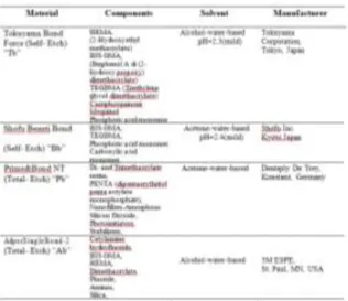
Two self-etching (Tokuyama Bond Force® “Tb”; Shofu Beautibond® “Bb”), and two total-etching (Dentsply Prime&Bond NT® “Pb”; 3M Adper Single Bond® “Ab”) dentin-bonding agents (DBA) were evaluated. Components of DBAs are listed in Table 1.
Cell survival assay using xCELLingence® system
Four DBA, namely Tb, Bb, Pb and Ab were diluted serially with the culture medium at a ratio of
221 Table 1. Principal components and manufacturers of the
dentin-bonding agents tested.
1:250, 1:500, 1:1000 (v:v) and filtered. Cytotoxicity was identified by plating the pulp cells into the disposable E-plate 96 of xCELLigence® system, at an initial density of 2×10⁴ cells/well. After around 7.5 h, different dilutions of Tb, Bb, Pb and Ab were added to the medium (DMEM with 10% FCS). Cells were further incubated for 48 h. Cell survival was evaluated and monitored every 30 minutes for 48 hours by the xCELLigence® system (Roche Diagnostics GmbH, Mannheim, Germany and ACEA Biosciences, Inc., San Diego, CA, USA), according to the instructions of the supplier (Figure 1). As described earlier, the xCELLigence® system consists of 4 main components: the impedance based real-time cell analyzer (RTCA), the RTCA single-plate station, the RTCA computer with integrated software, and a disposable E-plate 96. The RTCA single-plate station fits inside a standard tissue-culture incubator. In order to allow monitoring and detection of physiologic changes in the cells on the electrodes the electronic impedance of the sensor electrodes was measured. Approximately 20 mV (root mean square) voltage was applied to the electrodes during the RTCA measurement. The impedance measured between the electrodes in each well depends on the attachment of the cells to the electrodes, the electrode geometry, and ion concentration in the well. In the absence of cells, electrode impedance is mainly determined by the ion environment both at the electrode-solution interface and bulk solution. In the presence of cells, cells attached to the electrode sensor surfaces act as insulators, and thereby change the local ion
environment at the electrode-solution interface, leading to increased impedance. So, the obtained values of electrode impedance increase as the number of growing cells on the plate increases. The data presented in cell index units can be transferred to the Excel for any type of mathematical analysis.15
Figure 1. Dynamic monitoring of cell adhesion, survival and proliferation by the real-time cell analyzer (RTCA) softwareof xCELLigence® system.
Statistical analysis
Software of the xCELLigence® system was used for all calculations. Normalized cell index graphic which is curve-fitting to the experimental data points is performed by the RTCA software. The data are represented as mean (mmol/L) ± SD (n=5). Kolmogorov-Smirnov test was used as a normality test to compare the distribution of the data. For the proliferation experiments, the statistical analyses were performed by Kruskal-Wallis test, and multiple comparison tests. The value of P ˂ 0.01 was considered as statistically significant.
RESULTS
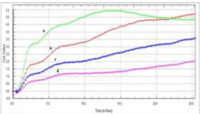
Firstly, the optimal concentration for cell proliferation and viability measurements was determined. For this purpose, 40,000, 20,000, 10,000, and 5000 cells/well were seeded in the E-Plate 96 and the impedance was determined. Cell cycle effects were seen in the concentrations between 5000 to 20,000 cells/well. On the other hand, the concentration of 40,000 cells/well was not suited for further experimentation, possibly because of a too high cell density resulting in contact inhibition. So, we conclude that the concentration of 20,000 cells/well was suited as optimal for our further experiments (Figure 2).
222 Figure 2. Dynamic monitoring of cell adhesion, and proliferation by the real-time cell analyzer (RTCA) software of xCELLigence® system. Dental pulp cells at a density of 40,000(a), 20,000 (b), 10,000 (c), 5000 (d) cells/well inE-Plates 96 were observed during 25 h
According to Kruskal-Wallis test, when evaluated at 24th and 48th hours, there were statistically significant differences between the cell indexes of the control and same dilutions of each DBA group (P < 0.001). Also, there were statistically significant differences between the cell indexes of different dilutions of each DBA group (P < 0.001). Table 2 and 3 represent the means and standard deviations for all groups and statistical comparisons.
Table 2: Means and standard deviations (SD) for all groups, and statistical comparisons between the cell indexes of the control and same dilutions of each DBA group.
Table 3. Means and standard deviations (SD) for all groups, and statistical comparisons between the cell indexes of different dilutions of each DBA group.
According to multiple comparison tests, when evaluated at the 24th hour, there were not any statistically significant differences between the cell indexes of the control and 1:1000 dilution of Ab (P = 0.126), and 1:500 dilution of Pb (P = 0. 988). Other multiple comparison tests showed statistically significant differences between the control and each dilution of all DBAs. However, cell index mean values of 1:1000 dilutions of Tb (0.925±0.096), Bb 144 (1.035±0.119), Pb (0.926±0.062) and Ab (0.805±0.035), 1:500 dilutions of Tb (0.944±0.063), 145 Bb (0.907±0.122) and Pb (0.807±0.062), and 1:250 dilution of Bb (0.854±0.049) were higher than the cell index mean value of the control group (0.791±0.172). When evaluated at the 48th hour, there were not statistically significant differences between the cell indexes of the control and 1:1000 dilution of Tb (p = 0.488), and 1:500 dilution of Bb (p = 0.179). In addition, although there was a statistically significant difference between the cell index mean values of the control and 1:1000 dilution of Bb (p < 0.001), mean value of Bb (1,086±0,034) was higher than the mean value of the control (1.001±0.021). Other multiple comparison tests showed statistically significant differences between the control and each dilution of all DBAs. Table 4 represents the means and standard deviations for all groups and multiple comparison tests.
Table 4. Means and standard deviations (SD) for all groups, and multiple comparison tests between the control and each dilution of all DBAs.
DISCUSSION
A dental adhesive system needs to be biocompatible since it provides connection with the biological tissue and allows for healing and tissue differentiation.16 There is no consensus over dentin bonding agents used as the direct pulp capping
223 material. Some researchers argue in favor that they are biocompatible and can be used for direct pulp capping,17,18 whereas others believe that dentin bonding agents are dangerous due to the continuous inflammation of dental pulp cells that do not seem to heal.19,20 Thus, in the light of these findings, we planned our current study according to the hypothesis that the use of adhesives without being polymerized and even by being directly applied as pulp capping materials will affect the dental pulp cytotoxicity.
In our study, xCELLigence RTCA technology® was used in order to get an accurate platform for non-invasive detection of cell viability and motility. The xCELLigence RTCA technology® has been introduced as an alternative non-invasive and label-free approach to assess cellular proliferation, migration and invasion in real time on a cell culture level.17 The viability of cultured cells can be monitored noninvasively by this system, which uses specially designed microtiter plates containing interdigitated gold microelectrodes providing electrical impedance as the readout. Also, it was reported by many studies that the system has strong correlations with conventional methods (MTT, WST-1 assays etc.).21-23
The acidic agents within adhesive systems may be responsible for the early cytotoxic effect. When adhesives with low pH are directly applied on the dental pulp, they may exhibit an excessive cytotoxic effect on the dental pulp cells. However, when an adhesive material is polymerized under clinical conditions, the acidic agents are diluted in time due to the circulation in the pulp, hence, their toxicity diminishes.24 Indeed, another study suggested that total-etching bond systems, when compared with self-etching adhesive systems, were more cytotoxic to the cells underneath the dentin discs.25 In our study, the cytotoxicity values of DBAs that we examined for 24 and 48 hours showed an increase depending on the duration. 1:250 dose of self-etching adhesive “Shofu Beautibond®” showed the least cytotoxic effect among the examined 1:250 dose of other DBAs. It did not exhibit any cytotoxicity in 24 hours, whereas this was just the other way around when it was subjected to 48 hours. Thus, we observed that although self-etching adhesive “Shofu Beautibond®” had an acidic content (mild, pH: 2.4), it did not show cytotoxicity in the case of long-lasting influence. Therefore, we are of the opinion that “Shofu Beautibond®” could be preferred in clinical practice as the least cytotoxic among the DBAs, which we used on dental pulp cells.
In addition, the ability of monomers to be diffused into the dental pulp in the course of applying adhesive systems on teeth can determine the toxicity of DBAs as well.26 According to the studies regarding the investigations as to why monomers are cytotoxic, it was observed that a synergistic action had occurred in the cases in which hydroxyethylmethacrylate (HEMA) and bisphenol A glycidyl methacrylate (BIS-GMA) contained within dentin bonding agents were used together. It was reported that resin monomers increase the mitochondrial activity and cause macrophage release by affecting inflammatory cells in the dental pulp tissue.4,27 Separately, it was reported that long-term exposure to these monomers had increased cytotoxicity.27 In general, the amount of monomer release ranges from micrograms to milligrams, which is responsible for a number of cytotoxic and metabolic conditions (such as tooth susceptibility) as well as local immunological effects and chronic inflammatory reactions of human pulp.28,29 According to the researchers, triethyleneglycol-dimethacrylate (TEGDMA) can penetrate into all the intracellular and extracellular biological areas including the cell nucleus (cystoblast) and cell membranes and may show cytotoxic effects.30 In contrast to this, high molecular-weighted hydrophobic materials like BIS-GMA, however, are released in very low amounts and can penetrate into the membranes at a rather low rate.18 This view seems to partially support our study. In our study; among the two self-etch bonding agents containing TEGDMA (M.W.= 286 g/mol), which we examined, “Shofu Beautibond®” without containing HEMA was found to be the least cytotoxic. We are of the opinion that BIS-GMA with a high molecular weight in “Shofu Beautibond®” may have been found to be non-cytotoxic, depending on its decreased rate of penetration into the membranes. TEGDMA, which is said to be found at a rate less than 5% in “Shofu Beautibond®”, can dissolve within acetone and it may become diluted after the quick evaporation of acetone. At this point, we are of the opinion that along with the significance of the monomers dissolving in solvents, the subject as to how solvents affect the cell membranes is also of importance. In our study, acetone-water-based total-etch “Dentsply Prime&Bond NT®”, and self-etch “Shofu Beautibond®” DBAs showed less cytotoxicity than alcohol-water-based total-etch “3M Adper Single Bond®”, and self-etch “Tokuyama Bond Force®” DBAs. We consider that the subject as to how acetone and alcohol affect different
224 cell membranes in dental pulp tissue, and/or monomers would be a necessary topic to be investigated. As a result, we think that in order to reach a conclusion regarding a precise factor related to each DBA's cytotoxic effect, the cytotoxicity effect of each content in DBAs on dental pulp cells should be investigated separately. This issue would be seen as a limitation of the current study.
CONCLUSIONS
Among the DBAs we used in our study, one bottle total-etch “3M Adper Single Bond” showed the most cytotoxic effect. Conversely, one bottle self-etch “Shofu Beatibond®” containing an acidic primer with a pH lower than that of phosphoric acid, showed the least cytotoxic effect. So, it may be more appropriate for clinical practices. On the other hand, within the limitations of the current study, we concluded that the monomer compositions and contents of DBAs, and their relationships with one another could be responsible for cytotoxicity. We are of the opinion that there may be a more complex mechanism likely to affect the monomers of acetone-ethanol-based solvents in DBAs. Besides, different types of dental pulp cells and their cell membrane permeabilities, there could be other factors affecting the cytotoxicity mechanism.31 In the future, more rigorous and specific studies regarding this issue should be planned.
Conflicts of Interest
The authors affirm that they do not presently have, nor have they had in the past, any direct financial interest in the subject or materials discussed in this manuscript, or any affiliation with any commercial organization with any such financial interest.
REFERENCES
1. Cox CF, Suzuki S. Re-evaluating pulp protection: calcium hydroxide liners vs cohesive hybridization. J Am Dent Assoc 1994;125:823–31.
2. Brannstrom M, Nyborg H. Pulp reaction to composite resin restorations. J Prosthet Dent 1972;27:181–9.
3. Hilton TJ, Ferracane JL, Mancl L, et al. Comparison of CaOH with MTA for direct pulp capping: a PBRN randomized clinical trial. J Dent Res 2013;92:16-22.
4. Matsuo T, Nakanishi T, Shimizu H, Ebisu S. A clinical study of direct pulp capping applied to carious exposed pulps. J Endod 1996;22:551-6.
5. Murray PE, Hafez A, Smith AJ, Cox CF. Hierarchy of pulp capping and repair activities responsible for dentin bridge formation. Am J Dent 2002;15:236-43.
6. Schröder U. Effect of calcium hydroxide-containing pulp-capping agents on pulp cell migration, proliferation and differentiation. J Dent Res 1985:64;541-8.
7. Tziafas D. Mechanisms controlling secondary initiation of dentinogenesis: a review. Int Endod J 1994;27:61-74.
8. Bergenholtz G. Evidence for bacterial causation of adverse pulpal responses in resin-based dental restorations. Crit Rev Oral Biol Med 2000;11:467- 80.
9. Chen RS, Liu CC, Tseng WY, Jeng JH, Lin CP. Cytotoxicity of three dentin bonding agents on human dental pulp cells. J Dent 2003;31:223-9. 10. Yevenes I, Baltra MO, Urzua I, Reyes J, Petrasic L.
Chemical stability of two dentin single-bottle adhesives as a function solvent loss. Rev Odonto Cienc 2008;23:220-4.
11. Eick JD, Gwinnett AJ, Pashley DH, Robinson SJ. Current concepts on adhesion to dentin. Crit Rev Oral Biol Med 1997;8:306-35.
12. Banava S, Najibfard K, Garcia-Godoy F, Saghiri MA, Ghahremani MH, Ostad N. Impact of dilution and polymerization on cytotoxicity of dentin adhesives to human gingival fibroblasts: early exposure time. J Dent Res Dent Clin Dent Prospect 2015;9:151-8.
13. Chang YC, Huang FM, ChengMH, Chou LSS, Chou MY. In vitro evaluation of the cytotoxicity and genotoxicity of root canal medicines on human pulp fibroblasts. J Endod 1998;24:604-6.
14. Huang FM, Chang YC. Cytotoxicity of dentine-bonding agents on human pulp cells in vitro. Int Endod J 2002;35:905-9.
15. Yalcin M, Barutcigil C, Umar I, Bozkurt BS, Hakki SS. Cytotoxicity of hemostatic agents on the human gingival fibroblasts. Eur Rev Med Pharmacol Sci 2013;17:984-8.
16. Yoshida Y, Van meerbeeK B, Nakayama Y, et al. Evidence of chemical bonding at biomaterial-hard tissue interfaces. J Dent Res 2000;79:709-14. 17. Olmez A, Oztas N, Basak F, Sabuncuoglu B. A
histopathologic study of direct pulp-capping with adhesive resins. Oral Surg Oral Med Oral Pathol Oral Radiol Endod 1998;86:98-103.
225 Biocompatibility of resin-based materials used as pulp-capping agents. Int Endod J 2003;36:831-9. 19. Pereira JC, Segala AD, Costa CA. Human pulpal
response to direct pulp capping with an adhesive system. Am J Dent 2000;13:139-47.
20. Koliniotou-Koumpia E, Tziafas D. Pulpal responses following direct pulp capping of healthy dog teeth with dentine adhesive systems. J Dent 2005;33:639-47.
21. Ke N, Wang X, Xu X, Abassi YA. The xCELLigence system for real-time and label-free monitoring of cell viability. Methods Mol Biol 2011;740:33-43. 22. Limame R, Wouters A, Pauwels B, et al.
Comparative analysis of dynamic cell viability, migration and invasion assessments by novel real-time technology and classic endpoint assays. Plos One 2012;7:e46536.
23. Atienza JM, Yu N, Kirstein SL, et al. Dynamic and label-free cell-based assays using the real-time cell electronic sensing system. Assay Drug Dev Technol 2006;4:597–607.
24. Costa CAS, Vaerten MA, Edwards CA, Hanks CT. Cytotoxic effects of current dental adhesive systems on immortalized odontoblast cell line MDPC-23. Dent Mater 1999;15:434–41.
25. Vajrabhaya LO, Pasasuk A, Harnirattisai C. Cytotoxicity evaluation of single component dentin bonding agents. Oper Dent 2003;28:440-4. 26. Pashley DH, Matthews WG. The effects of outward
forced convective flow on inward diffusion in human dentine in vitro. Arch Oral Biol 1993;38:577-82.
27. Noda M, Wataha JC, Kaga M, Lockwooh PE, Volkmann KR, Sano H. Component of dentinal adhesives modulate heat shock protein 72 expression in heat stressed TPH-1 human monocytes at sublethal concentrations. J Dent Res 2002;81:265-9.
28. Spagnuolo G, D’Anto V, Cosentino C, Schmalz G, Schweikl H, Rengo S. Effect of N-acetyl-L- cysteine on ROS production and cell death caused by HEMA in human primary gingival fibroblasts. Biomaterials 2006;27:1803-9.
29. Spagnuolo G, D’Anto V, Valletta R, et al. Effect of 2-hydroxyethyl methacrylate on human pulp cell survival pathways ERK and AKT. J Endod 2008;34:684-8.
30. Geurtsen W, Leyhausen G. Chemical- biological
interactions of the resin monomer
triethyleneglycol-dimethacrylate (TEDGMA). J Dent
Res 2001;80:2046-2050.
31. Zorba YO, Bayındır YZ, Yıldız M. GeçmiĢten günümüze dentin bonding ajanlar. Atatürk Üniv Dis Hek Fak Derg 2004;14:70-78.
YazıĢma Adresi Seyda Ersahan,DDS, PhD
Ġstanbul Medipol Üniversitesi, DiĢ Hekimliği Fakültesi, Endodonti Anabilim Dalı
Birlik Mah. Bahçeler Cad. No: 5 Esenler, Ġstanbul, TURKEY
Tel: +90 532 405 4088