Acquired Tolerance of Hepatocellular Carcinoma Cells to Selenium Deficiency:
A Selective Survival Mechanism?
1Meliha Burcu Irmak, Gulayse Ince,
2Mehmet Ozturk, and Rengul Cetin-Atalay
3Department of Molecular Biology and Genetics, Faculty of Science, Bilkent University, 06533 Ankara, Turkey
ABSTRACT
Selenium is essential to human health, and its deficiency is associated with different diseases including liver necrosis. Selenium is protective against viral hepatitis and hepatocellular carcinoma (HCC). The under-lying molecular mechanisms of selenium effects are not well known. In this study, in vitro response of HCC-derived cell lines to selenium deficiency is examined alone or in conjunction with Vitamin E and copper/zinc. Here, we show that in vitro selenium deficiency in a subset of HCC-derived cell lines causes oxidative stress and cytochrome c release with subsequent cell death by apoptosis. The oxidative stress and consequent cell death induced by selenium deficiency on these cells are reverted by the antioxidant effect of Vitamin E. However, most HCC cell lines (10 of 13) tolerate selenium deficiency. Consequently, they escape apoptosis. Moreover, nine of these tolerant cell lines have integrated hepatitis B Virus (HBV) DNA in their genomes, and some display p53-249 mutation, indicating past exposure to HBV or aflatoxins, established factors for oxidative stress and cancer risk in liver. An HBV-transfected clone (2.2.15) of the sensitive HepG2 cell line has gained tolerance to selenium deficiency. Our findings indicate that selenium deficiency induces apoptosis in some “hepatocyte-like” cells. However, most HCC cells, particularly HBV-related ones, tolerate sele-nium deficiency and escape its deadly consequences. Thus, as demon-strated by the gain of survival capacity of apoptosis-sensitive cell lines with Vitamin E, such malignant cells have acquired a selective survival advantage that is prominent under selenium-deficient and oxidative-stress conditions.
INTRODUCTION
Selenium is an essential trace element (1), and the necessity of selenium as a dietary factor has been known for almost 50 years. Low selenium status may contribute to the etiology of different disease conditions such as loss of immunocompetence, viral infections, re-productive deficiencies, thyroid and cardiovascular diseases, and pan-creatitis (2). In addition, epidemiological studies have suggested an inverse relationship between selenium levels and different cancers (3–7). Selenium is the protective factor against dietary “necrotic” liver degeneration in rats, based on seminal observations by Schwarz and Foltz (8). In humans, severe selenium deficiency was recognized initially in endemic Keshan and Keshan-Beck diseases (2). Selenium is considered to play an indirect antioxidant role as an essential constituent of a selenoprotein, namely Gpx,4
in elimination of cellular ROS. Cytosolic or classical Gpx, Gpx-1, is considered the most
critical enzyme for the protective effects of selenium. The major cellular site of ROS production is thought to be the mitochondria. During mitochondrial respiration about 2% of the oxygen fails to undergo complete reduction to H2O and is released as superoxide radical (9 –11). Superoxide radicals are converted to H2O2by super-oxide dismutase enzyme (12, 13). H2O2in turn is reduced to H2O by Gpx, which catalyzes the reduction of hydroperoxides as well as H2O2 to H2O and alcohols, which prevents the formation of highly toxic free radicals (1) and which is accompanied by the oxidation of GSH to GSGS (9, 14). Thus Gpx-1 is protective against oxidative stress, both in vitro and in vivo (15–17).
Decreased selenium levels were found in HBV and hepatitis C infection (6, 18), intrahepatic cholestasis (19), and post-viral or alco-holic cirrhosis (20). A recent epidemiological study among chronic carriers of HBV and/or hepatitis C showed that mean plasma selenium levels were significantly lower in patients who developed HCC than in the healthy HbsAg-positive controls, demonstrating an inverse correlation between plasma selenium levels and HCC (see Ref. 6). In addition, selenium supplementation showed a protective effect against HBV infection and HCC (18). These reports clearly indicate that selenium deficiency is associated with severe liver disease and that dietary selenium is protective against HCC.
Despite a large body of evidence linking selenium deficiency to different hepatic disease conditions, cell fate changes consequent to selenium deficiency and mechanisms of protective effects of selenium against HCC and other cancers are poorly known. Here, we report on an in vitro response of HCC-derived cell lines to selenium deficiency. We provide experimental evidence that selenium deficiency leads to apoptosis in some HCC-derived cell lines with or without copper/zinc. However, Vitamin E has a protective antioxidant effect against cell death in the absence of selenium. This apoptotic effect may represent the dietary necrotic liver degeneration, terminology often used to explain hepatic damage observed in animals and people under sele-nium deficiency. In this study, we also show that the apoptotic response to selenium deficiency is caused by oxidative stress. On the other hand, most HCC-derived cell lines tolerated selenium deficiency as well as oxidative stress-inducing conditions. On the basis of these in vitro observations, we hypothesize that selenium deficiency in liver confers growth advantage to malignant tumor cells, because they appear to adapt themselves to a selenium-deficient cellular environ-ment. Finally, our observations also indicate that the tolerance of HCC cells to selenium deficiency and oxidative stress treatment is associ-ated with the presence of HBV genome sequences in their host genomes, suggesting a possible link between HBV and adaptation to selenium deficiency.
MATERIALS AND METHODS
Cell Culture. Human hepatocellular cancer-derived cell lines (n ⫽ 14) were cultured routinely at 37°C under 5% CO2in the standard medium (2 mM
L-glutamine, 0.1 mMnonessential amino acids, 100 units/ml penicillin, 100 g/ml streptomycin in copper/zinc-free DMEM or HAM’s medium that con-tains 10Mcopper and 3 mMzinc), supplemented with 10% FCS (BioChrom). To study the effects of selenium deficiency, cells were grown either in selenium-deficient medium (standard medium with 0.01% FCS) or in a sele-nium-supplemented standard medium with 0.01% FCS and 0.1Msodium
Received 4/30/03; revised 7/16/03; accepted 7/28/03.
The costs of publication of this article were defrayed in part by the payment of page charges. This article must therefore be hereby marked advertisement in accordance with 18 U.S.C. Section 1734 solely to indicate this fact.
1This work was supported by the Turkish Academy of Sciences (to R. C-A., in the
framework of the young Scientist Award Program-RCA/TUBA-GEBIP/2001-2-3, and to M. O.) Partial funding was also provided by TUBITAK and Bilkent University Research Founds (Turkey).
2Present address: Johns Hopkins University School of Medicine, Department of
Neuroscience, 725 North Wolfe Street, Baltimore, MD 21205.
3To whom requests for reprints should be addressed, at Department of Molecular
Biology and Genetics, Faculty of Science, Bilkent University, 06533 Ankara, Turkey. E-mail: [email protected].
4The abbreviations used are: Gpx, glutathione peroxidase; ROS, reactive oxygen
species; HBV, hepatitis B; HCC, hepatocellular carcinoma; TUNEL, terminal de-oxynucleotidyl transferase-mediated dUTP nick end labeling; PBS-T, PBS with 0.1% Tween 20; GSH, reduced glutathione; PARP, poly(ADP-ribose) polymerase; DCF, di-chlorofluorescein; DCFH, dichlorodihydrofluorescein; DCFH-DA, 2⬘-7⬘-DCFH diacetate; NAPO, negative in apoptosis (assay).
selenite (Sigma) and 100MVitamin E (D-␣-tocopherol, Grandpherol; Ilsan Iltas A.S.) in 0.25% DMSO. For these experiments, cells were plated initially in a standard medium with 10% FCS, using 6-well tissue plates, or alterna-tively in 100-mm tissue culture dishes. The next day, after cells were washed three times with PBS, the culture medium was replaced by either a selenium-deficient or a selenium-supplemented medium. Cells were maintained in these media for up to 10 days by refeeding with fresh media every 2 days.
Detection of Apoptosis. Cells were seeded on autoclave-sterilized cover-slips in 6-well plates. After overnight culture in a standard medium with 10% FCS, cells were exposed to a selenium-deficient or a selenium-supplemented medium as described above, for up to 10 days. At times indicated in the legends of Figs. 1–9, the following tests were performed. Cell morphology changes were observed under inverted microscope. To determine nuclear condensation by Hoechst 33258 (Sigma) staining, coverslips were washed in ice-cold PBS twice and were fixed in 1 ml of 90% (v/v) cold ethanol for 10 min and then incubated with 3 g/ml Hoechst 33258 for 5 min in the dark. Coverslips were then rinsed with distilled water, mounted on glass microscopic slides in 50% glycerol, and examined under fluorescent microscope (Axio-skop, Zeiss). TUNEL assay was performed using In Situ Cell Death Detection kit (Roche), according to the manufacturer’s recommendations. The Annexin V assay was performed using Annexin V-PE reagent (PharMingen) on live cells according to the manufacturer’s recommendations, and the cells were fixed in ethanol before examination under fluorescent microscope. For NAPO assay (21) cells were fixed with 4% paraformaldehyde for 20 min at room temperature and then were rinsed and permeabilized with 0.1% Triton X-100 and 0.1% sodium-citrate in PBS on ice for 10 min. After the washing with PBS-T, cells were blocked with 3% BSA in PBS-T for 1 h at room temperature and then were incubated with anti-NAPO monoclonal antibodies for 1 h at room temperature. After being washed with PBS-T, the cells were incubated with FITC-conjugated antimouse secondary antibody (DAKO) for 1 h at room temperature, washed three times with PBS-T, and then mounted. Samples were analyzed by fluorescence microscopy.
Assay for Gpx Activity. The enzymatic activity of Gpx was measured on a native polyacrylamide gel, as described by Sun et al. (22), with minor modifications. This assay was reported to be specific for the visualization of the Gpx activity bands without interference from catalase and other peroxi-dases when the gel was soaked into GSH (Sigma) with concentrations higher than 1 mM(22). Briefly, cells were grown under selenium-supplemented or selenium-deficient media for 96 h. Cell pellets were obtained by scraping, stored overnight at⫺20°C, and lysed in a buffer containing 50 mMTris-HCl (pH 8.0), 300 mMNaCl, and 1⫻ (v/v) Complete Protease Inhibitor Cocktail (Roche) for 30 min on wet ice. Clear supernatants were obtained by centrif-ugation at 13,000 rpm for 15 min at 4°C. Freshly collected mouse liver tissue homogenate was used as a positive control. Equal quantities of proteins from cell lysate (50g) were separated by native gel electrophoresis. Wet gels were then washed three times in a buffer composed of 50 mMTris-HCl (pH 8.0) and 1.5 mMGSH (buffer 1) and then washed in buffer 1 containing 0.003% H2O2.
After rinsing with distilled water, the gel was stained in a reagent prepared by mixing equal volumes of freshly prepared 2% (w/v) solutions of potassium-ferricyanide and ferric-chloride (Sigma), until a dark green background ap-peared with yellow enzymatic activity bands.
Detection of HBV Virus DNA by PCR. Genomic DNA was extracted from cell lines, and the presence of HBV DNA was tested using two sets of primer pairs to amplify HBs and HBx gene sequences, as described previously (23). A plasmid containing head-to-tail dimer of HBV genome was used as a positive control (24).
In Vitro Cell Growth Assay. Cells (60,000) were plated into 6-well plates and were grown overnight in a control medium. The following day, cells were washed with PBS, fed with a selenium-supplemented or selenium-deficient medium, and further grown for 10 days. Attached cells were then stained with Giemsa (Sigma), and photographed.
Western Blot. Cells were lysed with a NP40 lysis buffer containing 150 mMNaCl, 50 mMTris-HCl (pH 8.0), and 1% NP40 (Igepal CA-630, Sigma); 1⫻ (v/v) Complete Protease Inhibitor Cocktail was added freshly. Briefly, cells were scraped with ice-cold PBS on ice, centrifuged at 13,000 rpm for 10 min, resuspended in four volumes of a NP40 lysis buffer, and incubated on ice for 30 min. The lysate was centrifuged at 13,000 rpm for 10 min at 4°C, and soluble proteins were quantified by Bradford assay (25) in supernatants. Proteins were separated by SDS-PAGE (8%) and were transferred onto
poly-vinylidene difluoride (Roche) membrane. The membrane was blocked 1 h at room temperature with 5% nonfat milk in PBS-T and incubated with anti-PARP monoclonal antibody or AKT polyclonal antibody (Santa Cruz Biotech-nology) in PBS-T with 2.5% nonfat milk for 1 h at room temperature. The unbound antibody was washed four times with PBS-T. The membrane was incubated with horseradish peroxidase-conjugated secondary antibody (DAKO) in PBS-T with 2.5% for 1 h at room temperature and washed, and then detected by Enhanced ChemiLuminescence (ECL) detection kit (Amer-sham-Pharmacia). Equal loading control was determined by reprobing the same membranes with anti-cytokeratin 18 monoclonal antibody JAR1 3 (kind-ly provided by D. Bellet, Institute Gustave Roussy).
Immunofluorescence Assay for Cytochrome c Release. Cytoplasmic cy-tochrome c was tested by immunofluorescence staining, as described by Achenbach et al. (26). Cells were grown on coverslips and fixed with 4% paraformaldehyde for 30 min at room temperature and then were rinsed and permeabilized in ice-cold acetone for 10 min. After washing with PBS, the cells were blocked with 3% BSA in PBS-T for 1 h at 37°C and then were incubated with anti-cytochrome c monoclonal antibody (PharMingen) over-night at 4°C. After washing with PBS-T, the cells were incubated with FITC-conjugated antimouse secondary antibody (DAKO) for 1 h at room temperature, washed three times with PBS-T, then mounted. Samples were analyzed by fluorescence microscopy. When released from mitochondria, cytochrome c gives a diffused and more intense fluorescent signal (26).
Detection of Oxidative Stress. Oxidative stress was detected by DCF assay (27). The nonfluorescent DCFH-DA (Sigma) is a cell-permeable com-pound that can enter into cells, in which it is deacetylated and entrapped as DCFH, the oxidation of which by ROS (hydroxyl radical, superoxide, hydro-gen peroxide . . . ) produces a highly fluorescent product, DCF, that can be visualized under a fluorescent microscope. Cells were cultured for 96 h, washed with PBS three times, and incubated for 15 min at 37°C in PBS containing 10 mMHEPES (pH 7.5), 10 mMglucose, and 1MDCFH-DA. Cells were washed again three times with PBS, mounted directly on a slide, and examined under a fluorescent microscope.
Hydrogen Peroxide Assay. Huh-7, Hep3B-TR, HepG2, and HepG2– 2.2.15 cells were cultured under selenium-supplemented and selenium-defi-cient conditions for 3 days. On the 3rd day of the culture, H2O2(0, 25, 50, 100,
and 200M) was added to the cell culture medium. Four h later, the live cells under oxidative stress were counted by DCF staining under inverted micro-scope, and the apoptotic cells were counted by Hoechst 33258 staining under a fluorescent microscope.
RESULTS
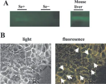
Experimental Induction of in Vitro Selenium Deficiency. He-patic selenium deficiency induced in vivo by feeding experimental animals is accompanied by a decrease in antioxidant selenoproteins, including Gpx and subsequent hepatic necrosis because of oxidative stress (8). It has also been reported that human hepatoma cells, when cultivated in vitro in a selenium-deficient medium, display a similar loss in Gpx activity (28), suggesting that such cells can serve as an in vitro model to study hepatocellular response to selenium deficiency. It has also been reported that Huh7 hepatoma cells can be maintained and expanded in a chemically defined serum-free culture medium, provided that it is supplemented with selenium (29). On the basis of these observations, we first studied the response of Huh7 cells to selenium deficiency by maintaining them in a selenium-deficient culture medium for up to 8 days. The cellular response to selenium deficiency was first assessed by testing cellular Gpx activity, using a specific in situ staining technique (22). Huh7 cells display positive Gpx activity when grown in standard culture medium that contains 10% FCS (data not shown). A similar positive Gpx activity was detected when the DMEM culture was depleted of FCS (0.1%), but complemented with selenium (0.1 M), as shown in Fig. 1A. As
expected, the removal of selenium from the latter provoked the loss of Gpx activity in these cells, which was no longer detectable after 4 days of culture (Fig. 1A).
The loss of Gpx activity in selenium-deficient cells impairs their antioxidant response, leading to an accumulation of ROS, which can be detected at the cellular level by the oxidation of nonfluorescent DCFH-DA into a fluorescent form (27, 30). As shown in Fig. 1B, Huh7 cell line, when cultivated in selenium-deficient HAM’s culture medium, which contains copper/zinc, or in DMEM culture (data not shown), displayed many fluorescent cells on examination under flu-orescence microscopy, indicating the accumulation of ROS in such cells. The fact that this response is attributable to selenium deficiency alone and in conjunction with copper/zinc was confirmed by the absence of fluorescent cells in the same cell line that was cultivated in selenium-supplemented medium (data not shown). Taken together, these observations indicated that selenium-deficient Huh7 cells dis-play a drop in their Gpx activity, accompanied by an intracellular accumulation of ROS, in a way quite similar to liver cells of animals subjected to in vivo selenium deficiency (31).
Impaired Survival of Selenium-Deficient Huh7 Cells Attribut-able to Apoptotic Cell Death. Next, we studied the growth of Huh7 cells under selenium-deficient conditions. Huh7 cells were examined under light microscopy at different intervals between 1 and 8 days. Cells showed no detectable change in cell morphology during the first 48 h of culture. However, starting at 72 h, individual Huh7 cells displayed morphological changes reminiscent of apoptotic cell death, with swelling and nuclear condensation (Fig. 2A). The cell death was time dependent and progressive, which was affecting more that 70% of cells at day 7, under selenium deficiency (Fig. 2). These changes were observed only in selenium-deficient cells, but not in those cultivated in the selenium-complemented medium (Fig. 2). Thus, the complementation of culture medium with 0.1Mselenium alone or in
addition to copper/zinc was sufficient to prevent not only oxidative stress but also the death of Huh7 cells.
Next, we characterized the nature of death that is observed in Huh7 cells under selenium deficiency. As shown in Fig. 3, selenium-defi-cient Huh7 cells displayed smaller and condensed nuclei with strong Hoechst 33258 staining (Fig. 3A, right panels) bearing apoptosis. The
apoptotic cell death was confirmed with three different tests; namely, TUNEL assay that detects DNA fragmentation (Fig. 3A, top panels) and Annexin V staining that detects cell membrane changes (data not shown), as well as by a recently described immunofluorescence tech-nique (15) detecting the loss of NAPO antigen in apoptotic cells (Fig. 3A, bottom panels). Control Huh7 cells were free of such described changes (data not shown).
Apoptotic cell death is often accompanied by the release of cyto-chrome c from the mitochondria (32, 33). Cytoplasmic cytocyto-chrome c triggers a cascade of caspase activation, initiated by the formation of apoptosome complex with Apaf-1 and caspase 9, ultimately leading to the activation of caspase 3, which cleaves a set of specific target proteins, including PARP (34). As shown in Fig. 3B, positive immu-nostaining for cytoplasmic cytochrome c was detectable among sele-nium-deficient cells but not among those of the control group. This indicated that mitochondrial cytochrome c was released into the cytoplasm of selenium-deficient cells. Fig. 3B clearly demonstrates that the diffuse cytoplasmic positive staining for cytochrome c corre-lates perfectly with the nuclear condensation of apoptotic cells, sug-gesting that cell death is imminent to the release of cytochrome c in these cells. To test the activation of caspase 3 in selenium-deficient Huh7 cells indirectly, we analyzed the levels of PARP by Western blot technique. Selenium-deficient cells were maintained in culture until more than 90% of the cells displayed apoptotic features, and then cell lysates were tested. PARP was no longer detectable in apoptotic Huh7 cells, and this loss appeared to be specific, because there was no detectable change in the levels of cytokeratin 18 under the same conditions (Fig. 3C).
The oxidative stress observed under selenium-deficient conditions triggered an apoptotic response in Huh7 cells. To further explore a cause-effect relationship among selenium deficiency, oxidative stress, and subsequent apoptosis, we cultivated Huh7 cells in either a sele-nium-deficient or a selenium-supplemented medium for 3 days and
Fig. 2. Death of Huh7 cells under selenium deficiency. A, analysis of Huh7 cell growth under selenium-supplemented (Se⫹) or selenium-deficient (Se⫺) HAM’s medium. Huh7 cells were examined under inverted light microscope on a daily basis, and pictures were taken at day 3.⫻100. B, the percentage of death cell nuclei was counted up to 8 days with Hoechst staining under a fluorescent microscope. Note the abundance of cell death in Huh7 cells in the selenium-deficient (Se⫺) DMEM cell culture. There is no cell death in selenium-supplemented (Se⫹) medium. The results are from three separate experiments.
Error bars, SD.
Fig. 1. In vitro response of Huh7 cells to selenium deficiency. Huh7 cells were grown in selenium-supplemented (Se⫹) or selenium-deficient (Se⫺) conditions. A, in vitro selenium deficiency was tested by the loss of Gpx activity. At 96 h, cells were solubilized as described in “Materials and Methods,” and 50g of soluble proteins from cell lysates were separated by native PAGE, followed by in situ staining for Gpx with a colorimetric assay. Mouse liver tissue lysate was used as a positive control. B, analysis of oxidative stress by oxidant-sensitive fluorescent dye, DCFH-DA. Huh7 cells were cultured under Se⫺ HAM’s medium for 72 h, and then DCF fluorescence was observed under an inverted fluorescent microscope. The presence of fluorescence indicates that Huh7 cells harbor oxidative stress (right panel). Inverted light microscopy photographs (left panel) of the corresponding area of fluorescent photographs of Huh7 cells under Se⫺ conditions were also shown.⫻200.
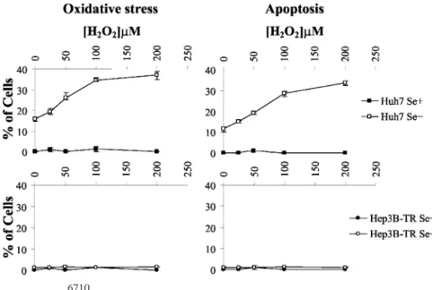
then treated them with increasing doses of hydrogen peroxide. The cells were left in culture for an additional 4 h, and the ratio of cells positive for ROS or apoptotic features were calculated. Huh7 cells cultivated in a selenium-supplemented medium were able to tolerate a treatment with at least 200Mhydrogen peroxide, with no sign of oxidative stress or apoptosis; in contrast, selenium-deficient Huh7 cells responded to hydrogen peroxide treatment by a dose-dependent increase in both oxidative stress and apoptotic cell death responses (Fig. 4, top panels). As little as 25 M hydrogen peroxide was
sufficient to bring an increase in oxidative stress with concomitant apoptosis. These responses were dose dependent and paralleled each other (compare Fig. 4, top left and right panels), strongly suggesting that selenium deficiency leads to sensitivity to oxidative stress, which in turn leads to apoptotic cell death.
Taken together, these observations indicate that in vitro selenium deficiency in Huh7 cells recapitulates hepatocellular injury observed in experimental animals (31) as well as in humans (loss of Gpx activity, oxidative stress, and cell death) exposed to selenium defi-ciency independent of copper/zinc, which are other important ions for oxidative stress. In addition, we provide experimental evidence that the cellular injury caused by selenium deficiency occurs in the form of programmed cell death, at least in vitro. However, epidemiological studies clearly indicate that selenium deficiency is associated with an increased risk of HCC development, particularly in patients with chronic liver disease because of HBV (6). At first glance, our obser-vations would be against the expectation that selenium deficiency will favor the expansion of HCC cells, rather than limiting their growth capacity by apoptotic cell death. Because individual cell lines may vary in their phenotypic characteristics, we aimed at performing similar studies in additional HCC cell lines.
Selenium-Deficient Hep3B-TR Cells Tolerate Oxidative Stress and Resist Apoptosis. It has been reported previously that well-differentiated human HCC-derived Hep3B cells respond to in vitro selenium deficiency by a progressive depletion of Gpx transcripts, as well as by a loss of their Gpx enzymatic activity (28). We reproduced selenium-deficient conditions for Hep3B-TR cells (35) and tested their response to H2O2treatment, as described for Huh7 cells. These cells displayed extreme tolerance to H2O2 treatment, without any indication of oxidative stress after exposure to 200MH2O2, even under deficient conditions. As shown in Fig. 4, selenium-deficient Hep3B-TR cells were more strongly resistant to exogenous H2O2treatment than were Huh7 cells. As a consequence of this higher tolerance, Hep3B-TR cell death was not observed under these condi-tions (Fig. 4, bottom right panel).
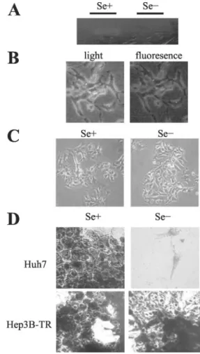
To test whether this tolerance of Hep3B-TR cells was caused by an unusual ability to maintain selenoprotein activity under our selenium starvation conditions, we assessed their Gpx activity. Hep3B-TR cells have lost their Gpx activity after 4 days of selenium starvation (Fig. 5A), similar to what we observed with Huh7 cells (Fig. 1A), and as reported previously by others (28). Thus, both Huh7 and Hep3B-TR cells became selenium deficient after 4 days of selenium starvation, but they differed sharply in their respective responses.
Selenium-Fig. 3. Characterization of Huh7 cell death as apoptosis. Huh7 cells were grown in a selenium-supplemented (Se⫹) and/or selenium-deficient (Se⫺) medium for 96 h. A, selenium-deficient Huh7 cells were tested by fluorescent TUNEL in DMEM and NAPO assays in HAM’s medium. Nuclear DNA was visualized by Hoechst 33258 counterstain-ing. Apoptotic Huh7 cells display positive TUNEL and negative NAPO staincounterstain-ing. B, demonstration of cytochrome c release under oxidative stress in Huh7 cells by immuno-staining of intracellular cytochrome c (left panels) selenium-supplemented (Se⫹) or selenium-deficient (Se⫺) DMEM. Under selenium-supplemented conditions, Huh7 cells show scattered mitochondrial cytochrome c stain, whereas they show diffuse and more intense stain under selenium-deficient conditions (white arrows). Notable are the con-densed nuclei of Huh7 cells that release cytochrome c under selenium-deficient conditions (right panel). C, immunoblot analysis of PARP protein in DMEM. Selenium deficiency induces total proteolysis of PARP in apoptosis-sensitive Huh7 cell line when there was more than 90% apoptosis. On the other hand, Huh7 has intact PARP protein under Se⫹ conditions. Each well was loaded with 10g of protein in 8% SDS-PAGE. Cytokeratin 18 probing was performed as an equal loading control.
Fig. 4. Dose-dependent association of external oxidative stress with onset of apoptosis. Huh7 and Hep3B-TR cells were cultured under Se⫹ and Se⫺ conditions in DMEM for 72 h and then treated with the indicated concentrations of H2O2. Four h later, cells under
oxidative stress were counted by fluorescent microscope, and apo-ptotic cells were counted by Hoechst 33258 staining. With increas-ing concentrations of H2O2, the percentage of Huh7 cells under
oxidative stress and apoptosis increased accordingly under Se⫺ conditions. On the other hand, Hep3B-TR cells did not display any oxidative stress and apoptosis even in the presence of external oxidative stress. The results are from three separate experiments.
Error bars, SD.
deficient Hep3B-TR cells were free of oxidative stress as indicated by a total lack of oxidation of nonfluorescent DCFH-DA in reactive cells (Fig. 5B, right panel), and they appear healthy under these conditions when the medium was supplemented with copper/zinc (Fig. 5C). As expected from these observations, they displayed no sign of apoptosis (data not shown).
The ability of Hep3B-TR cells to tolerate selenium deficiency prompted us to compare their survival capacity to that of Huh7 cells. As shown in Fig. 5D, selenium-deficient Hep3B-TR cells survived under these conditions for at least 10 days (longest time tested), at which time virtually no Huh7 cells were alive.
In addition, we examined the activation of AKT a downstream component of the phosphatidylinositol 3-kinase (PI3K) pathway, which is important in regulating cell survival and apoptosis and which influences cell survival on oxidative injury (36). AKT protein levels were viewed by Western blot analysis under selenium-deficient or
-supplemented conditions with Huh7 and Hep3B-TR cells. We ob-served no difference in either unphosphorylated or active phospho-rylated AKT protein levels in these apoptosis-sensitive (Huh7) and apoptosis-resistant (Hep3B-TR) cell lines (data not shown). In addi-tion to our observaaddi-tions with DCFH assay our data with AKT protein status indirectly indicates that under selenium-deficient conditions, resistant Hep3B-TR cells do not display oxidative stress.
Taken together, our observations with Hep3B-TR cells provided strong evidence that these cells tolerate selenium deficiency. It ap-peared that these malignant cells have acquired a capacity to survive and proliferate under long periods of selenium deficiency. This ca-pacity is accompanied by and probably attributable to their high tolerance to ROS, as evidenced by our H2O2treatment studies. It is expected that the metabolic activity of these cells that is maintained under selenium-deficient conditions will continue to produce ROS in their mitochondria. The lack of oxidative stress under these condi-tions, even in the presence of an excess of H2O2, clearly indicates that they adapted themselves to eliminate such reactive species by seleni-um-independent means. This capacity provides them a selective abil-ity for survival under selenium-deficient and oxidative stress condi-tions.
Rescue of Selenium-Deficient Huh7 Cells from Oxidative Stress and Cell Death with Vitamin E. Vitamin E has a protective scav-enger effect against free-radical damage. In the presence of metal ions, hydroxyl radical (HO•) is produced from H2O2. Vitamin E that is integrated in cellular membranes has been shown to reduce HO• and, in this way, protects peroxidation of lipids (37). Because sele-nium deficiency leads to the accumulation of ROS as evidenced by the DCFH assay in Huh7 cells leading to apoptosis (Fig. 1B), we wanted to test whether Vitamin E has a protective scavenger effect on sele-nium deficiency-induced oxidative stress and the apoptosis in Huh7 cells. Indeed, as shown in Fig. 6, Vitamin E has a protective antiox-idant effect on selenium deficiency-induced ROS. As a consequence, we observed neither the oxidative stress nor the apoptosis in Huh7 cells in the presence of Vitamin E under selenium-deficient condi-tions. Vitamin E had an indifferent effect on Hep3B-TR cells under the same conditions, because we did not observe any oxidative stress (data not shown).
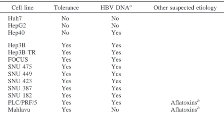
Most Hepatocarcinoma Cell Lines Tolerate Selenium Defi-ciency. Contrasting abilities of two HCC cell lines to survive under selenium-deficient conditions led us to extend our investigations to other hepatocellular cancer cell lines. Twelve HCC-derived and one hepatoblastoma-derived cell lines were tested. All but three cell lines were able to survive under selenium-deficient conditions, in a way quite similar to the Hep3B-TR cell line (experimental data not shown; see Table 1 for a summary). In total, 10 (77%) of 13 cell lines tolerated selenium deficiency. Included in this tolerant group was the Hep3B cell line from which Hep3B-TR was derived. In addition to Huh7, HCC-derived Hep40 and hepatoblastoma-derived HepG2 cell lines died by apoptosis under selenium deficiency (data not shown for Hep40; see Figs. 7–9 for HepG2). It is noteworthy that only one (33%) of these three sensitive cell lines had integrated HBV se-quences in its genome. In contrast, 9 (90%) of 10 tolerant cell lines displayed integrated HBV sequences, indicating past exposure to this virus. Keeping in mind that both selenium deficiency and HBV infection have a synergic effect on HCC risk (18), the association of tolerance to selenium deficiency with a history of HBV infection suggested to us that these two could be related. For example, selenium deficiency could favor the risk of HBV infection. Alternatively, HBV infection could facilitate the ability of malignant cells to gain toler-ance to selenium deficiency.
Fig. 5. Analysis of Hep3B-TR cells under supplemented and selenium-deficient conditions. Hep3B-TR cells were grown in selenium-supplemented (Se⫹) or selenium-deficient (Se⫺) conditions. A, in vitro selenium deficiency of Hep3B-TR cells in DMEM was tested by the loss of Gpx activity. At 96 h, cells were solubilized, and equal amounts of soluble proteins from cell lysates were separated by native PAGE, followed by in situ staining for Gpx with a colorimetric assay. B, analysis of oxidative stress by oxidant-sensitive fluorescent dye, DCFH-DA. Hep3B-TR cells were cultured under sele-nium-deficient (Se⫺) HAM’s medium for 96 h. Hep3B-TR cells did not exhibit a positive fluorescent signal, indicating that, unlike Huh7 cells (Fig. 1B), they do not harbor oxidative stress (right). Light microscopy photographs of the corresponding area of fluorescent photographs of Hep3B-TR cells under selenium-deficient (Se⫺) conditions were also shown (left).⫻200. C, Hep3B-TR cells grown in a selenium-supplemented (Se⫹) or selenium-deficient (Se⫺) HAM’s medium, examined under light microscope on a daily basis, with pictures taken at day 5. ⫻100. D, comparison of the survival of HBV-negative Huh7 and HBV-positive Hep3B-TR cells. Cell lines were cultured under selenium-supplemented (Se⫹) and selenium-deficient (Se⫺) DMEM cell culture for 10 days, and attached cells were visualized by Giemsa staining. Whereas Hep3B-TR cells were able to expand under selenium-deficient conditions, Huh7 cells did not survive. Survival of the both cell lines was not affected under selenium-supplemented conditions.
⫻100.
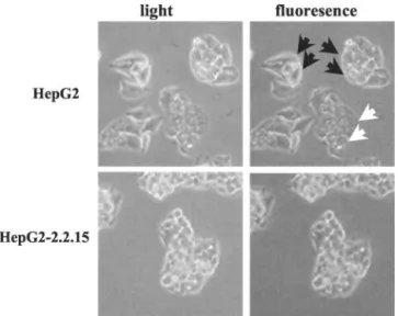
HBV Confers Tolerance to Selenium Deficiency in HepG2 Cells. To test whether the introduction of HBV into malignant cells is associated with acquired tolerance to selenium deficiency, we took advantage of the availability of the HBV-transfected 2.2.15 clone of the HepG2 cell line that produces infectious HBV virions (38). First, these two isogenic cell lines were cultivated under selenium-deficient and selenium-supplemented conditions for 10 days. Both cell lines showed similar growth rates under selenium-supplemented condi-tions. However, their responses to selenium deficiency were different. As reported in Table 1, parental HepG2 cells were sensitive to selenium deficiency, whereas the 2.2.15 clone was tolerant. This acquired tolerance was accompanied with the following changes in 2.2.15 clone cells. When exposed to 200MH2O2, under selenium deficiency, the 2.2.15 clone, but not the parental HepG2 cells, re-sponded with a lack of progressive oxidative stress that was associated with a lack of cell death (Fig. 7). Accordingly, selenium deficiency in the HepG2 cell line resulted in the accumulation of cells with oxida-tive stress in selenium-deficient copper/zinc-containing HAM’s me-dium (Fig. 8). The release of cytochrome c from mitochondria (Fig. 9A, bottom right panels), and apoptosis (Fig. 9B) were also observed in the HepG2 cell line (as in the case of the Huh7 cell line) but not in the HepG2-2.2.15 cell line. The 2.2.15 clone resisted selenium defi-ciency, so that the effects of selenium starvation observed in the parental HepG2 were no longer detectable in the cells of this clone (Figs. 8 and 9). Moreover, Vitamin E had the same protective anti-oxidant effect on HepG2 cells under selenium deficiency (data not shown) as Huh7 cells (Fig. 6). These major differences between 2.2.15 and HepG2 cells were detectable only under selenium-deficient conditions, because the in vitro survival and proliferation capacities of the two cell lines did not differ from each other under selenium-supplemented conditions (data not shown).
DISCUSSION
Despite increasing awareness about the importance of selenium to human health, its fundamental roles in cell biology remain poorly understood. Moreover, cellular mechanisms involved in the
pathogen-esis of selenium-deficiency syndromes and the molecular basis of selenium protection are not well known. Previous studies on selenium deficiencies relied on in vivo whole animal models, mostly using rodents (39, 40). Here, we developed an in vitro experimental model based on human cell lines, rather than rodent tissues, so that biological responses may be a better representation of related changes in hu-mans. In addition, in vitro models are easy to perform, allow a better control of selenium supply, and provide clear-cut end results because of the homogeneity of the cell populations studied. Finally, cell lines are readily available for in vitro modifications to change experimental parameters. The caveat is that the extrapolation of in vitro observa-tions to in vivo situaobserva-tions requires extreme care and prudence. With these considerations in mind, we believe that our findings provide some insights on selenium deficiency and its implications in hepato-cellular malignancy in human.
It has been known for a long time that rats that are fed with a selenium-deficient diet develop so called dietary “necrotic” liver degeneration (see Ref. 8). Although in vivo hepatic response to selenium deficiency has not been characterized specifically as apo-ptotic cell death, it is now well established that selenium deficiency causes oxidative stress (see Refs. 1 and 2), and apoptosis is a common cellular response to oxidative stress (41). Our extensive observations with Huh7 and HepG2 indicate that these “hepatocyte-like” malignant cells undergo a delayed apoptotic cell death that is detectable after 3– 4 days of culture in a selenium-deficient medium. This apoptotic response is associated with the depletion of Gpx activity in the cells and correlates with oxidative stress and the release of cytochrome c from the mitochondria, in a time-dependent manner. The depletion of selenium-dependent Gpxs, which act as antiapoptotic factors under oxidative stress by catalyzing the reduction of phospholipid hydroper-oxides as well as hydrogen peroxide (42), may contribute to apoptosis induced by selenium deficiency in our cell lines. There are several studies demonstrating that H2O2leads to the release of cytochrome c from the electron transport chain, consequently from mitochondria to cytoplasm leading to apoptosis (43– 46). In the absence of the sele-nium-dependent Gpx activity, H2O2, which is produced from a su-peroxide radical by susu-peroxide dismutase enzyme, is converted to a hydroxyl radical (HO•) by ferrous (Fe2⫹) iron-dependent Fenton’s
reaction. The release of cytochrome c from mitochondria has been previously linked to the peroxidation of cardiolipin, a mitochondrion-membrane-specific phospholipid, by ROS (42, 47). In our study, we also support this mechanism by preventing cell death in selenium deficiency-induced apoptosis-sensitive Huh7 and HepG2 cell lines with Vitamin E, which is incorporated into cellular membranes as a potent scavenger of HO•. In the absence of a ROS scavenger such as
Table 1 Tolerance of HCCs to in vitro selenium deficiency Cell line Tolerance HBV DNAa Other suspected etiology
Huh7 No No HepG2 No No Hep40 No Yes Hep3B Yes Yes Hep3B-TR Yes Yes FOCUS Yes Yes SNU 475 Yes Yes SNU 449 Yes Yes SNU 423 Yes Yes SNU 387 Yes Yes SNU 182 Yes Yes
PLC/PRF/5 Yes Yes Aflatoxinsb
Mahlavu Yes No Aflatoxinsb aPresence of HBV DNA was tested by PCR amplification, as described previously
(17). HBV status of these cell lines has been reported previously (58, 59).
bCell line displaying p53-codon 249 mutation that has been associated to aflatoxin
exposure (50, 51). Fig. 6. Resistance of Huh7 cells to selenium deficiency-induced oxidative stress with
vitamin E. Huh7 cells were cultured under selenium-deficient (Se⫺) DMEM that con-tained a total of 100Mvitamin E for 72 h (on the first day, 50Mand, on the second and third days, 25MVitamin E was added to culture medium) then tested for oxidative stress by oxidant-sensitive fluorescent dye DCFH-DA under an inverted fluorescent microscope. Huh7 cells harbor oxidative stress (black arrows) and apoptosis (white
arrows) in the absence of vitamin E (bottom left panel). Absence of fluorescence in the
presence of vitamin E (bottom right panel) indicates that Huh7 cells have acquired resistance to oxidative stress. Inverted light microscopy photographs (top panels) of the corresponding area of fluorescent photographs of Huh7 cells under Se⫺ conditions are also shown. Notable is the absence of cell death in Se⫺ medium supplemented with vitamin E (right panels).⫻200.
Vitamin E under selenium deficiency, accumulated ROS, as illus-trated by the observations with Huh7 and HepG2 cell lines, may have an effect on the peroxidation of cardiolipin leading to the release of cytochrome c. Cytoplasmic cytochrome c is the immediate trigger of apoptosis via activation of caspase 9, followed by activation of caspase 3, as evidenced here by the degradation of PARP, a known substrate for caspase 3 (34). On the other hand, the apoptotic response observed here is p53 independent because of the fact that this gene is mutated in Huh7 cells (48).
The second, but probably the most challenging, of our observations is that a large group of HCC cell lines [10 (77%) of 13] tolerate selenium deficiency. Malignant cells belonging to this group are able to survive and keep their proliferative capacity for a long period (at least 10 days in culture) under selenium-deficient conditions. As illustrated by the observations with Hep3B-TR cell lines from this group, the in vitro survival of HCC cells under selenium deficiency depends on their ability to counteract oxidative stress-generating ROS, as evidenced by their resistance to oxidative stress challenge by
exogenously supplied H2O2. Because they do not display a positive reaction with an oxidative stress indicator and they do not release cytochrome c from their mitochondria under selenium deficiency, it is highly likely that these HCC cells are also able to counteract meta-bolically generated ROS during mitochondrial respiration (see Ref. 49). Moreover the fact that apoptosis-sensitive Huh7 and HepG2 cells became resistant to selenium deficiency in the presence of Vitamin E demonstrates that these malignant cells adapt by a mechanism to scavenge ROS produced by electron transport chain in the absence of selenium. This adaptation could not be explained by a slow rate of proliferation, because the proliferation rates of these cell lines are not affected under selenium-deficient conditions when compared with their growth in selenium-supplemented conditions, even though they grow much better in 10% FCS-containing medium (data not shown). Our studies with HepG2-derived 2.2.15 clone indicate that the
Fig. 7. Tolerance of HBV-positive 2.2.15 clone of HepG2 cells to selenium deficiency and oxidative stress. HepG2 and 2.2.15 cells were cultured under selenium-supplemented (Se⫹) and selenium-deficient (Se⫺) DMEM for 72 h and were treated with the indicated concentrations of H2O2. Four h later, cells under oxidative stress were
counted under a fluorescent microscope, and apoptotic cells were counted by Hoechst 33258 staining. With in-creasing concentrations of H2O2, the percentage of
HBV-negative HepG2 cells under oxidative stress and apoptosis increased accordingly. However, HBV-positive HepG2-2.2.15 cells did not display oxidative stress even in the presence of external oxidative stress source H2O2.
The results are from three separate experiments. Error
bars, SD.
Fig. 8. Analysis of oxidative stress in HepG2-2.2.15 cells under selenium deficiency. HepG2 and 2.2.15 cells were cultured under selenium-deficient (Se⫺) HAM’s medium for 72 h. DCF fluorescence as an indicator of oxidative stress was observed under an inverted fluorescent microscope. HepG2 cells harbored oxidative stress (black arrows) and apop-tosis (white arrows), whereas 2.2.15 cells did not.⫻200.
Fig. 9. Apoptosis as a result of oxidative stress in HepG2 cells but not in HepG2-2.2.15 cells. A, immunostaining of cytochrome c protein under selenium-supplemented (Se⫹) and selenium-deficient (Se⫺) conditions. Under selenium-supplemented conditions, HepG2 and HepG2-2.2.15 cells show punctuate mitochondrial cytochrome c stain. HepG2 cells, but not HepG2-2.2.15 cells, showed diffuse cytoplasmic cytochrome c stains (white
arrows) under selenium-deficient conditions. Corresponding nuclei of all cells were
counterstained with Hoechst 33258. Notable are the condensed nuclei of HepG2 cells that release cytochrome c. B, HepG2 and HepG2-2.2.15 cells were compared by fluorescent NAPO immunostaining. Nuclear DNA was visualized by Hoechst 33258 counterstaining. HepG2-2.2.15 cells were negative for the assay, indicating that unlike HepG2 cells, they did not enter apoptosis under selenium-deficient conditions.
tolerance of HCC cells to selenium deficiency is an acquired capa-bility. Because selenium is an essential nutrient in animals, our normal cells cannot tolerate selenium deficiency under normal physiological conditions. Therefore, it would not be surprising that tolerance to selenium deficiency is an acquired capability of these malignant cells. It is interesting to note that, this tolerance correlates closely with the presence of HBV sequences in the genomes of these cells. The tolerance displayed by an HBV-positive HepG2-2.2.15 clone of the sensitive HepG2 cell line may provide additional evidence that this virus may contribute to the tolerance to selenium deficiency.
Presently, it is unclear whether 10 HCC cell lines (Table 1) have acquired their tolerance to selenium deficiency in vivo or after in vitro establishment. It is tempting to speculate that this occurred in vivo at least in some cell lines. Of these 10 cell lines, 9 have integrated HBV sequences in their genomes, and the remaining cell line displays an aflatoxin-related p53 mutation (codon 249; G3T; Refs. 50 and 51). These aberrations can be considered genetic fingerprints of past exposure of respective patients to these agents. Both chronic HBV infection and aflatoxin exposure are known to induce ROS in hepa-tocytes (52–54). Oxidative stress is considered to play an important role in liver malignancy (55). Because both selenium depletion and the excess of ROS are common in chronic liver diseases serving as a reservoir for hepatic malignancies (6, 20, 56, 57), we cannot dismiss the possibility that the acquired tolerance of HCC cells to selenium deficiency and ROS may occur in vivo. Acquired tolerance to sele-nium deficiency-induced ROS may not be restricted to past exposure of HCC patients to viral or aflatoxin exposures. Oxidative stress because of selenium deficiency may play an important role not only in liver cancer but also in other malignancies. Finally we wanted to extend our study to cell lines of other cancer types. We tested a total of 27 different cancer cell lines (breast, 14; colon, 10; melanoma, 3). We found that 11 of 14 breast cancer cell lines, 8 of 10 colon cancer cell lines, and all of 3 melanoma cell lines were resistant to selenium deficiency-induced cell death. Our preliminary data may suggest that escape from deadly consequences of selenium deficiency may play a role in other cancer types. In addition, it brings new insight to the scientific debate on dietary selenium supplementation for cancer pa-tients, which may eventually be further investigated.
Independent of their causes, the tolerance of most HCC cell lines to selenium deficiency and ROS provides them a “cryptic survivor phenotype.” We call it “cryptic” because this phenotype is not ex-pressed under normal culture conditions, and this is probably why it remained unnoticed until now. This phenotype could provide a novel survival mechanism for a subset of HCCs.
ACKNOWLEDGMENTS
We thank B. Carr (University of Pittsburgh, Pittsburgh, PA) for providing Hep3B-TR and Hep40 cell lines and D. Bellet (Institute Gustave Roussy, Paris, France) for providing anti-cytokeratin 18 monoclonal antibody JAR1 3.
REFERENCES
1. Flohe, L., Andreesen, J. R., Brigelius-Flohe, R., Maiorino, M., and Ursini, F. Sele-nium, the element of the moon, in life on earth. IUBMB Life, 49: 411– 420, 2000. 2. Rayman, M. P. The importance of selenium to human health. Lancet, 356: 233–241,
2000.
3. Willett, W. C., Polk, B. F., Morris, J. S., Stampfer, M. J., Pressel, S., Rosner, B., Taylor, J. O., Schneider, K., and Hames, C. G. Prediagnostic serum selenium and risk of cancer. Lancet, 2: 130 –134, 1983.
4. Knekt, P., Aromaa, A., Maatela, J., Alfthan, G., Aaran, R. K., Hakama, M., Hakulinen, T., Peto, R., and Teppo, L. Serum selenium and subsequent risk of cancer among Finnish men and women. J. Natl. Cancer Inst. (Bethesda), 82: 864 – 868, 1990. 5. Yoshizawa, K., Willett, W. C., Morris, S. J., Stampfer, M. J., Spiegelman, D., Rimm, E. B., and Giovannucci, E. Study of prediagnostic selenium level in toenails and the risk of advanced prostate cancer. J. Natl. Cancer Inst. (Bethesda), 90: 1219 –1224, 1998.
6. Yu, M. W., Horng, I. S., Hsu, K. H., Chiang, Y. C., Liaw, Y. F., and Chen, C. J. Plasma selenium levels and risk of hepatocellular carcinoma among men with chronic hepatitis virus infection. Am. J. Epidemiol., 150: 367–374, 1999.
7. Mark, S. D., Qiao, Y. L., Dawsey, S. M., Wu, Y. P., Katki, H., Gunter, E. W., Fraumeni, J. F., Jr., Blot, W. J., Dong, Z. W., and Taylor, P. R. Prospective study of serum selenium levels and incident esophageal and gastric cancers. J. Natl. Cancer Inst. (Bethesda), 92: 1753–1763, 2000.
8. Schwarz, K., and Foltz, C. M. Selenium as an integral part of factor 3 against dietary necrotic liver degeneration. Nutrition, 15: 255, 1999.
9. Chance, B., Sies, H., and Boveris, A. Hydroperoxide metabolism in mammalian organs. Physiol. Rev., 59: 527– 605, 1979.
10. Turrens, J. F. Superoxide production by the mitochondrial respiratory chain. Biosci. Rep., 17: 3– 8, 1997.
11. Turrens, J. F., and Boveris, A. Generation of superoxide anion by the NADH dehydrogenase of bovine heart mitochondria. Biochem. J., 191: 421– 427, 1980. 12. Kira, Y., Sato, E. F., and Inoue, M. Association of Cu,Zn-type superoxide dismutase
with mitochondria and peroxisomes. Arch. Biochem. Biophys., 399: 96 –102, 2002. 13. Zelko, I. N., Mariani, T. J., and Folz, R. J. Superoxide dismutase multigene family: a comparison of the CuZn-SOD (SOD1), Mn-SOD (SOD2), and EC-SOD (SOD3) gene structures, evolution, and expression. Free Radic. Biol. Med., 33: 337–349, 2002.
14. Cadenas, E., and Davies, K. J. Mitochondrial free radical generation, oxidative stress, and aging. Free Radic. Biol. Med., 29: 222–230, 2000.
15. Mirault, M. E., Tremblay, A., Beaudoin, N., and Tremblay, M. Overexpression of seleno-glutathione peroxidase by gene transfer enhances the resistance of T47D human breast cells to clastogenic oxidants. J. Biol. Chem., 266: 20752–20760, 1991. 16. Geiger, P. G., Thomas, J. P., and Girotti, A. W. Lethal damage to murine L1210 cells by exogenous lipid hydroperoxides: protective role of glutathione-dependent sel-enoperoxidases. Arch. Biochem. Biophys., 288: 671– 680, 1991.
17. Cheng, W. H., Ho, Y. S., Valentine, B. A., Ross, D. A., Combs, G. F., Jr., and Lei, X. G. Cellular glutathione peroxidase is the mediator of body selenium to protect against paraquat lethality in transgenic mice. J. Nutr., 128: 1070 –1076, 1998. 18. Yu, S. Y., Zhu, Y. J., and Li, W. G. Protective role of selenium against hepatitis B
virus and primary liver cancer in Qidong. Biol. Trace Elem. Res., 56: 117–124, 1997. 19. Reyes, H. Intrahepatic cholestasis. A puzzling disorder of pregnancy. J. Gastroenterol.
Hepatol., 12: 211–216, 1997.
20. Guarini, P., Stanzial, A. M., Olivieri, O., Casaril, M., Galvani, S., Pantalena, M., and Corrocher, R. Erythrocyte membrane lipids and serum selenium in post-viral and alcoholic cirrhosis. Clin. Chim. Acta, 270: 139 –150, 1998.
21. Sayan, B. S., Ince, G., Sayan, A. E., and Ozturk, M. NAPO as a novel marker for apoptosis. J. Cell Biol., 155: 719 –724, 2001.
22. Sun, Y., Elwell, J. H., and Oberley, L. W. A simultaneous visualization of the antioxidant enzymes glutathione peroxidase and catalase on polyacrylamide gels. Free Radic. Res. Commun., 5: 67–75, 1988.
23. Unsal, H., Yakicier, C., Marcais, C., Kew, M., Volkmann, M., Zentgraf, H., Isselbacher, K. J., and Ozturk, M. Genetic heterogeneity of hepatocellular carcinoma. Proc. Natl. Acad. Sci. USA, 91: 822– 826, 1994.
24. Blum, H. E., Zhang, Z. S., Galun, E., von Weizsacker, F., Garner, B., Liang, T. J., and Wands, J. R. Hepatitis B virus X protein is not central to the viral life cycle in vitro. J. Virol., 66: 1223–1227, 1992.
25. Bradford, M. M. A rapid and sensitive method for the quantitation of microgram quantities of protein utilizing the principle of protein-dye binding. Anal. Biochem.,
72: 248 –254, 1976.
26. Achenbach, T. V., Muller, R., and Slater, E. P. Bcl-2 independence of flavopiridol-induced apoptosis. Mitochondrial depolarization in the absence of cytochrome c release. J. Biol. Chem., 275: 32089 –32097, 2000.
27. Richter, C., Gogvadze, V., Laffranchi, R., Schlapbach, R., Schweizer, M., Suter, M., Walter-Royall, J. A., and Ischiropoulos, H. Evaluation of 2⬘,7⬘-dichlorofluorescin and dihydrorhodamine 123 as fluorescent probes for intracellular H2O2in cultured
endo-thelial cells. Arch. Biochem. Biophys., 302: 348 –355, 1993.
28. Baker, R. D., Baker, S. S., LaRosa, K., Whitney, C., and Newburger, P. E. Selenium regulation of glutathione peroxidase in human hepatoma cell line Hep3B. Arch. Biochem. Biophys., 304: 53–57, 1993.
29. Nakabayashi, H., Taketa, K., Miyano, K., Yamane, T., and Sato, J. Growth of human hepatoma cells lines with differentiated functions in chemically defined medium. Cancer Res., 42: 3858 –3863, 1982.
30. Ueda, Y., Matsumoto, K., and Endo, K. Evidence of hepatic endogenous hydrogen peroxide in bile of selenium-deficient rats. Biochem. Biophys. Res. Commun., 271: 699 –702, 2000.
31. Cheng, W. H., Zheng, X., Quimby, F. R., Roneker, C. A., and Lei, X. G. Low levels of glutathione peroxidase 1 activity in selenium-deficient mouse liver affect c-Jun N-terminal kinase activation and p53 phosphorylation on Ser-15 in pro-oxidant-induced aponecrosis. Biochem. J., 370: 927–934, 2003.
32. Liu, X., Kim, C. N., Yang, J., Jemmerson, R., and Wang, X. Induction of apoptotic program in cell-free extracts: requirement for dATP and cytochrome c. Cell, 86: 147–157, 1996.
33. Li, P., Nijhawan, D., Budihardjo, I., Srinivasula, S. M., Ahmad, M., Alnemri, E. S., and Wang, X. Cytochrome c and dATP-dependent formation of Apaf-1/caspase-9 complex initiates an apoptotic protease cascade. Cell, 91: 479 – 489, 1997. 34. Kaufmann, S. H., Desnoyers, S., Ottaviano, Y., Davidson, N. E., and Poirier, G. G.
Specific proteolytic cleavage of poly(ADP-ribose) polymerase: an early marker of chemotherapy-induced apoptosis. Cancer Res., 53: 3976 –3985, 1993.
35. Hasegawa, K., Wang, Z., Inagaki, M., and Carr, B. I. Characterization of a human hepatoma cell line with acquired resistance to growth inhibition by transforming growth factor 1 (TGF-1). In Vitro Cell Dev. Biol. Anim., 31: 55–61, 1995.
36. Ostrakhovitch, E. A., Lordnejad, M. R., Schliess, F., Sies, H., and Klotz, L. O. Copper ions strongly activate the phosphoinositide-3-kinase/Akt pathway independent of the generation of reactive oxygen species. Arch. Biochem. Biophys., 397: 232–239, 2002. 37. Claycombe, K. J., and Meydani, S. N. Vitamin E and genome stability. Mutat. Res.,
475: 37– 44, 2001.
38. Sells, M. A., Chen, M. L., and Acs, G. Production of hepatitis B virus particles in HepG2 cells transfected with cloned hepatitis B virus DNA. Proc. Natl. Acad. Sci. USA, 84: 1005–1009, 1987.
39. McConnell, K. P., Hsu, J. M., Anthony, W. L., and Bieri, J. G. Selenium deficiency and protein, RNA and DNA synthesis in rat pancreas and liver. Proc. Soc. Exp. Biol. Med., 147: 575–577, 1974.
40. Aviado, D. M., Drimal, J., Watanabe, T., and Lish, P. M. Cardiac effects of sodium selenite. Cardiology, 60: 113–120, 1975.
41. Chandra, J., Samali, A., and Orrenus, S. Triggering and modulation of apoptosis by oxidative stress. Free Radic. Biol. Med., 29: 323–333, 2000.
42. Nomura, K., Imai, H., Koumura, T., Kobayashi, T., and Nakagawa, Y. Mitochondrial phospholipid hydroperoxide glutathione peroxidase inhibits the release of cytochrome
c from mitochondria by suppressing the peroxidation of cardiolipin in
hypoglycae-mia-induced apoptosis. Biochem. J., 351: 183–193, 2000.
43. Stridh, H., Kimland, M., Jones, D. P., Orrenius, S., and Hampton, M. B. Cytochrome
c release and caspase activation in hydrogen peroxide- and tributyltin-induced
apo-ptosis. FEBS Lett., 429: 351–355, 1998.
44. Cai, J., Yang, J., and Jones, D. P. Mitochondrial control of apoptosis: the role of cytochrome c. Biochim. Biophys. Acta, 1366: 139 –149, 1998.
45. Morales, A., Garcia-Ruiz, C., Miranda, M., Mari, M., Colell, A., Ardite, E., and Fernandez-Checa, J. C. Tumor necrosis factor increases hepatocellular glutathione by transcriptional regulation of the heavy subunit chain of␥-glutamylcysteine synthe-tase. J. Biol. Chem., 272: 30371–30379, 1997.
46. Atlante, A., Calissano, P., Bobba, A., Azzariti, A., Marra, E., and Passarella, S. Cytochrome c is released from mitochondria in a reactive oxygen species (ROS)-dependent fashion and can operate as a ROS scavenger and as a respiratory substrate in cerebellar neurons undergoing excitotoxic death. J. Biol. Chem., 275: 37159 – 37166, 2000.
47. Shidoji, Y., Hayashi, K., Komura, S., Ohishi, N., and Yagi, K. Loss of molecular interaction between cytochrome c and cardiolipin due to lipid peroxidation. Biochem. Biophys. Res. Commun., 264: 343–347, 1999.
48. Hsu, I. C., Tokiwa, T., Bennett, W., Metcalf, R. A., Welsh, J. A., Sun, T., and Harris, C. C. p53 gene mutation and integrated hepatitis B viral DNA sequences in human liver cancer cell lines. Carcinogenesis (Lond.), 14: 987–992, 1993.
49. Richter, C., Gogvadze, V., Laffranchi, R., Schlapbach, R., Schweizer, M., Suter, M., Walter, P., and Yaffee, M. Oxidants in mitochondria: from physiology to diseases. Biochim. Biophys. Acta, 1271: 67–74, 1995.
50. Bressac, B., Kew, M., Wands, J., and Ozturk, M. Selective G to T mutations of p53 gene in hepatocellular carcinoma from southern Africa. Nature (Lond.), 350: 429 – 431, 1991.
51. Hsu, I. C., Metcalf, R. A., Sun, T., Welsh, J. A., Wang, N. J., and Harris, C. C. Mutational hotspot in the p53 gene in human hepatocellular carcinomas. Nature (Lond.), 350: 427– 428, 1991.
52. Yang, C. F., Liu, J., Wasser, S., Shen, H. M., Tan, C. E., and Ong, C. N. Inhibition of ebselen on aflatoxin B1-induced hepatocarcinogenesis in Fischer 344 rats.
Carci-nogenesis (Lond.), 21: 2237–2243, 2000.
53. Shen, H. M., Ong, C. N., and Shi, C. Y. Involvement of reactive oxygen species in aflatoxin B1-induced cell injury in cultured rat hepatocytes. Toxicology, 99: 115–123,
1995.
54. Shen, H. M., Shi, C. Y., Shen, Y., and Ong, C. N. Detection of elevated reactive oxygen species level in cultured rat hepatocytes treated with aflatoxin B1. Free Radic.
Biol. Med., 21: 139 –146, 1996.
55. Toyokuni, S., Okamoto, K., Yodoi, J., and Hiai, H. Persistent oxidative stress in cancer. FEBS Lett., 358: 1–3, 1995.
56. Lieber, C. S. Role of oxidative stress and antioxidant therapy in alcoholic and nonalcoholic liver diseases. Adv. Pharmacol., 38: 601– 628, 1997.
57. Ozturk, M. Genetic aspects of hepatocellular carcinogenesis. Semin. Liver Dis., 19: 235–242, 1999.
58. Park, J. G., Lee, J. H., Kang, M. S., Park, K. J., Jeon, Y. M., Lee, H. J., Kwon, H. S., Park, H. S., Yeo, K. S., Lee, K. U., et al. Characterization of cell lines established from human hepatocellular carcinoma. Int. J. Cancer, 62: 276 –282, 1995. 59. Simon, D., and Carr, B. I. Integration of hepatitis B virus and alteration of the 1p36
region found in cancerous tissue of primary hepatocellular carcinoma with viral replication evidenced only in noncancerous, cirrhotic tissue. Hepatology, 22: 1393– 1398, 1995.