Radial Artery Pseudoaneurysm After Transradial Coronary
Angiogram: A Mostly Overlooked Complication
Sıklıkla Atlanan Komplikasyon: Transradyal Koroner Anjiografi Sonrası Gelişen Radial Arter Psödoanevrizması
Veysel Özgür Barış
1, Özgür Ulaş Özcan
1, İrem Müge Akbulut
1, Eralp Tutar
11 Ankara University School of Medicine, Department of Cardiology
Radial arter, tanısal ve girişimsel kalp kateterizasyon işlemleri için önerilen güvenli girişim yeridir. Bu yazı-mızda, antikoagülan tedavi altında transradial anjiografi olduktan 10 gün sonra radial arter psödoanevrizması gelişen 73 yaşında kadın olguyu takdim ettik. Ultrasonografi aracılı kompresyonun başarısız olması nedeniyle, hastada cerrahi tedavi uygulandı. Antikoagülan tedavi altında transradial anjiografi yapılan hastalarda, işlem sonrası dikkatli takip önermekteyiz.
Anahtar Sözcükler: Radyal Arter, Psödoanevrizma, Koroner Anjiografi, Komplikasyon
Radial artery is suggested as a safe access for diagnostic and interventional cardiac catheterization.We pre-sent a 73 year old female who was suffered a radial artery pseudoaneurysm 10 days after transradial coronary angiography while under anticoagulation therapy. Prolonged ultrasound guided compression was unsuc-cessful and the patient was treated with surgical repair. We suggest careful observation after transradial cat-heterization for patients under anticoagulant therapy.
Key Words: Radial Artery, Pseudoaneurysm ,Coronary Angiogram, Complication
Transradial access is recommended during percutaneous coronary diagnostic and interventional procedures, especially among patients with acute coronary syndromes and patients under anticoa-gulation therapy. Radial artery is more favorable for vascular access than fe-moral artery because of owning some advantages in terms of lower incidence of bleeding events and earlier time for ambulation (1, 2). However, there also are complications related to transradial access (3). Radial artery pseudoa-neurysm is an extremely rare complica-tion with an incidence of 0.048% (3, 4).
Case Report
A 73 year old female was presented to our clinic with refractory angina pectoris. Transthoracic echocardiogram revealed hypokinesis of mid and apical segments of anterior wall. She was under warfarin treatment for paroxysmal atrial fibrillation. The procedure was performed after warfarin had been ceased for 3 days. The patency of dual hand circulation was confirmed with positive modified Allen test. The International Normalized Ratio (INR)
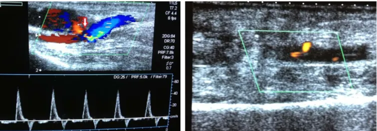
at the time of angiography was 1.5. Coronary angiography performed via the left radial artery using a 5Fr arrow introducer sheath (Arrow International, Reading, PA). Heparin (3000 IU) and verapamil (5 mg) was given to prevent thrombosis and spasm respectively. Coronary angiogram revealed non-significant atherosclerotic plaques. The vascular sheath was removed immediately after the procedure and the puncture site was manually compressed for 15 minutes. Afterwards, a compressive dressing was applied for 24 hours. The next day, warfarin therapy was initiated again because of paroxysmal atrial fibrillation with a CHA2DS2-VASc score of 6. No sign of hematoma was determined on radial artery and the patient was discharged. Ten days after the radial artery puncture, the patient was noted a painful swelling with a small hematoma that appeared over the site of the radial access. She seeked attention 5 days after the symptom onset. Her INR level was 2.4 at that time. Colour Doppler ultrasound (Toshiba Diagnostic Ultrasound System, Model SSA-770A) revealed a 17x10 mm sized pseudoaneurysm with
Received : June 28,2016 • Accepted: Oct 03,2016 Corresponding Author:
Barış, Veysel Özgür MD
E-mail: veyselozgurbaris@yahoo.com Phone: +90312-508 25 16
Ankara University School of Medicine Ankara İbn-i Sina Hospital 12th Floor Sıhhiye, Ankara
Ankara Üniversitesi Tıp Fakültesi Mecmuası 2016, 69 (3)
DOI: 10.1501/Tıpfak_000000946 DAHİLİ BİLİMLERİ/ MEDICAL SCIENCES
Ankara Üniversitesi Tıp Fakültesi Mecmuası 2016, 69 (3)
Radial Artery Pseudoaneurysm After Transradial Coronary Angiogram: A Mostly Overlooked Complication 210
a 1,6 mm neck communicating between the radial artery and the false aneurysm
(Figure 1). Prolonged
ultrasound-guided compression was attempted. However, it was unsuccessful. Therefore, the patient underwent surgical repair with ligation and excision of the pseudoaneurysm. After the surgical repair there was trombus formation in the radial artery (Figure 2)
and no evidence of pseudoaneurysm was detected. Radial pulse and distal perfusion was preserved.
Discussion
Transradial access for percutaneous coro-nary procedure is considered as safe
and convenient approach compared to the transfemoral access. However, the vascular complications associated with transradial access are not rare. It is es-sential for the interventional cardiolo-gists to be aware of these complica-tions. Symptomatic radial artery occlu-sion, non-occlusive radial artery injury and radial artery spasms are commonly reported complications of this appro-ach (3). Pseudoaneurysm formation and radial artery perforation are rarely reported complications of transradial approach (5). In this case, we present a patient with radial artery pseudoa-neurysm following percutaneous car-diac catheterization. There are many
risk factors associated with pseudoa-neurysm formation. These include; pe-netrating injury of the arterial wall du-ring cannulation, multiple puncture at-tempts, catheter infection, aggressive anticoagulation therapy, and the use of large sized sheaths (3). In our case, none of these risk factors was present. The anticoagulation therapy was restar-ted the day after catheterization. Even
suggested as a safe procedure transra-dial approach may warrant more care-ful observation particularly among pa-tients on anticoagulant therapy and in-terventional cardiologists should be aware of the potential for such late ac-cess site complication.
Figure 1: Arrow indicates 17x10 mm sized pseudoaneurysm with a
1.6 mm neck communicating between the radial artery and the false aneurysm.
Figure 2: After the surgical repair there was trombus formation in the
ra-dial artery.
REFERENCES
1. Jolly SS, Yusuf S, Cairns J, et al. Radial ver-sus femoral Access for coronary angiog-raphy and intervention in patients with acute coronarys yndromes (RIVAL): a ran-domised, parallelgroup, multicentretrial. Lancet. 2011;377:1409-1420.
2. Romagnoli E, Biondi-Zoccai G, Sciahbasi A, et al. Radial versus femoral randomized investigation in ST-segment elevation
acute coronary syndrome: the RIFLE-STEACS (Radial Versus Femoral Rando-mized Investigation in ST-Elevation Acute Coronary Syndrome) study. J Am Coll Cardiol.2012;60:2481-2489. 3. Kanei Y, Kwan T, Nakra NC, et al.
Trans-radial cardiac catheterization: a review of access site complications. Catheter Cardi-ovasc Interv. 2011;78:840-846.
4. Collins N, Wainstein R, Ward M, et al. Pseudoaneurysm after transradial cardiac catheterization: case series and review of the literature. Catheter Cardiovasc Interv. 2012;80:283-287.
5. Bhat T, Bhat H, Teli S, et al. Pseudoa-neurysm a rare complication of transradial cardiac catheterization: a case report. Vas-cular. 2013 Mar 18.
