Skeletal deformities of cultured sharpsnout seabream (Diplodus
puntazzo) larvae during early life development
Şükrü YILDIRIM1, Deniz ÇOBAN2, Cüneyt SÜZER1, Kürşat FIRAT1, Şahin SAKA1 1 Ege University, Faculty of Fisheries, Aquaculture Department, 35100 Bornova, İzmir; 2Adnan Menderes Univerity, Faculty of
Agriculture, Aquaculture and Fisheries Engineering Department, Aydın, Turkey.
Summary: In this study, skeletal deformities were investigated during early life stages in sharpsnout seabream, Diplodus puntazzo, larvae during larval development period. Whole larval period (1-42 days after hatching, DAH) was examined in three stages (preflexion, flexion and postflexion) based on notochord curve. Totally, 1210 specimens were examined during the experiment but 689 deformities were recorded in 256 larvae. Also, deformity rate was calculated as 21.2% at the end of the experiment. In terms of notochord flexion at stage-specific, no differences were found between preflexion (19.7%) and postflexion (15.8%) stages (p>0.05) while there was a significant difference during flexion stage (32.8%) in comparison with other groups (p<0.05). Correspondence analysis (CA) was performed in order to describe of skeletal deformities mainly depend on notochord flexion. According to CA analysis, it was determined that deformities were closely related with both flexion stages quantitatively and also postflexion stages qualitatively.
Keywords: D. puntazzo, larval development, skeletal deformities, notochord flexion.
Kültür koşullarındaki sivriburun karagöz (Diplodus puntazzo) larvalarında erken dönem iskelet deformasyonları
Özet: Bu çalışmada, yetiştiriciliği yapılan sivriburun karagöz (Diplodus puntazzo) larvalarının erken hayat döngüsü içerisinde iskelet sisteminde meydana gelen deformasyonlar incelenmiştir. Deneme boyunca larval dönem (1-42 gün) notokorda da meydana gelen değişimlere göre üç bölüme ayrılmıştır (bükülme öncesi, bükülme sırası ve bükülme sonrası). Deneme boyunca toplamda 1210 larva incelenmiş ve 256 deforme larvada 689 iskelet deformasyonu tespit edilmiştir. Deneme sonunda deformasyon oranı %21,2 hesaplanmıştır. Larval dönemdeki safhalara göre deformasyon oranları bükülme öncesi (%19,7) ile bükülme sonrası (%15,8) önemsiz bulunurken (p>0,05), bükülme sırası (%32,8) evresi önemlilik arz etmiştir (p<0,05). Notokordaya göre iskelet deformasyonları Korrespond Analiz yöntemi kullanılarak yapılmıştır. Bu analiz yöntemine göre iskelet deformasyonları safhalar arasında hem niteliksel olarak farklılıkları ortaya koymuş hem de bükülme sonrası dönemde niceliksel ayrımı belirtmiştir.
Anahtar sözcükler: D. puntazzo, larval gelişim, iskelet deformasyonu, notokorda bükülmesi.
Introduction
It is commonly known that morpho-anatomical abnormalities in marine fish under culture conditions usually have negative effects not only on biological performance but also on marketing image and commercial value (1, 3). Description of deformities during larval and juvenile stages could provide fish farms a tool to predict anomalies at the adult stages or could allow making changes to abiotic or biotic factors to improve the quality of specimens during growth (4,5).
Total rate of species-specific deformations in cultured marine organism are usually changed from 15 to 50% under culture conditions, however, these rates are mainly recorded as vertebral deformities between 30 and 60%. Besides, they were observed called as scoliosis,
lordosis and kyphosis, and also observed Z, V and ˄ type curve of vertebra (6,8). To this date, although larval phase was evaluated until juvenile stage (2,3,9,10), recent studies were focused on skeletogenesis mainly depend on bone and cartilage developmental stages with notochordal ontogeny (11,12). However, it is well recorded that factors effecting skeletal development in larval fish are malnutrition and/or sudden changes in biotical and abiotical factors under culture conditions (8,11,12).
In this study, therefore, it is aimed that bone and cartilage deformities depend on developmental phase were investigated in terms of flexional stages (preflexion, flexion and postflexion) of the notochord during early ontogeny of D. puntazzo larvae.
Materials and methods
D. puntazzo broodstocks were selected from the
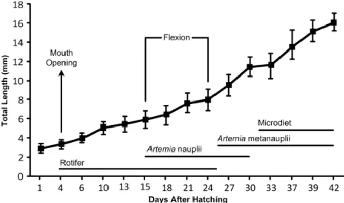
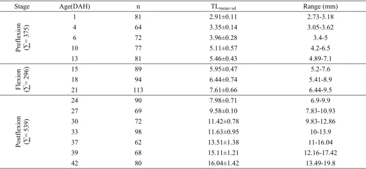
wild breeders and also maturation and spawning occurred spontaneously. Larval rearing and feeding protocol were applied in triplicate according to Suzer et al. (13). Larval feeding regime is summarized in Figure 1. A total of 1210 larvae of D. puntazzo were randomly sampled from tank at 3 day intervals from hatching until 42 DAH. In order to determine deformities in bone and cartilage tissues, double staining method by alizarin red S and alcian blue was used as described by Potthoff (14), Favaloro and Mazzola (5) and Çoban et al. (11). Then, stained larvae were examined under the light microscope and photos were taken from the left side by a digital camera (Nikon Coolpix 5000, Japan) (Figure 2). Deformation analyses were classified with a letter indicating the region and a number indicating the anomaly as listed in Table 1. Also, for determination of effective period of deformities during larval stages, this period was divided into 3 stages and classified as preflexion, flexion and postflexion mainly depending on notochord flexion (15). Each stage with more than 51% of the specimens presenting a certain characteristics was used as the reference point in the description of the flexional stages.
Figure 1. Total length (mean±sd) and feeding regime of D. puntazzo larvae.
Şekil 1. Sivriburun karagöz larvalarına ait total boy (Ort±ss) ve besleme rejimi.
Table 1. Regional deformations and their symbols in D. puntazzo larvae (16).
Tablo 1. D. puntazzo larvalarında gözlenen bölgesel deformas-yonlar ve sembolleri (16). Region Abbreviations External Skeleton Caudal Fin A1 Dorsal Fin A2 Anal Fin A3 Pectoral Fin A4 Appendicular Skeleton B
Axial Skeleton (Syncranium) C Axial Skeleton (Vertebral Column)
Cephalic D1 Pre-hemal D2 Hemal D3 Caudal D4 Anomalies Lordosis 1 Kyphosis 2 Vertebral fusion 3 Vertebral malformation 4
Malformed neural arch and/or spine 5 Malformed hemal arch and/or spine 6 Malformed ray (deformed, absent, fused,
supernumerary) 7 Malformed pterygophores (deformed, absent,
fused, supernumerary) 8
Malformed hypural (deformed, absent, fused,
supernumerary) 9 Malformed epural (deformed, absent, fused,
supernumerary) 10
Supernumerary vertebra 11
Dislocation of glossohyal 12
Malformed dentale 13
Malformed maxillary and/or pre-maxillary 14
Swim-bladder anomaly 15
Figure 2. Double stained D. puntazzo larvae (7.1 mm TL).
In this study, deformation rate and ratio between deformed larva and total larva were found and also the number of deformations in per deformed larvae was calculated by ratio of number of total deformities to total deformed larvae (16). Deformation frequency was evaluated by duration of larval stage with individual differences of sampled larvae (16). Also, chi-square test was used in order to compare of deformations observed in three larval stages and deformation rates in different skeletal regions. Then, for determination of relations between larval stages and regional deformations in larvae, Correspondence analysis was performed.
Results
Notochord flexion was observed between 15 and 24 DAH and also larval development, total number of larvae and related anomalies according to different larval stages are demonstrated in Table 2.
During the study, 689 anomalies of 15 different types were detected in 256 specimens of 1210 larvae and deformity rate was calculated as 21.2%. In overall
results, the number of deformities per whole observed larvae and the number of deformities per abnormal larvae were estimated as 0.57 and 2.69, respectively. Characterization of abnormalities and also obtained results from different larval stages regarding notochord flexion were presented in Table 3.
Observed abnormalities according to the skeletal system: In this section, abnormalities were evaluated
regarding regions of skeletal system in each flexion stage. After that, frequencies of observed deformity were equalized and compared in between flexion stages for each region.
External skeleton: The major deformity in this region
was observed as malformed ray (type 7). Fin deformities which were observed in flexion and postflexion stages were 8.6% and 11.1% of overall deformities detected in larvae. Also, there were no significant differences between flexion and postflexion stages (p>0.05). Additionally, no deformations were recorded in the preflexion stage due to non-formation of fin rays in this stage at external skeletal system. The highest deformity
Table 2. Stage-specific growth of D. puntazzo larvae:Total length(TL) by means(±SD) and ranges. Tablo 2. D. puntazzo larval dönem-büyüme verileri. Toplam boy ortalama (±SS) değişimleri.
Stage Age(DAH) n TLmean+sd Range (mm)
Preflexion (∑ = 375) 1 81 2.91±0.11 2.73-3.18 4 64 3.35±0.14 3.05-3.62 6 72 3.96±0.28 3.4-5 10 77 5.11±0.57 4.2-6.5 13 81 5.46±0.43 4.89-7.1 Fle xion (∑ = 296) 15 89 5.95±0.47 5.2-7.6 18 94 6.44±0.74 5.41-8.9 21 113 7.61±0.66 6.44-9.5 Postfle xion (∑ = 539) 24 90 7.98±0.71 6.9-9.9 27 69 9.58±0.10 7.83-10.93 30 72 11.42±0.78 9.83-12.86 33 98 11.63±0.95 10-13.9 37 62 13.51±1.38 11-16.04 39 68 15.11±1.21 12.16-17.42 42 80 16.04±1.42 13.49-19.8
Table 3. Characterization of abnormalities and abnormal larvae regarding notochord flexion time in D. puntazzo larvae. Superscript * was refer to significant differences by (p<0.05).
Tablo 3. Notokorda bükülme zamanına göre gözlenen deformasyonların ve deforme D. puntazzo larvalarının özellikleri. İstatisitiki önemli farklılıklar üssel * gösterilmiştir.
Preflexion (2.91-5.95 mm TL) Flexion (5.05-7.98 mm TL) Postflexion (7.98-16.04 mm TL)
Total number of observed larvae (A) 375 296* 539
Total number of deformed larvae (B) 74 97* 85
Total number of observed deformation (C) 179 292* 218
Deformation rate (%), (B/A) 19.7 32.8* 15.8
The number of deformity per whole observed larvae, (C/A) 0.48 0.99* 0.40 The number of deformity per abnormal larvae, (C/B) 2.42 3.01* 2.56
rate was observed in pectoral rays for both flexion stages (flexion 4.8%; postflexion 6.8%).
Appendicular skeleton: Several deformities were
recorded in this skeletal system as malformed pterygyophore (type 8), malformed hypural (type 9) and malformed epural (type 10). Although some deformities which occurred in appendicular systems that supported the fins were not observed in preflexion stage, they were detected as 10.3% for flexion and 10.6% for postflexion stages (p>0.05). For both flexion stages, type 8 was detected as the dominant deformity type for dorsal fin (flexion 6.8%, postflexion 6.3%). Also, saddleback syndrome was not recorded in the current study.
Axial skeleton: Some abnormalities observed in the
syncranium were dislocation of glossohyal (type 12), malformed dentale (type 13) and malformed maxillary and/or pre-maxillary (type 14). The highest rate of deformity was calculated in the postflexion stage as 6.8%, and followed by preflexion (6.2%) and flexion stages (3.1%). There were no significant differences among stages (p>0.05).
Vertebral column: According to results, the highest
rate of malformation was detected in this region compared to the other groups. A similar result was obtained in the frequency of abnormalities for this region as the highest rate. These malformations were observed as kyphosis (type 1), lordosis (type 2), vertebrae fusion and deformation (types 3 and 4), malformed neural arch and/or spine (type 5), malformed hemal arch and/or spine (type 6), and supernumerary vertebra (type 11). Deformity rates in preflexion, flexion and postflexion stage were estimated as 83.6%, 71.2% and 57.5%, respectively. Also, there were significant differences among notochord flexion stages (p<0.05). Swim-bladder anomalies (type 15) were detected as 10.2%, 6.8% and 13.5% for preflexion, flexion and postflexion stages, respectively. Also, no significant differences were found among notochord flexion stages (p>0.05).
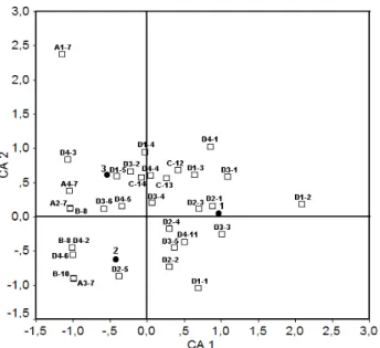
Correspondence Analysis: In this analysis, according to overall results from table 4, differences among skeletal systems were evaluated entirely but swim-bladder anomaly was not included in the CA analysis (Figure 3). Fin deformities (especially A1-7, A2-7, A3-7, and A4-7) were located on negative CA1 due to the fact that fins were not formed in the preflexion stage and also they were presented relatively close relations with flexion and postflexion stages. Additionally, deformities observed in the appendicular system were situated on negative CA1 due to in similar with external skeleton and also exhibited similar close relations with flexion and postflexion stages. For both flexion stages (flexion and postflexion), dorsal and pectoral fin deformations and malformed pterygiophores were detected at positive CA2 and these deformities were closely related with postflexion. Moreover, anal fin deformation and
malformed epural and hypural were determined at negative CA2 and it was more effective in the flexion stage. Also, it was determined that whole caudal fin deformities were closely related with postflexion stage. Besides, deformities in syncranium were located at positive CA2 which suggested that deformities in the flexion stage were less effective than the other stages.
Figure 3. Correspondence analyses applied to types of abnormalities, the skeletal regions and larval period. For abnormality and region abbreviations, refer to Table 4. □ abnormalities and skeletal regions; ● larval period.
Şekil 3. Larval periyot süresince iskelet bölgeleri ve deformayon tipine bağlı uygulaqnan Korrespon analizi. Defromasyon ve bölgesel kısaltmalar Tablo 4’teki gibidir. □ deformasyonlar ve iskelet bölgeleri; ● larval periyot.
Vertebral column deformities were distributed to the whole area of both axis (CA1 and CA2). Several deformities that were observed in vertebra such as lordosis, kyphosis in prehemal and caudal, vertebral fusion and malformations, malformed neural arch and/or spines in hemal region, and differences on supernumerary vertebra were located at positive CA1 and were also closely related with preflexion and postflexion stages. Other than the mentioned deformities above, vertebral column deformities were situated at negative CA1 and were entirely related with the flexion stage.
Discussion and Conclusion
This study examined skeletal anomalies and deformities in the early stages of life of sharpsnout sea bream larvae. In previous studies, skeletal deformities and meristic characteristics of sub adults (173 DAH) and adults of sharpsnout sea bream were investigated by Favalora and Mazzola (17). They reported that the number of anomalies strongly varied with age. Also, in another study, same authors pointed out that wild specimen have low malformation rate than the cultured
population of this species (17). Additionally, Boglione et al. (18) studied on skeletal abnormalities of wild and cultured D. puntazzo and P. erythrinus post-larva and juveniles. According to their results, rate of deformed fish in cultured D. puntazzo was 95.2%, in cultured P.
erythrinus 72.9%, while in cultured gilthead sea bream, it
varied from 98.3% to 100% (18).
Vertebral deformities were a slight deformation of the centra with a low frequency (7.7%) compared to other hatchery-reared species where larval deformities of 24% to 44% have been reported (10,19,20). Boglione et al. (16) reported that only 4% of wild caught S. aurata shows body deformations, in contrast to the very high values observed in hatchery-reared larvae (21). Additionally, deformity rates in wild specimens of D.
puntazzo juveniles changed between 19.6% and 21.4%
while it ranged from 88.2% to 100% for the cultured juveniles. In the current study, deformity rate was calculated as 21.5% during larval development and also the highest rate of deformity was observed as 32.8% in the flexion stage.
It is well documented that dramatical changes in abiotical factors in both intensive and extensive culture conditions could cause several deformations mainly in fins of the larvae (20). In addition to this, fin rays are more sensitive to thermal fluctuations especially during early ontogeny (8). As reported by Sfakianakis et al. (22), development of fin and fin supports initiate in notochord flexion and continue development during postflexion stages in D. puntazzo larvae. In this study, no deformations were recorded in external and appendicular skeletal systems during the preflexion stage, but several deformities were noted during of flexion and postflexion stages.
It is well described that malformed hypurals, epurals and pterygyphores are identified as developmental deformities and are also closely related to culture conditions such as stocking density (17). These type anomalies were reported for D. labrax (9), D. dentex (20), S. aurata (16), D. puntazzo larvae and juveniles (17). In this study, these anomalies were noted and also the rate of malformed pterygyphores was found to be relatively higher than rates of malformed epural and hypural.
Vertebral deformities, especially lordosis, kyphosis, scoliosis, are the most important skeletal malformations (7,20,21). Also, it is reported that pre-haemal kyphosis in
D. labrax develops during the larval stage and in the
range of 10–17 mm in TL (7). In the current study, the rates of lordosis and kyphosis were found to be relatively higher in the preflexion stage than both flexion and posflexion stages. Also, in preflexion stage, vertebral fusion, vertebral malformation and malformed neural and haemal arch and/or spine were relatively higher than the other stages. Sfakianakis et al. (22) indicated that
formation of neural (4.9 mm TL) and haemal arch (5.6 mm TL) is initiated during the preflexion stage in D.
puntazzo larvae. It is possible that obtained relatively
higher rates of deformations from this study could be related with differences in culture conditions and feeding protocol during the osteological development of larvae. Moreover, Koumoundouros et al. (7) reported that kyphosis was significantly associated with deformations of the branchiostegal rays, but not with lordosis or swim bladder non-inflation.
It has been found that there was negative correlation between deformities of vertebral column and larval developmental stages and that the rate of deformity concurrently decreased by larval age. There might be two reasons for this situation: firstly deformed larvae could be improved their health, secondly they could be died. In previous studies, it is clearly reported that deformities observed in this region did not recover and caused to high mortality rates (7). Concisely, it is recommended that all deformity types observed in the vertebral column should be monitored throughout each flexional stage. Also, deformities occurred in preflexion stage will be kept to continue during the other stages.
Development of haemal and neural archs on vertabra occurred between 4.5 mm and 6.4 mm TL in D.
puntazzo larvae (22). In addition, development of digestive
system and enzymatic ontogeny were completed in the same period (13). Moreover, we observed that specific activity of digestive proteases decreased between 10 and 15 DAH and could be related to some morphological and physiological changes in larvae (23). Malnutrition of larvae due to insufficient enrichment of live food by DHA/EPA and fatty acids could also cause vertebral column deformities (24). Additionally, histological analysis agreed with this phenomenon that lack of several vitamins, especially Vitamin C, minerals and various food nutrients might trigger increase of skeletal deformities during the early ontogeny (25, 26).
In the current study, the CA was useful for evaluation of characteristics of deformities in detail with stage and region. According to CA, deformities were found in relation with the flexion stage quantitatively whereas they demonstrated close relation with the postflexion stage qualitatively. In previous studies, relationships between fish origin and deformation frequency was analysed by using CA for both D. labrax (1) and S. aurata (16). Also, Ferreri et al. (27) used CA for comparison of meristic characteristics of wild and cultured Danio rerio. It is considered that skeletal deformities depend on notochord flexion time occurring in early larval stages could be easily analysed by CA for the monitoring and sustainability of larval quality in intensive culture.
This study investigates skeletal deformations in respect to notochord flexional stages in D. puntazzo
larvae during early ontogeny for the first time. Moreover, it strongly pointed out that stress originating from malnutrition of larvae by DHA/EPA, osteological development, metamorphosis and organogenesis of larvae could trigger larval deformity (23, 28). Boglione et al. (4) reported that investigation of larval quality should be crucial between 50 and 100 DAH in cultured species in commercial hatcheries. However, deformities occurred in axial skeletal system should be monitored from preflexion stage whereas deformations in external and appendicular skeletal system should be examined from flexion stage in D. puntazzo larvae. As a result, commercial larval producers could gain an economic profit by investigation of larval deformities during the flexion and preflexion stages. For further studies, relationships between broodstock quality and larval deformities should be clearly described, to decrease deformity rates during early larval stages, which will be useful to aquaculturists and commercial producers.
Acknowledgments
The authors would like to express their sincere gratitude to the staff of the Teknomar Sea Fish Broodstock Centre where the experiments were conducted (Akuvatur Mediterranean Sea Foods, Izmir, TURKEY) for their most efficient technical assistance.
References
1. Boglione C, Marino G, Ferreri F, Finoa MG, Scardi M, Fresi E, Cataudella S ( 1994): Anatomical aspects for seed quality assessment in sea bass (Dicentrarchus labrax): hatchery and wild populations. In: Kestemont P, Muir J, Sevila F, Williot P (Eds), Measures for Success: Metrology and Instrumentation in Aquaculture Management. Bordeaux Aquaculture 1994. Eur. Aquacult. Soc., Ghent, Belgium, Spec. Pub. 21, 191–197.
2. Koumoundouros G, Gagliardi F, Divanach P, Boglione C, Cataudella S, Kentouri M (1997a): Normal and abnormal osteological development of caudal fin in Sparus aurata L fry. Aquaculture, 149, 215–226.
3. Koumoundouros G, Oran G, Divanach P, Stefanakis S, Kentouri M (1997b): The opercular complex deformity in intensive gilthead sea bream (Sparus aurata L.) larviculture. Moment of apparition and description. Aquaculture, 156, 165–177.
4. Afonso JM, Montero D, Robaina L, Astorga N, Izquierdo MS, Gines R (2000): Association of a lordosis-scoliosis-kyphosis deformity in gilthead seabream (Sparus aurata) with family structure. Fish Physiology and Biochemistry, 22, 159–163.
5. Favaloro E, Mazzola A (2006): Meristic character counts and incidence of skeletal anomalies in the wild Diplodus puntazzo (Cetti, 1777) of an area of the south-eastern Mediterranean Sea. Fish Physiol. Biochem., 32, 159–166. 6. Favaloro E, Mazzola A (2003): Meristic variation and
skeletal anomalies of wild and reared sharpsnout seabream juveniles (Diplodus puntazzo, Cetti 1777) off
coastal Sicily, Mediterranean Sea. Aquaculture Research, 34, 575–579.
7. Koumoundouros G, Maingot E, Divanach P, Kentouri M (2002): Kyphosis in reared sea bass (Dicentrarchus labrax L.): ontogeny and effects on mortality. Aquaculture, 209, 49–58.
8. Sfakianakis DG, Koumoundouros G, Divanach P, Kentouri M (2004): Osteological development of the vertebral column and the fins in Pagellus erythrinus (L. 1758). Temperature effect on developmental plasticity and morpho-anatomical abnormalities. Aquaculture, 232, 407– 424.
9. Marino G, Boglione C, Bertolini B, Rossi A, Ferreri F, Cataudella S (1993) : Observations on the development and anomalies in the appendicular skeleton of sea bass, D. labrax L. 1758, larvae and juveniles. Aquacult. Fish Manage., 24, 445–456.
10. Gavaia PJ, Dinis MT, Cancela ML (2002): Osteological development and abnormalities of the vertebral column and caudal skeleton in larval and juvenile stages of hatchery-reared Senegal sole (Solea senegalensis). Aquaculture, 211, 305–323.
11. Çoban D, Suzer C, Kamacı HO, Saka Ş, Fırat K (2009): Early osteological development of the fins in the hatchery-reared red porgy, Pagrus pagrus (L. 1758). Journal of Applied Ichthyology, 25, 26-32.
12. Çoban D, Suzer C, Kamacı HO, Yıldırım Ş, Saka Ş, Fırat K (2010): Sivriburun karagöz (Diplodus puntazzo) larvalarında omurga ve kaudal yüzgecin osteolojik gelişimi. Ankara Üniv. Vet. Fak. Derg., 57, 119-124. 13. Suzer C, Aktülün S, Çoban D, Kamacı HO, Saka Ş,
Fırat K, Alpbaz A (2007): Digestive enzyme activities in larvae of sharpsnout seabream (Diplodus puntazzo). Comparative Biochemistry and Physiology, Part A, 148, 470–477.
14. Potthoff T (1983): Ontogeny and systematics of fisheries. American Society of Ichthyologists and Herpetogists, Special publication, 1, 223-229.
15. Ahlstrom EH, Butler JL, Sumida Y (1976): Pelagic stromateoid fishes (Pisces, Perciformes) of the Eastern Pacific: kinds, distributions, and early life histories and observations on five of these from the Northwest Atlantic. Bulletin of Marine Science, 26, 285-402.
16. Boglione C, Gagliardi F, Scardi M, Cataudella S (2001): Skeletal descriptors and quality assessment in larvae and post-larvae of wild-caught and hatchery-reared gilthead sea bream (Sparus aurata L. 1758). Aquaculture, 192, 1–22.
17. Favaloro E, Mazzola A (2000): Meristic character analysis and skeletal anomalies during growth in reared sharpsnout seabream. Aquaculture International, 8, 417–430.
18. Boglione C, Costa C, Di Dato P, Ferzini G, Scardi M,
Cataudella S (2003): Skeletal quality assessment of reared
and wild sharpsnout sea bream and pandora juveniles. Aquaculture, 227, 373–394.
19. Zambonino Infante JL, Cahu CL, Péres A (1997): Partial substitution of di- and tripeptides for native proteins in sea bass diet improves Dicentrarchus labrax larval development. J. Nutr., 127, 608–614.
20. Koumoundouros G, Divanach P, Kentouri M (2001): The effect of rearing conditions on development of
saddleback syndrome and caudal fin deformities in Dentex dentex (L.). Aquaculture, 200, 285–304.
21. Divanach P, Boglione C, Menu M, Koumoundouros G, Kentouri M, Cataudella S (1996): Abnormalities in finfish mariculture: an overview of the problem, causes and solutions. Sea-bass and Sea Bream Culture: Problems and Prospects, Verona, Italy, October 16–18. European Aquaculture Society, Oostende, Belgium, 45–66.
22. Sfakianakis DG, Doxa CK, Kouttouki S, Koumoundouros G, Maingot E, Divanach P, Kentouri M (2005): Osteological development of the vertebral column and of the fins in Diplodus puntazzo (Cetti, 1777). Aquaculture, 250, 36–46.
23. Suzuki T, Srivastava AS, Kurokawa T (2000): Experimental induction of jaw, gill and pectoral fin malformations in Japanese flounder, Paralichthys olivaceus, larvae. Aquaculture, 185, 175-187.
24. Gapasin RSJ, Duray MN (2001): Effects of DHA-enriched live food on growth, survival and incidence of opercular deformities in milkfish (Chanos chanos). Aquaculture, 193, 49-63.
25. Soliman AK, Jauncey K, Roberts RJ (1986): The effect of varying forms of dietary ascorbic acid on the nutrition of juvenile tilapias (Oreochromis niloticus). Aquaculture, 52, 1-10.
26. Kamacı HO, Suzer C, Çoban D, Saka Ş, Fırat K (2010): Organogenesis of exocrine pancreas in sharpsnout sea bream (Diplodus puntazzo) larvae: characterization of trypsin expression. Fish Physiol. Biochem., 36, 993-1000. 27. Ferreri F, Nicolais C, Boglione C, Bertolini B (2000) :
Skeletal characterization of wild and reared zebrafish: anomalies and meristic characters. Journal of Fish Biology, 56, 1115–1128.
28. Sawada Y, Hattori M, Sudo N, Kato K, Takagi Y, Ura K, Kurata M, Okada T, Kumai H (2006): Hypoxic conditions induce centrum defects in red sea bream Pagrus major (Temminck and Schlegel). Aquaculture Research, 37, 805-812.
Geliş tarihi: 02.01.2014 / Kabul tarihi: 15.04.2014 Address for correspondence:
Dr. Şükrü Yıldırım Ege University, Faculty of Fisheries, Aquaculture Department, 35100 Bornova, İzmir, TURKEY e-mail: [email protected]