TURKISH JOURNAL of
DOI: 10.5152/TurkJOrthod.2018.17061
CASE REPORT
Lingual Treatment of an Adult Patient with a Simplified
Extraction Protocol
ABSTRACT
Successful orthodontic treatment of adult cases depends on the biological, mechanical, and esthetic requirements of patients. While customized lingual appliance systems meet the esthetic expectations of the patients, they provide improved patient comfort, have three-dimensional mechanical control, and can be used for the treatment of all types of malocclusions. This report demonstrated the use of fully customized lingual orthodontic brackets for treating an adult case with extraction.
Keywords: Customized lingual orthodontics, adult orthodontics, fixed lingual orthodontics
INTRODUCTION
Visible orthodontic appliances are challenging for adult patients. In a recent study, 33% and 62% adults refused orthodontic treatment using a visible appliance (1). With the increasing esthetic demands of adult patients, lingual orthodontics and clear aligners have recently become popular (2). Lingual orthodontics provides the best esthetic option for complex cases with three-dimensional control (3). Customized lingual appliance systems have improved patient comfort, provided accurate bracket positions, and produced similar treatment outcomes as labial ortho-dontics.
The objective of this case report was to present the treatment results of an adult patient treated with extraction using fully customized lingual brackets.
CASE PRESENTATION
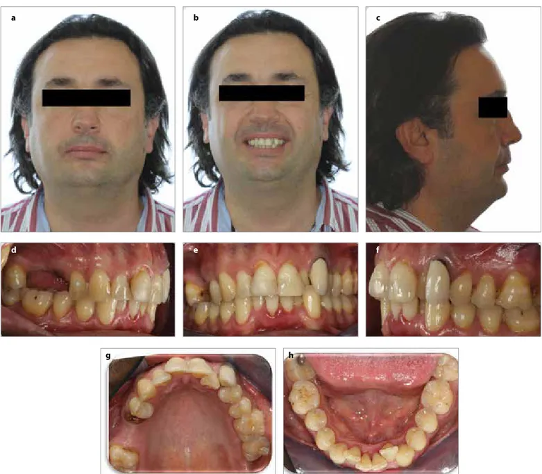
A male patient 43 years 4 months of age was referred to our clinic with the chief compliant of dental crowding. Diagnostic records showed that he demonstrated Class II molar and canine relationships on the left side and Class I canine relationship on the right side with retrusive upper, protrusive lower incisors, normal overjet, and mildly increased overbite (Figure 1, 2). Upper right first molar had previously been extracted. There was an ectopic canine tooth with an unesthetic veneer crown on the upper left quadrant. According to dental cast analysis, dental arch discrepancies were measured as 9.2 mm in maxilla and 4 mm in mandibula. Cephalometric measurements are presented in Table 1.
Treatment goals were to eliminate dental crowding, obtain Class I canine relationship on both sides, and also achieve ideal overjet and overbite. The treatment plan was to extract the buccally positioned upper left canine and use upper left first premolar as canine substitution. The color and shape of the premolar were noted as suitable for the canine substitution. A dental implant was planned for replacing the upper right first molar. Written informed consent was obtained from the patient.
Ömür Polat Özsoy1 , Hande Pamukçu2
1Department of Orthodontics, Cyprus Health and Social Sciences University School of Dentistry, Güzelyurt, Cyprus 2Department of Orthodontics, Başkent University School of Dentistry, Ankara, Turkey
Address for Correspondence: Hande Pamukçu, Department of Orthodontics, Başkent University School of Dentistry, Ankara, Turkey E-mail: [email protected]
©Copyright 2018 by Turkish Orthodontic Society - Available online at turkjorthod.org ORCID IDs of the authors: Ö.P.Ö. 0000-0002-8555-1367; H.P. 0000-0003-4242-5114.
Cite this article as: Polat Özsoy Ö, Pamukçu H. Lingual Treatment of an Adult Patient with a Simplified Extraction Protocol. Turk J Orthod 2018; 31: 62-6.
Received: 3 January 2018 Accepted: 25 February 2018
Treatment Sequence
Fully customized lingual brackets (Incognito, TOP service, 3M Unitek, Bad Essen, Germany) were manufactured according to the patient’s impressions and a digital setup was created (Figure 3). After the bonding of brackets, upper left canine was extract-ed. The arch-wire sequence was .014” SE (super-elastic) nickel titanium for levelling and alignment; .016”×.022” SE nickel ti-tanium, .018”×.025” SE nickel titanium for correcting rotations and providing initial torque control; .016”×.024” stainless steel for torque control, and .018”×.018” TMA (titanium-molybdenum alloy) for finishing. Interproximal reduction was performed for the crowding of the mandibular anterior teeth during the treat-ment. An attempt was made to achieve mesial movement of the upper right second molar, but it failed due to pneumatization of the maxillary sinus. The patient refused to undergo a sinus lift surgery.
At completion of the treatment, Class II molar relationship on the left side and Class I canine relationships on both sides were ob-tained; also, a balanced and ideal occlusion was achieved (Figure 4). Post-treatment cephalometric radiographs are shown in Figure 5. Total treatment duration was 2 years and 8 months. Throughout this period, the attempt for molar mesialization took 10 months. After debonding, fixed retainers were bonded, and additional essix plates were fabricated for both arches. The cephalometric parameters for pre- and post-treatment are shown in Table 1. The superimposi-tion of pre- and post-treatment lateral cephalometric radiographs showed the extrusion of upper molar and proclination of lower in-cisor (Figure 6).
DISCUSSION
The demand for adult orthodontic treatment has progressively
in-Figure 1. a-h. Pretreatment (T0) extraoral and intraoral images: Pretreatment extraoral frontal rest image (a); pretreatment extraoral frontal
smile image (b); pretreatment extraoral profile image (c); pretreatment intraoral right lateral image (d); pretreatment intraoral frontal image (e); pretreatment intraoral left lateral image (f); pretreatment intraoral upper occlusal image (g); pretreatment intraoral lower occlusal image (h)
a d g b e c f h
63
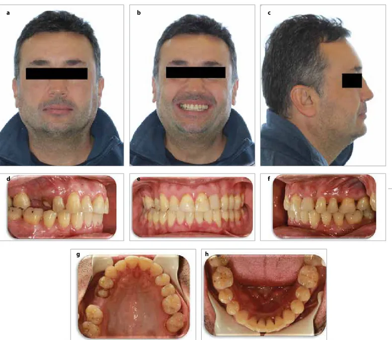
Figure 2. a, b. Pretreatment (T0) radiographic records: Pretreatment
lateral cephalometric radiograph (a); pretreatment panoramic radiograph (b) a b c b a
Figure 3. a-d. In-progress intraoral images: In-progress intraoral right lateral
image (a); in-progress intraoral left lateral image (b); in-progress intraoral upper occlusal image (c); in-progress intraoral lower occlusal image (d)
d Table 1. Pre- (T0) and post-treatment (T1) cephalometric
measure-ments Norm Values T0 T1 Sagittal Analysis SNA (°) 80±2 78.5 77.5 SNB (°) 78±2 74.6 74.2 ANB (°) 2±2 3.9 3.1 GoGnSN (°) 32±6 27.5 28.5 Gonial Angle (°) 130±7 111 111 Dental Analysis U1 -NA (mm) 4 3 4 U1-NA (°) 22±5 12.5 16 L1-NB (mm) 4 5.1 5.3 IMPA (°) 90±3 102 106 Overjet (mm) 3 3.5 2.8 Overbite (mm) 3 4 2.8
Soft Tissue Analysis
Upper Lip-E Line (mm) -4 -6 -5
Lower Lip-E Line (mm) -2 -6 -4.4
SNA: Sella-nasion-A point angle; SNB: Sella-nasion-B point angle; ANB: A point, nasion, B point; GoGnSN: angle that is measured at the junction of the planes Gonion to Gnathion and Sella-Nasion; IMPA: incisor mandibular plane angle; U1-NA (°): angle between upper incisor inclination and NA plane; L1-NB (°): angle between lower incisor inclination and NB plane
64
Turk J Orthod 2018; 31: 62-6 Polat Özsoy and Pamukçu. Lingual Treatment with Simplified Extraction Protocol
creased in recent years, and reportedly 25% of orthodontic patients were adults in United States (4). A previous study showed that 90% of adult orthodontic treatments required fixed appliances (5). Customized lingual orthodontics is the most esthetic option for adult patients with three-dimensional control, and it is suitable for all types of malocclusions. Improved digital technology of custom-ized lingual systems helps create a virtual set-up, customcustom-ized brack-et positioning, arch-wire, and brackbrack-et fabrication. These steps facili-tate improvement in the of the treatment outcomes.
Adults generally have restored or endodontically treated teeth, which can complicate the treatment plan. In the patient in the pres-ent study, we extracted the buccally positioned upper left canine using endodontic treatment for correcting the dental crowding. We used upper left first premolar as canine. In literature, premolars are often used instead of canines, and it was suggested that premolars
would effectively enhance esthetics in cases of orthodontic space closure (6, 7).
The upper molar was attempted to move mesially but because of the pneumatization of the maxillary sinus, this movement was not completed. Teeth can be moved if there is adequate bone in the di-rection of movement and it is challenging to move teeth through anatomic limitations such as maxillary sinus, sutural, or cortical bar-riers.
CONCLUSION
The treatment of adult cases with high esthetic concerns can be ef-fectively performed using customized lingual brackets. Customized lingual appliance systems have the ability to treat complex cases, and advanced digital technology can help clinicians plan all the treatment steps.
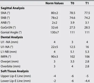
Figure 4. a-h. Post-treatment (T1) extraoral and intraoral images: Post-treatment extraoral frontal rest image (a); post-treatment extraoral frontal smile
image (b); treatment extraoral profile image (c); treatment intraoral right lateral image (d); treatment intraoral frontal image (e); post-treatment intraoral left lateral image (f); post-post-treatment intraoral upper occlusal image (g); post-post-treatment intraoral lower occlusal image (h)
a d g b e c f h
65
Informed Consent: Written informed consent was obtained from the pa-tient who participated in this study.
Peer-review: Externally peer-reviewed.
Author Contributions: Concept - Ö.P.Ö., H.P.; Design - Ö.P.Ö., H.P.; Supervi-sion - Ö.P.Ö., H.P.; Resources - Ö.P.Ö., H.P.; Materials - Ö.P.Ö., H.P.; Data Collec-tion and/or Processing - Ö.P.Ö., H.P.; Analysis and/or InterpretaCollec-tion - Ö.P.Ö., H.P.; Literature Search - Ö.P.Ö., H.P.; Writing Manuscript - Ö.P.Ö., H.P.; Critical Review - Ö.P.Ö., H.P.
Conflict of Interest: The authors have no conflict of interest to declare. Financial Disclosure: The authors declared that this study has received no financial support.
REFERENCES
1. Meier B, Wiemer KB, Miethke R-R. Invisalign Patient Profiling. J Oro-fac Ortho 2003; 64: 352-8. [CrossRef]
2. Fritz U, Diedrich P, Wiechmann D. Lingual technique - Patients’ char-acteristics, motivation and acceptance. Interpretation of a retro-spective survey. J Orofac Orthop 2002; 63: 227-33. [CrossRef]
3. Echarri P. Revisiting the History of Lingual Orthodontics: A Basis for the Future. Semin Orthod 2006; 12: 153-9. [CrossRef]
4. Gottlieb EL, Nelson AH, Vogels DS. 1991 JCO orthodontic practice study. 2. Practice success. J Clin Orthod 1991; 25: 740-7.
5. Khan RS, Horrocks EN. A study of adult orthodontic patients and their treatment. Br J Orthod 1991; 18: 183-94. [CrossRef]
6. Zachrisson BU, Rosa M, Toreskog S. Congenitally missing maxillary lateral incisors: Canine substitution. Am J Orthod Dentofac Orthop 2011; 139: 434-44. [CrossRef]
7. Sharma PK, Sharma P. Interdisciplinary management of congeni-tally absent maxillary lateral incisors: Orthodontic/prosthodontic perspectives. Semin Orthod 2015; 21: 27-37. [CrossRef]
Figure 5. a, b. treatment (T1) radiographic records:
Post-treatment lateral cephalometric radiograph (a); post-Post-treatment panoramic radiograph (b)
a
b
Figure 6. Superimposition of pre- and post-treatment lateral
cephalometric films
Pre-treatment (T0): black line; post-treatment (T1): red line
66
Turk J Orthod 2018; 31: 62-6 Polat Özsoy and Pamukçu. Lingual Treatment with Simplified Extraction Protocol