A survey of feline neoplasms in Ankara from 1971 to 2005
Yılmaz AYDIN1, Zafer ÖZYILDIZ2, Latife BEYAZ31Department of Pathology, Faculty of Veterinary Medicine, Ankara University, Ankara; 2Department of Pathology, Faculty of Veterinary Medicine, Kafkas University, Kars; 3 Department of Pathology, Faculty of Veterinary Medicine, Erciyes University, Kayseri.
Summary: In this study, cat tumor cases from the archives of Pathology Department of Veterinary Medicine Faculty of Ankara University that were encountered in the last 35 years from January 1971 to December 2005 were investigated. During this period 265 cases were received; 244 of these cases were obtained as biopsy while the rest 21 cases were from necropsy materials. Parameters regarding to age, sex, tumor localizations of the organs or body systems, origin, malignancy and the years encountered were indicated with Tables. Of the cases 131 were localized in the cutaneous system which is composed of skin and subcutaneus connective tissue. Seventy-five were female genital system tumors mostly localized in mammary glands. The rest were observed in alimentary, skeletal, hematopoietic, respiratory, and neuronal, endocrine, urinary systems as 22, 19, 8, 4, and 2, 2, 2 respectively. Tumors occurred in cats were compared due to type, localization, frequency and prevalence of the tumors and age, breed, sex of the cases. The data obtained were investigated and evaluated statistically.
Key words: Feline neoplasms, frequency, localisation, statistical analysis, survey, tumor type.
1971-2005 yılları arasında Ankara ve yöresinde kedilerde karşılaşılan tümörler
Özet: Bu çalışmada, Ankara Üniversitesi Veteriner Fakültesi Patoloji Anabilim Dalı arşiv kayıtları esas alınarak, Ocak 1971-Aralık 2005 yılları arasındaki 35 yıllık sürede Ankara ve çevresinde kedilerde görülen tümör olguları araştırıldı ve sayısal olarak karşılaştırıldı. Bu amaçla, 35 yıllık sürede tümör tanısı konan kedilere ait olgular, arşiv kayıtları esas alınarak incelendi ve toplu olarak değerlendirildi. Bu süre içinde kedilerde, 244’ü biyopsi ve 21’i nekropsi materyaline ait olmak üzere, toplam 265 adet tümör olgusu ile karşılaşıldığı saptandı. Ayrımı yapılan olguların yaş, ırk ve cinsiyet dağılımı ile görüldüğü yıllar, görüldüğü vücut sistemleri ve lokalizasyonu ile epitelyal-mezenşimal ve benign-malign oranları Tablolarla gösterildi. Sonuçta olgulardan 131’inin deri ve eklentileri ile deri altı yumuşak dokulardan kaynaklanan kutanöz sistem, 75’inin çoğu meme dokusu orijinli dişi genital sistem, 22’sinin sindirim sistemi, 19’unun iskelet sistemi, 8’inin hematopoietik sistem, 4’ünün solunum sistemi ve geriye kalan 2’şer adedinin üriner, sinir ve endokrin sistem orijinli tümör olduğu belirlendi. Bu çalışma ile kedilerde ne oranda ve hangi tiplerde tümörlere rastlandığı, son yıllarda hangi tip tümörlerin ortaya çıktığı ve görülme sıklıkları, tümörlerin yaş, ırk ve cinsiyete göre ayrımı, hangi vücut sistemlerinin daha fazla etkilendiği vb konularında istatistiki veriler elde edilmeye çalışıldı ve bunlar tablolarla açıklandı.
Anahtar sözcükler: İstatistiki analiz, kedi tümörleri, lokalizasyon, prevalans, survey, tümör tipi.
Introduction
From time to time, disease and tumor cases of domestic animals in a region around the world are investigated statistically overall to assess the prevalence of these cases in that region (9,10,12-14). Also in our country, disease and tumor cases of various domestic animals were investigated periodically based on the archived records (1-5, 11, 15-19, 21, 23-25, 27, 28). In a previous study (23) emphasized that “In a country, unless the diseases and their prevalence are not known it is impossible to take measures for the fight of these diseases. Such knowledge can be gained by published statistical data. It would be helpful to make occasional analysis of the records of the department. In this way, we can gain a dear knowledge of the animal diseases
occurring in the vicinity of Ankara. Therefore, the department of Pathology-Anatomy is determined to investigate the diseases occurring in various animal species”.
As a result of this effort, tumor cases observed in cats have been reviewed up to 1971 and published (15, 23, 24) totally in different dates. Although it is reported individual cases (6-8, 20, 26), it is clear that these early efforts made in our country are not sufficient and new investigations are still needed.
As a continual of the previous studies, in the present study, cat tumor cases that are encountered between 1971 and 2005 were reevaluated and local new data are obtained.
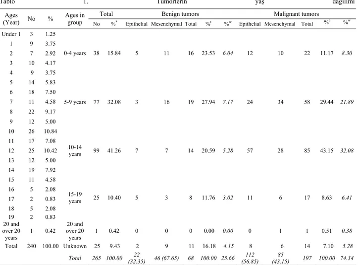
Table 1. Age distribution of feline tumors*
Tablo 1. Tümörlerin yaş dağılımı*
Total Benign tumors Malignant tumors
Ages
(Year) No %
Ages in
group No %* Epithelial Mesenchymal Total %t %w Epithelial Mesenchymal Total %t %w
Under 1 3 1.25 1 9 3.75 2 7 2.92 3 10 4.17 4 9 3.75 0-4 years 38 15.84 5 11 16 23.53 6.04 12 10 22 11.17 8.30 5 14 5.83 6 18 7.50 7 11 4.58 8 22 9.17 9 12 5.00 5-9 years 77 32.08 3 16 19 27.94 7.17 24 34 58 29.44 21.89 10 26 10.84 11 17 7.08 12 25 10.42 13 12 5.00 14 19 7.92 10-14 years 99 41.26 7 7 14 20.59 5.28 57 28 85 43.15 32.08 15 11 4.58 16 5 2.08 17 2 0.83 18 5 2.08 19 2 0.83 15-19 years 25 10.40 5 3 8 11.76 3.02 11 6 17 8.63 6.41 20 and over 20 years 1 0.42 20 and over 20 years 1 0.42 0 0 0 0.00 0.00 0 1 1 0.51 0.38 Total 240 100.00 Unknown 25 9.43 2 9 11 16.18 4.15 8 6 14 7.10 5.28 Total 265 100.00 (32.35) 22 46 (67.65) 68 100.00 25.66 (56.85) 112 (43.15) 85 197 100.00 74.34 *
There are except for unkonwn ages in percentage of ratios. %t: ratios in epithelial or mesenchymal tumors. %w: ratios in total tumors *Oranların hesaplanmasında yaşı bilinmeyenler hariç tutulmuştur. %t: epitelyal veya mezenşimal tümörlerin kendi içindeki oran. %w: toplam tümör içindeki oran.
Materials and Methods
The study materials of this survey were feline biopsy samples or necropsy materials, which submitted to our department from private veterinary clinics or the clinics of Veterinary Medicine Faculty of Ankara University. Cat tumor cases recorded between January 1971 and December 2005 were included in the study.
The age and sex of the cats with tumors, and the submission year, type and anatomical sites of tumors were investigated. Frequently observed cases were subdivided, and their frequency in total number of cases was calculated and the data of these tumors were tabulated. All cases were subdivided into epithelial or mesenchymal in origin, and benign or malignant in behavior.
The data in the Table 1 was evaluated in the age groups and between the age groups with statistical analysis methods. In this evaluation; Chi square analysis was utilised for the malignant and the benign tumors within the age groups and within these groups the epithelial and the mesenchymal tumors was evaluated.
Tumors were divided into four major categories based on the year they are diagnosed; first group (between 1971 and 1979) when no regular records were kept and there was no access to tissue blocks, second group, the years between 1980 and 1989; third group, the years between 1990 and 1999, and fourth group, the years between 2000 and 2005.
Tumor types were classified according to the Meuten’s classification system (22). In the classification of the tumors original diagnosis were kept as much as possible, however, in needed, additional tissue sections were cut from the paraffin blocks and were stained immunohistochemically with tumor markers for better diagnosis. The nomenclature used for malignant mesenchymal tumors of cutaneous system and mammary gland carcinomas of female genital system was simplified for clarification.
Results
Between the years of January 1971 and December 2005, there were total of 265 tumors cases, 244 of which
were biopsy material and 21 necropsy cases. The data about the age of animal, origin of tumor, tumor type, ratios of epithelial to mesenchymal and benign to malignant cases were shown in Tables 1-3.
Statistical analysis revealed no significant difference between 0-4, 10-14 and 15-19 years. However, there was a statistically significant difference for the 5-9 age group (p<0.05). This difference was originated from low number of the benign epithelial tumors versus mesenchymal malignant tumors. For the analysis of tumor cases between the age groups correlation analysis was used. It was tried to determine if there is an increase in the tumor numbers with increasing age. When all the four age groups evaluated together there was no statistically significant difference for the 15-19 age group because of the limited case number. However, high correlation was found when 15-19 age group excluded and analysis performed on only three age groups. The findings showed a statistically significant increase for the malignant tumors with increasing age (p<0.05).
Total tumor cases and the numbers of the benign and the malignant tumors within these cases and origins of these tumors were investigated with Chi square test in the table 2. Statistically significant differences were found when the distribution mentioned above searched
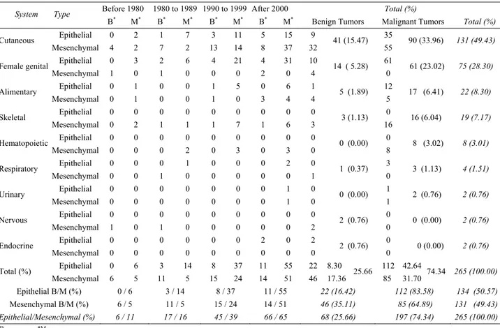
within the total case number (p<0.001). This significance was originated from higher case numbers of the malignant epithelial tumors. Separate evaluation of the female genital system and alimentary system revealed significant differences (p<0.05). In both systems malignant epithelial tumor numbers was statistically higher than other tumors. In the skin, the difference was close to the statistical significance (p<0.057). Although this difference was accepted as statistically not significant, it is obvious that the number of the cutaneous mesenchymal tumors is higher than other type tumors. There was no statistically significant difference in the other systems for the distibution of the tumor types (p>0.05). Yearly changes of the total distribution of tumor cases of the different systems were evaluated by Chi square test. Sytems excluding skin and female genital systems were put together under the "other" because of limited number of cases and these total 265 cases was evaluated under this three headings. Analysis revealed no statistically significant difference among these cases (p>0.05).Yearly increase was evaluated for the skin and female genital systems because of adequate number of cases. Both systems revealed yearly significant increase (p<0.05) and this increase was supported with presence of strong correlation (r=0.98). Table 2. Origin systems of the benign and the malignant tumors encountered according to years with ten each in cats.
Tablo 2. Yaklaşık 10 yıllık periyotlar halinde karşılaşılan benign ve malign tümörlerin sistemlere göre dağılımı.
Before 1980 1980 to 1989 1990 to 1999 After 2000 Total (%)
System Type
B* M* B* M* B* M* B* M* Benign Tumors Malignant Tumors Total (%)
Epithelial 0 2 1 7 3 11 5 15 9 35 Cutaneous Mesenchymal 4 2 7 2 13 14 8 37 32 41 (15.47) 55 90 (33.96) 131 (49.43) Epithelial 0 3 2 6 4 21 4 31 10 61 Female genital Mesenchymal 1 0 1 0 0 0 2 0 4 14 ( 5.28) 0 61 (23.02) 75 (28.30) Epithelial 0 1 0 0 1 5 0 6 1 12 Alimentary Mesenchymal 0 1 0 0 1 0 3 4 4 5 (1.89) 5 17 (6.41) 22 (8.30) Epithelial 0 0 0 0 0 0 0 0 0 0 Skeletal Mesenchymal 0 2 1 1 1 7 1 6 3 3 (1.13) 16 16 (6.04) 19 (7.17) Epithelial 0 0 0 0 0 0 0 0 0 0 Hematopoietic Mesenchymal 0 0 0 2 0 3 0 3 0 0 (0.00) 8 8 (3.02) 8 (3.01) Epithelial 0 0 0 1 0 0 0 2 0 3 Respiratory Mesenchymal 0 0 1 0 0 0 0 0 1 1 (0.37) 0 3 (1.13) 4 (1.51) Epithelial 0 0 0 0 0 0 0 1 0 1 Urinary Mesenchymal 0 0 0 0 0 0 0 1 0 0 (0.00) 1 2 (0.76) 2 (0.76) Epithelial 0 0 0 0 0 0 0 0 0 0 Nervous Mesenchymal 1 0 1 0 0 0 0 0 2 2 (0.76) 0 0 (0.00) 2 (0.76) Epithelial 0 0 0 0 0 0 2 0 2 0 Endocrine Mesenchymal 0 0 0 0 0 0 0 0 0 2 (0.76) 0 0 (0.00) 2 (0.76) Epithelial 0 6 3 14 8 37 11 55 22 8.30 112 42.64 Total (%) Mesenchymal 6 5 11 5 15 24 14 51 46 17.36 25.66 85 31.70 74.34 265 (100.00) Epithelial B/M (%) 0 / 6 3 / 14 8 / 37 11 / 55 22 (16.42) 112 (83.58) 134 (50.57) Mesenchymal B/M (%) 6 / 5 11 / 5 15 / 24 14 / 51 46 (35.11) 85 (64.89) 131 (49.43) Epithelial/Mesenchymal (%) 6 / 11 17 / 16 45 / 39 66 / 65 68 (25.66) 197 (74.34) 265 (100.00) *B Benign, *M Malignant
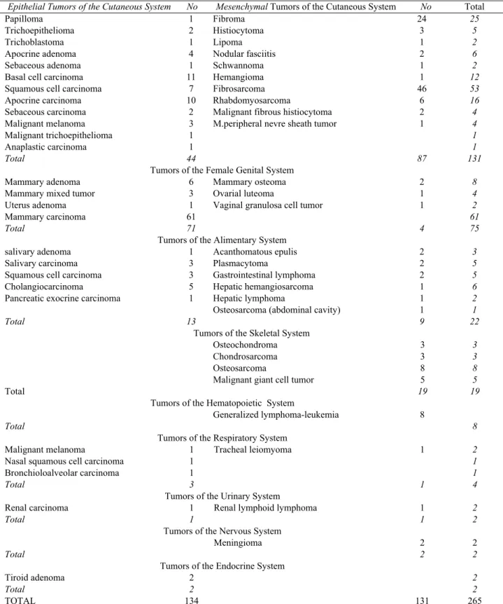
Table 3. Origin systems of the tumors encountered according to types of the tumor. Tablo 3. Karşılaşılan tümör tiplerinin orijin sistemleri.
Epithelial Tumors of the Cutaneous System No Mesenchymal Tumors of the Cutaneous System No Total
Yearly increases was also seen in the cases evaluated under the "others" heading (p<0.05) and this was supported with strong correlation (r=0.98). Yearly increase on the malignant tumors; cutaneous (p<0.05) r= 0.96; female genital (p<0.05) r= 0.97; others (p=0.052) r= 0.95.
Only cutaneous and "others" were compared because of the inadequate data. No significant difference was found between cutaneous and "others" for the distribution of the cases (p>0.05). Yearly increase was significant for the "others" and not significant for the
Papilloma 1 Fibroma 24 25
Trichoepithelioma 2 Histiocytoma 3 5
Trichoblastoma 1 Lipoma 1 2
6
Apocrine adenoma 4 Nodular fasciitis 2
2
Sebaceous adenoma 1 Schwannoma 1
12
Basal cell carcinoma 11 Hemangioma 1
53
Squamous cell carcinoma 7 Fibrosarcoma 46
16
Apocrine carcinoma 10 Rhabdomyosarcoma 6
4
Sebaceous carcinoma 2 Malignant fibrous histiocytoma 2
4
Malignant melanoma 3 M.peripheral nevre sheath tumor 1
1
Malignant trichoepithelioma 1
1
Anaplastic carcinoma 1
Total 44 87 131
Tumors of the Female Genital System
8
Mammary adenoma 6 Mammary osteoma 2
4
Mammary mixed tumor 3 Ovarial luteoma 1
2
Uterus adenoma 1 Vaginal granulosa cell tumor 1
61
Mammary carcinoma 61
Total 71 4 75
Tumors of the Alimentary System
3
salivary adenoma 1 Acanthomatous epulis 2
5
Salivary carcinoma 3 Plasmacytoma 2
5
Squamous cell carcinoma 3 Gastrointestinal lymphoma 2
6
Cholangiocarcinoma 5 Hepatic hemangiosarcoma 1
Pancreatic exocrine carcinoma 1 Hepatic lymphoma 1 2
1
Osteosarcoma (abdominal cavity) 1
Total 13 9 22
Tumors of the Skeletal System
Osteochondroma 3 3
Chondrosarcoma 3 3
Osteosarcoma 8 8
5
Malignant giant cell tumor 5
19 19
Total Tumors of the Hematopoietic System
Generalized lymphoma-leukemia 8
Total 8
Tumors of the Respiratory System
2
Malignant melanoma 1 Tracheal leiomyoma 1
1
Nasal squamous cell carcinoma 1
1
Bronchioloalveolar carcinoma 1
Total 3 1 4
Tumors of the Urinary System
2
Renal carcinoma 1 Renal lymphoid lymphoma 1
Total 1 1 2
Tumors of the Nervous System
Meningioma 2 2
Total 2 2
Tumors of the Endocrine System
2
Tiroid adenoma 2
Total 2 2
cutaneous system. However, correlation level was rather significant (r=0.95; r=0.97).
Chi square test was used for the evaluation of the cases in the table 3. Statistically significant difference was present for the female genital and alimentary systems. The statistical difference was close to the significance for the cutaneous system (p<0.057). The number of the malignant tumors was higher than others in all the three systems (p<0.05). Statistical analysis could not be performed for the other remaining sytems because of the limited number of cases.
Age incidence: Of the 265 cats, while the ages of
240 (90.57 %) animals was known in 25 (9.43 %) cases were not. The ages of the animals varied from 0 to 22 years with a mean value of 9.14. The number of cases with a four year interval period of ages was as follow; 1-4 years, 35 (11-4.58 %), 5-9 years, 77 (32.08 %), 10-11-4 years, 99 (41.25 %), 15-19 years, 25 (10.42 %). Only 3 cases (1.25 %) were under 1 year old and 1 case (0.42 %) was over 20 years old.
Sex incidence: Of the 265 cats, 181 (68.30 %) were
female, 63 (23.77 %) were male, and 21 (7.93 %) were unknown. Distribution of tumor types in each sex group was as fallow; of the 181 female cats, 76 (42.70 %) cutaneous tumors; 70 (37.64 %) genital system tumors, 16 (8.99 %) tumors of alimentary system; 6 (3.37 %) tumors of skeletal system; 5 (2.81 %) hematopoietic system tumors; 3 (1.69 %) tumors of respiratory system; 2 (1.12 %) tumors of endocrine system; 2 (1.12 %) tumors of urinary system; and 1 (0.56 %) nervous system tumor. Of the 63 male cats 40 (63.49 %) was cutaneous tumors, 7 (11.11 %) was tumors of skeletal, 6 (9.52 %) was tumors of alimentary tract, 5 (7.94 %) was genital system tumors, 3 (4.76 %) was hematopoietic system tumors, 1 (1.59 %) was tumor of respiratory system, and 1 (1.59 %) was nervous system tumor. Of the 21 cats with unkonown sex; 15 (75.00 %) was cutaneous tumors and 6 (25.00 %) was skeletal system tumors.
Tumor types and ratios of benign to malignant: Of
the 265 tumors, 134 (50.57 %) were epithelial and 131 (49.43 %) were mesenchymal and histologically, 197 (74.34 %) were malignant, 68 (25.66 %) were benign.
The distribution of the tumors according to the origin of organ system was as follow: cutaneous, 131(49.43 %); female genital, 75 (28.30 %); alimentary, 22 (8.30 %); skeletal, 19 (7.17 %); hematopoietic, 8 (3.01 %); respiratory, 4 (1.51 %); nervous, 2 (0.76 %); endocrine, 2 (0.76 %), and urinary, 2 (0.76 %).
Benign Tumors: Of the 68 benign tumors
encountered, 22 were epithelial (32.35 %) and 46 were mesenchymal (67.65 %). Localizations of the 22 epithelial tumors were as follow; 10 genital, 9 cutaneous, 2 endocrine, and 1 alimentary system. Localizations of
the 46 mesenchymal tumors were as follow; 32 cutaneous, 4 genital, 4 alimentary system, 3 skeletal, 2 nervous, and 1 respiratory system.
The origins of the 22 benign epithelial tumors encountered were as follow respectively; 10 female genital (6 mammary adenomas, 3 mammary benign mixed tumor, uterus adenoma), 9 cutaneous (papilloma, 2 trichoepitheliomas, trichoblastoma, 4 apocrine adenomas, sebaceous adenoma), 2 endocrine (tiroid adenomas), and 1 alimentary (salivary adenoma).
The origins of the 46 benign mesenchymal tumors encountered also were as follow; 32 cutaneous (24 fibromas, 3 cutaneous histiocytomas, 2 nodular fasciitis, lipoma, hemangioma, and schwannoma), 4 female genital (2 mammary gland osteoma, ovarian lutheoma, and vaginal granulosa cell tumor), 4 alimentary (2 oral extramedullar plasmacytoma, 2 acanthomatous epulis), 3 skeletal (osteochondromas), 2 nervous (meningiomas), and 1 respiratory system (tracheal leiomyoma).
Malignant Tumors: Of the 197 malignant tumors
encountered, 112 were epithelial (56.85 %) and 85 were mesenchymal (43.15 %). Localizations of the 112 epithelial tumors were as follow; 61 (54.47 %) genital, 35 (31.25 %) cutaneous, 12 (10.71 %) alimentary, 3 (2.68 %) respiratory, and 1 (0.89 %) urinary system. Localizations of the 85 mesenchymal tumors were as follow; 55 (64.71 %) cutaneous, 16 (18.82 %) skeletal, 8 (9.41 %) hematopoietic, 5 (5.88 %) alimentary, and 1 (1.18 %) urinary system (Table 2).
Frequency of Malignancy
Epithelial Tumors: Of the 112 carcinomas, 61 were
originated from mammary gland (various carcinomas), 35 cutaneous (18 epidermis-11 basal cell, 7 squamous cell, 13 adnexal-10 apocrine, 2 sebaceous carcinomas, and 1 malignant trichoepithelioma, 3 melanocytic tumors, and 1 anaplastic carcinoma), 12 alimentary (5 cholangiocellular carcinomas, 3 squamous cell carcinomas, 3 salivary gland carcinomas, and 1 pancreatic exocrine carcinoma), 3 respiratory (nasal squamous cell carcinoma, bronchioalveolar carcinoma, and malignant melanoma), and 1 urinary system (renal carcinoma).
Mesenchymal Tumors: Of the 85 sarcomas, 55 were
cutaneous (46 fibrosarcomas, 6 rhabdomyosarcomas, 2 malignant fibrous histiocytomas, and malignant peripheral nerve sheath tumor), 16 were skeletal (8 osteosarcomas, 5 malignant giant cell tumors, and 3 chondrosarcomas), 5 alimentary (2 gastrointestinal lymphomas, hepatic lymphoma, hepatic hemangiosarcoma, and osteosarcoma of the abdominal cavity), 8 hematopoietic (all of them were lymphoma and leukemia that were located in various tissues), and 1 urinary system (renal lymphoid lymphoma).
Discussion and Conclusion
The first histopathological data about the tumors encountered on domestic animals in Turkey is around the years of 1940s. In the paper published in Ankara Ziraat Enstitüsü Dergisi by Akçay (1), it was pointed out that cancer is a long known phenomenon in our country and stated, “… however publishing a paper on cancer diagnosis using histopathological means was first accomplished by us. Today ...cancer is among the routine inspections in the field”. After Akçay’s first report on tumors, Pamukçu (23) published a paper investigating tumor cases in cats in Ankara between the years of 1938 and 1953. In this publication, 366 necropsy and 13 biopsy samples in Ankara University, Faculty of Veterinary Medicine were investigated and 15 tumor cases with mostly skin and mammary gland origin were reported. Similarly, 13 biopsy samples from cats were investigated in Ankara and its vicinity, and various tumors were detected in 11 of these cases with the 1:3 sarcoma to carcinoma ratio (15). It was stated that the number of cases investigated was not big enough to make a comparison and the results gave only broad information.
In our department, after the year of 1971, cat tumor cases were not investigated in toto except the individual case reports (6-8, 20, 27, 28).
In a previous study on cats (23), 15 tumor cases were investigated in a 16-year period and the tumor incidence was reported as 3.98%. However, since there was no record about ages of most cats, age distribution of the tumors could not be determined. In the same study, 80 % of the cases were malignant, the ratio of benign to malignant was 1:4 and the ratio of sarcoma to carcinoma was 1:1.4. In that study, it was concluded that tumors could occur in all organ systems, however, skin and mammary tumors are the most frequently seen ones, and the frequencies of malignant tumors and carcinomas are higher than others. In an investigation on domestic animals conducted by Akçay (1), similar results were reported and stated that higher incidences of cancer occur in animals with ages 7-8.
According to the results of the current investigation, between the years 1971 and 1980 in when regular records were not kept the number of yearly cat tumor cases was not more than 4, and in years between 1980 and 1997 this number was not more than 9. However, the number of tumors encountered after the year 1998 seems to increase. While there was no significant change in the number of tumor cases after this date the number of biopsy cases and the tumor cases encountered in biopsy materials increased.
In inspection of tumors according to the distribution in years, a significant increase in the number was
observed after the year 1980. It was seen that the late 1980s in and especially late 1990s the number of tumors almost doubled. This increase was determined to be due to malignant mesenchymal tumors (sarcomas).
The most interesting finding of this study was that before the year of 1980 number of cats submitted were 1-4 annually. Between 1980 when the regular records were started to be kept and late 1997 a slight increase in the number of tumors (9/year) was recorded. After 1998 a significant increase in the number of tumors was determined and this finding is noteworthy. While the number of cat necropsy materials was higher than the biopsy materials between 1980 and 1997, and after 1998, this ratio changed in favour of biopsy material. This is a noteworthy result and can be explained by various factors such as; a decrease in death ratio due to diseases in cats; treatment of the cases with diseases and hence delaying death; a successful application of terapy and eradication techniques, especially with the help of private veterinary clinics; animals have become home pets rather than free-living-stray street animals; in most cases, necropsy is not performed in death cats and rather they are buried by the owners; moreover, the owners have brought animals to clinics when they had recognized tumors.
This list can be improved. The important thing is to analyze the factors that cause this pattern. First things that come to mind might be developing a better love of animal by the public and better husbandry conditions. Tumors that develop on the skin or in general on the body surface can be detected easily by the owners and required steps can be taken early after the diagnosis. Moreover, recent development in treatment and eradication studies have given results causing less death and hence less necropsy material. Increased number of biopsy cases may be explained by increased exposure of animals with various carcinogenic substances by direct contact, aerosol and ingestion vith increased animal population.
Acknowledgements
The authors wish to thank faculty clinics and special veterinary clinics for the materials of this study, academic staff for diagnosis of contribute efforts in some accomplishment, and Dr. Mehmet N. Orman for statistical analysis.
References
1. Akçay Ş (1944): İnsan, hayvan ve nebatlarda kanser ve
sarkom. Ankara Ziraat Enst Derg, 1, 1-29.
2. Alçığır G, Berkin Ş (1988): 1971-1986 yılları arasında
incelenen 248 kedinin postmortem bulgularının değerlendirilmesi. Ankara Üniv Vet Fak Derg, 35,
3. Alçığır G, Berkin Ş, Altınsaat S, Atasever A (1996):
1973-1996 yılları arasında incelenen 234 tek tırnaklı hayvanın postmortem bulguları üzerinde survey çalışma.
Ankara Üniv Vet Fak Derg, 43, 169-176.
4. Alibaşoğlu M, Yalçıner Ş (1965): 1933-61 yılları
arasında Ankara ve yöresinde atlarda görülen hastalıklara toplu bir bakış. Ankara Univ Vet Fak Derg, 12, 98-111.
5. Aydın Y, Atasever A, Köküuslu C (1991): 1974-1991
yıllarında incelenen kanatlı hayvan hastalıkları ve tümörleri. Ankara Univ Vet Fak Derg, 38, 352-358.
6. Aydın Y, Vural SA, Öznur N (2003): Bir kedide dev
hücreli malign fibröz histiyositom. Ankara Univ Vet Fak
Derg, 50, 247-249.
7. Aydın Y, Börkü MK, Kutsal O, Atalay Ö, Beyaz L (2003): Poorly differentiated pancreatic carcinoma
associated with partial alopecia in a cat. Turk J Vet Anim
Sci, 27, 481-488.
8. Aydın Y, Bilir B (2005): Vaccine-associated feline
sarcoma (VAFS) with multiple recurrens in a Turkish Van cat. Turk J Vet Anim Sci, 29, 927-931.
9. Bastianello SS (1982): A survey on neoplasia in domestic
species over a 40-year period from 1935 to 1974 in the Republic of South Africa. II. tumors occurring in sheep.
Onderstepoort J Vet Res, 49, 205-209.
10. Bastianello SS (1983): A survey on neoplasia in domestic
species over a 40-year period from 1935 to 1974 in the Republic of South Africa. VI. tumors occurring in dogs.
Onderstepoort J Vet. Res, 50,199-220.
11. Berkin Ş, Alçığır G (1986): 1973-1984 periyodunda
incelenen 523 köpeğin post-mortem bulguları üzerinde survey çalışma. Ankara Univ Vet Fak Derg, 33, 153-164.
12. Cotchin E (1954): Neoplasia in the dog. Vet Rec, 66, 879-885.
13. Cotchin E, (1957): Neoplasia in the cat. Vet Rec, 69, 425-434.
14. Dorn CR, Taylor DON, Schneider R., Hibbard HH, Klauber MR (1968): Survey of animal neoplasm in
Alameda and Contra costa counties, California. II. cancer morbidity in dogs and cats alameda county. J Nat Cancer
Inst, 40, 307-318.
15. Ertürk E, Tanzer F, Bulucu M (1971): Patolojik anatomi
kürsüsünde 1964-1970 yıllarında incelenen köpek ve kedi tümörleri. Ankara Üniv Vet Fak Derg, 18, 383-390.
16. Ertürk E, Tanzer F (1972): 1961-1970 periyodunda
Ankara ve yöresinde kedilerde görülen hastalıklar. Ankara
Üniv Vet Fak Derg, 19, 127-131.
17. Ertürk E, Tanzer F (1973): 1961-1972 yılları arasında
Ankara ve yöresinde köpeklerde görülen hastalık olaylarına kısa bir bakış. Ankara Üniv Vet Fak Derg, 20,
277-280.
18. Ertürk E, Pamukçu M, Tanzer F (1973): 1933-1974
yılları arasında Ankara ve yöresinde ruminantlarda görülen tümör çeşitleri. Ankara Üniv Vet Fak Derg, 20, 635-641.
19. Ertürk E, Pamukçu AM (1974): 1933-1974 yılları
arasında Ankara ve yöresinde kanatlı hayvanlarda rastlanan hastalık ve tümör olayları. Ankara Üniv Vet Fak
Derg, 21, 13-20.
20. Güvenç T, Kutsal O, Kul O (2001): Köpek ve kedilerde
oral ekstramedullar plazmasitom. Ankara Üniv Vet Fak
Derg, 48, 219-221.
21. Köküuslu C, Akkayan C (1972): Ankara’da 1968-1972
yılları arasında incelenen kedi ve köpeklerin primer meme tümörleri. Ankara Üniv Vet Fak Derg, 19, 502-516.
22. Meuten, DJ (2002): Tumors in Domestic Animals. Iowa State Press, Ames, Iowa, 2002.
23. Pamukçu AM (1954): 16 sene zarfında Ankara’da
kedilerde rastlanan tümör vakalarına toplu bakış. Ankara
Üniv Vet Fak Derg, 2, 1-19.
24. Pamukçu M, Ertürk E (1961): 1933-1960 yılları arasında
Ankara ve yöresinde köpeklerde görülen hastalıklara toplu bir bakış. Ankara Üniv Vet Fak Derg, 8, 323-346.
25. Pamukçu M, Ertürk E (1962): Ankara’da köpeklerde
görülen tümör çeşitleri (1933-1960 yılları arasında).
Ankara Üniv Vet Fak Derg, 9, 1-9.
26. Urman HK, Tekeli S (1960): Kedide pankreas
adenocarcinom’u ve haemosiderozis. Ankara Üniv Vet
Fak Derg, 7, 124-130.
27. Vural SA, Aydın Y (2001): Ankara’da 1973-1998 yılları
arasında incelenen köpek meme tümörleri. Turk J Vet
Anim Sci, 25, 233-239.
28. Vural SA, Aydın Y (2001): Köpeklerin mast hücre
tümörleri: 19 olguya ait patolojik survey. Turk J Vet Anim
Sci, 25, 887-893.
Geliş tarihi: 01.12.2006 / Kabul tarihi: 27.03.2007
Address for correspondance
Prof Dr Yılmaz Aydın Department of Pathology, Faculty of Veterinary Medicine, Ankara University